. 2010 Oct 1;24(19):2180-93.
doi: 10.1101/gad.1966210.

TULP3 bridges the IFT-A complex and membrane phosphoinositides to promote trafficking of G protein-coupled receptors into primary cilia

Affiliations

TULP3 bridges the IFT-A complex and membrane phosphoinositides to promote trafficking of G protein-coupled receptors into primary cilia

Saikat Mukhopadhyay et al. Genes Dev. .

Abstract

Primary cilia function as a sensory signaling compartment in processes ranging from mammalian Hedgehog signaling to neuronal control of obesity. Intraflagellar transport (IFT) is an ancient, conserved mechanism required to assemble cilia and for trafficking within cilia. The link between IFT, sensory signaling, and obesity is not clearly defined, but some novel monogenic obesity disorders may be linked to ciliary defects. The tubby mouse, which presents with adult-onset obesity, arises from mutation in the Tub gene. The tubby-like proteins comprise a related family of poorly understood proteins with roles in neural development and function. We find that specific Tubby family proteins, notably Tubby-like protein 3 (TULP3), bind to the IFT-A complex. IFT-A is linked to retrograde ciliary transport, but, surprisingly, we find that the IFT-A complex has a second role directing ciliary entry of TULP3. TULP3 and IFT-A, in turn, promote trafficking of a subset of G protein-coupled receptors (GPCRs), but not Smoothened, to cilia. Both IFT-A and membrane phosphoinositide-binding properties of TULP3 are required for ciliary GPCR localization. TULP3 and IFT-A proteins both negatively regulate Hedgehog signaling in the mouse embryo, and the TULP3-IFT-A interaction suggests how these proteins cooperate during neural tube patterning.


Figures
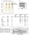
Figure 1.
Figure 1.
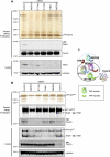
TULP3 interacts with the IFT-A complex. (A) Tandem affinity purification of the LAP (GFP-TEV/PreScission-Stag) cassette from control LAP line (without any insert), LAPTULP3 (from two stable clonal lines with varying levels of LAPTULP3), and IFT140LAP in RPE cells. Eluates were resolved on a 4%–12% Bis-Tris NuPAGE gel, and silver-stained. Stagfusion proteins in each lane are marked by arrowheads. The IFT-A complex proteins are marked based on LC MS/MS analysis of gel slices. (B) Western blot of LAP only, LAPTULP3, and IFT140LAP tandem purification eluates using indicated antibodies. The anti-THM1 antibody cross-reacts with Stagfusion proteins, and IFT140Stag protein is shown with an arrowhead. (C) Table showing the peptide spectral count and percent coverage of selected proteins in LAP only, LAPTULP3, and IFT140LAP purification eluates identified by mass spectrometry from RPE cells. (D) Table showing the homologs of IFT-A complex subunits in other organisms. (E) TULP3 partially cofractionates with the IFT-A complex protein THM1. RPE cell lysate was fractionated by size exclusion chromatography, and fractions were immunoblotted with indicated antibodies. See also Supplemental Figure S1.
Figure 2.
Figure 2.
Organization of the IFT-A complex into “core” and accessory subunits. (A) TULP3-associated proteins were purified from LAPTULP3 RPE cells as in Figure 1A, except following 72 h depletion of the indicated proteins by siRNA. Silver-stained gel (top panel) and Western blots (bottom panels) of tandem affinity purifications and total lysates are shown. The IFT-A complex proteins are labeled as in Figure 1A. (B) As in A except RPE IFT140LAP instead of LAPTULP3 cells were used. (C) A summary cartoon of the components of the IFT-A complex and their association with TULP3. The black dotted line encircles the “core” complex subunits, while the blue dashed line contains the accessory subunits. See also Supplemental Figure S2.
Figure 3.
Figure 3.
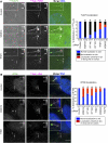
TULP3 localizes to the primary cilium dependent on the IFT-A complex. (A) RPE LAPTULP3 line A cells were transfected with siRNAs (indicated to the left of the micrograph panels) for 72 h and serum-starved for the last 24 h before fixing and staining for pericentrosomal pericentrin (PCNT, magenta), the axonemal marker Ac-tubulin (AcTub, magenta), and DNA (blue). White arrows indicate the ciliary base. Bars, 5 μm. Quantification of localization of LAPTULP3 to the cilia in similar assays is shown to the right. (*) P < 0.0001 with respect to control, using a χ2 test. (B) RPE cells were transfected with the indicated siRNAs for 72 h and serum-starved for the last 24 h before fixing and staining for IFT88 (green), pericentrosomal γ-tubulin (γTub, magenta), the axonemal marker Ac-tubulin (AcTub, magenta), and DNA (blue). White arrows indicate the ciliary base. Bars, 5 μm. Quantification and statistical significance are as in A.
Figure 4.
Figure 4.
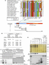
Fine mapping of the IFT-A interaction domain of TULP3. (A) A cartoon depicting different domains of TULP3 is shown below the alignments, along with the different sets of mutations evaluated and their binding properties. (B) Table showing the peptide spectral count and percent coverage of selected proteins in LAPTUB isoform b, and LAPTULP2 tandem affinity purification eluates identified by mass spectrometry. (C) Coimmunoprecipitation of IFT140 with TULP3 in HEK293T cells. Cells were cotransfected with IFT140LAP or LAP empty cassette and the indicated Myc-tagged constructs for 72 h. GFP immunoprecipitates (left panels), and total lysates (right panels) were immunoblotted for GFP and Myc tag. Cross-reacting bands in the anti-Myc blots are marked with asterisks. (D) TULP3-associated proteins were purified from RPE cells expressing different LAPTULP3 fragments, as in Figure 1A, and tandem affinity purification eluates were silver-stained (top panel) or immunoblotted (bottom panels). The IFT-A complex proteins are labeled as in Figure 1A. Arrowheads and asterisk indicate StagTULP3 full-length or fragment forms and a cross-reacting band (in the anti-THM1 blot), respectively. See also Supplemental Figure S3.
Figure 5.
Figure 5.
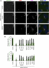
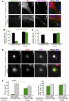
TULP3 and IFT-A coregulate localization of ciliary GPCRs. (A) RPE MCHR1GFP stable cells were transfected with the indicated siRNAs for 78 h and serum-starved for the last 30 h before fixing and staining for pericentrin (PCNT, red), Ac-tubulin (AcTub, magenta), and DNA (blue). White arrows indicate cilia. Bars, 5 μm. (B) Percentages of total cilia and GFP-positive cilia in assays similar to A were counted in two to three independent experiments with RPE MCHR1GFP and RPE SSTR3GFP stable cells. Error bars represent SEM. (*) P < 0.05; (**) P < 0.001 with respect to control in each group. See also Supplemental Figure S5.
Figure 6.
Figure 6.
TULP3 N-terminal fragment regulates localization of ciliary GPCRs. (A) RPE MCHR1GFP cells stably expressing indicated myc-tagged N-terminal TULP3 fragments were serum-starved for 30 h before fixing and immunostaining for myc (red), pericentrin (PCNT, magenta), axonemal marker Glu-tubulin (GluTub, magenta), and DNA (blue). White arrowheads and arrows indicate cilia in myc-positive cells and nonexpressing cells, respectively. Bar, 5 μm. (B) Percentages of total cilia and GFP-positive cilia in myc-positive cells in assays similar to A were counted in two independent experiments. Error bars represent SD. (*) P < 0.05. (C) RPE SSTR3GFP stable cells were transfected with the indicated myc-tagged constructs for 78 h and serum-starved for the last 30 h before fixing and staining as in A. Percentages of total cilia and GFP-positive cilia in myc-positive cells were counted in two independent experiments. Error bars represent SD. (*) P < 0.05. (D) Effect of transfecting indicated GFP-tagged constructs on endogenous Sstr3 localization in mouse primary hippocampal neurons. DIV5 primary hippocampal neurons cultured from E16 mice were transfected. Three days later, neurons were fixed and immunostained for endogenous Sstr3 (red), ACIII (magenta), and DNA (blue). White arrows indicate cilia. Bar, 10 μm. (E) Percentages of ACIII-positive cilia coexpressing Sstr3 (left) or neurons with ACIII-positive cilia (right) in transfected (GFP+) or untransfected (GFP) primary hippocampal neurons in assays similar to D were counted in three independent experiments. Error bars represent SEM. (*) P < 0.01.
Figure 7.
Figure 7.
Both IFT-A- and phosphoinositide-binding properties of TULP3 regulate GPCR-trafficking. (A) The TULP3 N terminus prevents recruitment of full-length TULP3 to an intact preformed IFT-A complex. PreScission eluates from TULP3 siRNA-treated IFT140LAP RPE cells were added to MBPTULP3 or MBPTULP3mut12 beads in the presence of HisTULP3 (1–183) or HisTULP3 (1–183) mut12. MBPTULP3 or MBPTULP3mut12-bound proteins and corresponding flowthroughs were immunoblotted for THM1 and TULP3. The anti-THM1 antibody cross-reacts with Stagfusion proteins, and allowed us to detect the IFT140Stag protein. For details, see the Supplemental Material. (B) RPE MCHR1GFP stable cells were transfected with the indicated myc-tagged TULP3 constructs and serum-starved for 30 h before fixing and immunostaining for myc, pericentrin, axonemal marker Glu-tubulin, and DNA. Percentages of total cilia and GFP-positive cilia in myc-positive cells were counted in at least three independent experiments. Error bars represent SEM. (*) P < 0.05 with respect to MCHR1GFP-positive cilia in MycTULP3 (1–183) mut12, MycTULP3, or MycTULP3mut12KR-expressing cells; (#) P < 0.05 with respect to MCHR1GFP-positive cilia in MycTULP3 (1–183) mut12-expressing cells. All other values are not significant with respect to each other. (C) Summary table of different TULP3 constructs and their effects on IFT-A binding, phosphoinositide binding, and GPCR trafficking. The mutant domains (for the image key, see D) are shown in black. (D) Model depicting the role of IFT-A and TULP3 in GPCR trafficking. IFT-A “core” complex associates with and provides ciliary access to TULP3. TULP3, in turn, is required for trafficking of certain ciliary-localized GPCRs. GPCR trafficking may be facilitated by loading of IFT-A onto preciliary vesicles (see also Sedmak and Wolfrum 2010) via the association of TULP3 with membrane phosphoinositides or novel interacting proteins. Dynein-1 also binds to the IFT-A complex, and may have roles in preciliary transport. Knocking down the IFT-A “core” complex proteins prevents TULP3 from localizing to the cilia, thereby inhibiting GPCR trafficking. Knocking down TULP3 affects ciliary localization of GPCRs, but not of IFT-A or IFT-B. Expression of the TULP3 N-terminal fragment prevents endogenous TULP3 from being recruited to the IFT-A complex, and possibly prevents loading of the IFT-A complex to the preciliary vesicles. See also Supplemental Figure S6.

Similar articles

Cited by

References

    1. Berbari NF, Lewis JS, Bishop GA, Askwith CC, Mykytyn K 2008. Bardet-Biedl syndrome proteins are required for the localization of G protein-coupled receptors to primary cilia. Proc Natl Acad Sci 105: 4242–4246 - PMC - PubMed
    1. Bishop GA, Berbari NF, Lewis J, Mykytyn K 2007. Type III adenylyl cyclase localizes to primary cilia throughout the adult mouse brain. J Comp Neurol 505: 562–571 - PubMed
    1. Blacque OE, Li C, Inglis PN, Esmail MA, Ou G, Mah AK, Baillie DL, Scholey JM, Leroux MR 2006. The WD repeat-containing protein IFTA-1 is required for retrograde intraflagellar transport. Mol Biol Cell 17: 5053–5062 - PMC - PubMed
    1. Boggon TJ, Shan WS, Santagata S, Myers SC, Shapiro L 1999. Implication of tubby proteins as transcription factors by structure-based functional analysis. Science 286: 2119–2125 - PubMed
    1. Cameron DA, Pennimpede T, Petkovich M 2009. Tulp3 is a critical repressor of mouse hedgehog signaling. Dev Dyn 238: 1140–1149 - PubMed

MeSH terms

Substances