. 2010 Dec 16;116(25):5638-49.
doi: 10.1182/blood-2010-04-281600. Epub 2010 Oct 1.

C/EBPα regulated microRNA-34a targets E2F3 during granulopoiesis and is down-regulated in AML with CEBPA mutations

Affiliations

C/EBPα regulated microRNA-34a targets E2F3 during granulopoiesis and is down-regulated in AML with CEBPA mutations

John A Pulikkan et al. Blood. .

Abstract

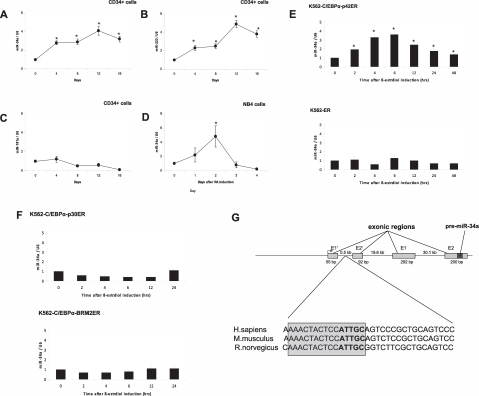
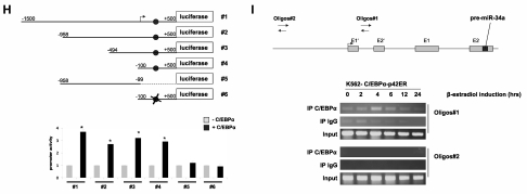
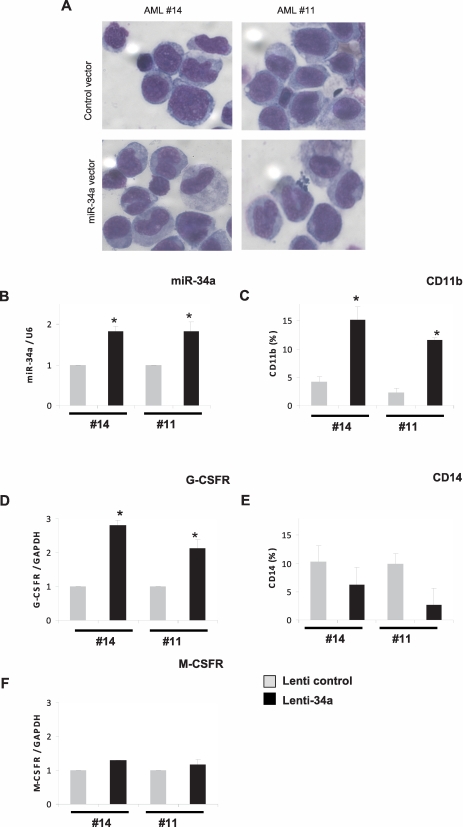
The transcription factor, CCAAT enhancer binding protein alpha (C/EBPα), is crucial for granulopoiesis and is deregulated by various mechanisms in acute myeloid leukemia (AML). Mutations in the CEBPA gene are reported in 10% of human patients with AML. Even though the C/EBPα mutants are known to display distinct biologic function during leukemogenesis, the molecular basis for this subtype of AML remains elusive. We have recently showed the significance of deregulation of C/EBPα-regulated microRNA (miR) in AML. In this study, we report that miR-34a is a novel target of C/EBPα in granulopoiesis. During granulopoiesis, miR-34a targets E2F3 and blocks myeloid cell proliferation. Analysis of AML samples with CEBPA mutations revealed a lower expression of miR-34a and elevated levels of E2F3 as well as E2F1, a transcriptional target of E2F3. Manipulation of miR-34a reprograms granulocytic differentiation of AML blast cells with CEBPA mutations. These results define miR-34a as a novel therapeutic target in AML with CEBPA mutations.


Figures

Figure 1
Figure 1
C/EBPα-p42 regulates miR-34a during granulopoiesis. (A-C) Hematopoietic CD34+ cells were cultured as discussed in “Cell cultures.” Total RNA was isolated at different time points and analyzed by quantitative real-time RT-PCR with oligos for miR-34a (A), miR-223 (B), and miR-181a (C). Data are represented as mean ± SD from 3 independent experiments. *P < .05. (D) NB4 cells were induced with retinoic acid (1μM) for respective time points. Total RNA was analyzed by quantitative real-time RT-PCR with oligos for miR-34a. Data are represented as mean ± SD from 3 independent experiments. *P < .05. (E) K562-C/EBPα-p42-ER and K562-ER cells were induced with β-estradiol (5μM) for respective time points. Total RNA was analyzed by quantitative real-time RT-PCR with oligos for miR-34a. Data are represented as mean ± SD from 3 independent experiments. *P < .05. (F) K562-C/EBPα-p30-ER and K562-C/EBPα-BRM2-ER cells were induced with β-estradiol (5μM) for respective time points. Total RNA was analyzed by quantitative real-time RT-PCR with oligos for miR-34a. Data are represented as mean ± SD from 3 independent experiments. (G) Schematic representation of the miR-34a genomic region and phylogenic conservation of genomic region in the first intron of miR-34a in humans, mice, and rats. The conserved region is shown by sequences in gray box, and the C/EBPα site is shown in bold letters. (H) Luciferase reporter assays were performed in Kasumi-6 cells using indicated reporters and C/EBPα. Cells were transfected with corresponding firefly luciferase vectors (miR-34 Promoter constructs #1 to #6, Renilla luciferase reporter construct as control vector, and C/EBPα-pCDNA3 vector. Luciferase activity was measured 24 hours later. Bars represent promoter activity for the corresponding vectors. Data are represented as mean ± SD from 3 independent experiments. *P < .05. (I) Chromatin derived from K562-C/EBPα-p42-ER cells was immunoprecipitated with anti-C/EBPα and IgG antibodies. Recovered DNA was PCR amplified with primers specific for C/EBPα-binding amplicon (oligos 1) and the nonbinding amplicon (oligos 2).
Figure 1
Figure 1
C/EBPα-p42 regulates miR-34a during granulopoiesis. (A-C) Hematopoietic CD34+ cells were cultured as discussed in “Cell cultures.” Total RNA was isolated at different time points and analyzed by quantitative real-time RT-PCR with oligos for miR-34a (A), miR-223 (B), and miR-181a (C). Data are represented as mean ± SD from 3 independent experiments. *P < .05. (D) NB4 cells were induced with retinoic acid (1μM) for respective time points. Total RNA was analyzed by quantitative real-time RT-PCR with oligos for miR-34a. Data are represented as mean ± SD from 3 independent experiments. *P < .05. (E) K562-C/EBPα-p42-ER and K562-ER cells were induced with β-estradiol (5μM) for respective time points. Total RNA was analyzed by quantitative real-time RT-PCR with oligos for miR-34a. Data are represented as mean ± SD from 3 independent experiments. *P < .05. (F) K562-C/EBPα-p30-ER and K562-C/EBPα-BRM2-ER cells were induced with β-estradiol (5μM) for respective time points. Total RNA was analyzed by quantitative real-time RT-PCR with oligos for miR-34a. Data are represented as mean ± SD from 3 independent experiments. (G) Schematic representation of the miR-34a genomic region and phylogenic conservation of genomic region in the first intron of miR-34a in humans, mice, and rats. The conserved region is shown by sequences in gray box, and the C/EBPα site is shown in bold letters. (H) Luciferase reporter assays were performed in Kasumi-6 cells using indicated reporters and C/EBPα. Cells were transfected with corresponding firefly luciferase vectors (miR-34 Promoter constructs #1 to #6, Renilla luciferase reporter construct as control vector, and C/EBPα-pCDNA3 vector. Luciferase activity was measured 24 hours later. Bars represent promoter activity for the corresponding vectors. Data are represented as mean ± SD from 3 independent experiments. *P < .05. (I) Chromatin derived from K562-C/EBPα-p42-ER cells was immunoprecipitated with anti-C/EBPα and IgG antibodies. Recovered DNA was PCR amplified with primers specific for C/EBPα-binding amplicon (oligos 1) and the nonbinding amplicon (oligos 2).
Figure 2
Figure 2
E2F3 is a direct target of miR-34a during granulopoiesis. (A) Schematic representation of miR-34a binding site in the human E2F3 3′UTR. The numbers (+2730 to +2736) represent the nucleotides relative to the termination codon of human E2F3. (B) Conservation of miR-34a binding site in E2F3 3′UTR in human, mouse, and rat genomes. (C) Sequences of predicted miR-34a binding site of E2F3. (D) Luciferase assays in Kasumi-6 cells transfected with E2F3 3′UTR constructs (wild type and mutant) and miR-34a-pCDNA. Bars represent luciferase activity for the corresponding vectors. Data are represented as mean ± SD from 3 independent experiments. *P < .05. (E) Kasumi-6 cells were transfected with control and miR-34a-pCDNA vectors. Total protein was analyzed by Western blot analysis with anti-E2F3 antibody. Values below the gel image indicate the E2F3 protein levels normalized to β-tubulin. (F) K562-C/EBPα-p42-ER, K562-ER, K562-C/EBPα-p30-ER, and K562-C/EBPα-BRM2-ER cells were induced with ß-estradiol (5μM) for respective time points. Total protein was analyzed by Western blot analysis with anti-E2F3 antibody. Values below the gel image indicate the E2F3 protein levels normalized to β-tubulin.
Figure 3
Figure 3
miR-34a functions as a tumor suppressor in AML with CEBPA mutations. (A) Quantitative real-time RT-PCR for miR-34a was carried out using bone marrow cells derived from AML patients. Values were normalized with U6. AML-NK, AML with normal karyotype; CB, cord blood; PB, peripheral blood. Data are represented as mean from 3 experiments. (B) Western blot analysis for E2F3 and E2F1 were carried out using bone marrow cells derived from AML patients. Values below the gel image indicate the E2F3 and E2F1 protein levels normalized to actin. (C) Growth curve of Kasumi-6 cells transfected with control or miR-34a.pCDNA vectors. Data are represented as mean ± SD from 3 independent experiments. (D) Flow cytometry of propidium iodide–stained Kasumi-6 cells transfected with control or miR-34a.pCDNA vectors from a representative experiment. (E) Cell-cycle profile of Kasumi-6 cells 2 days after transfection with control or miR-34a.pCDNA vectors. Data are represented as mean ± SD from 3 independent experiments. *P < .05, **P < .001.
Figure 3
Figure 3
miR-34a functions as a tumor suppressor in AML with CEBPA mutations. (A) Quantitative real-time RT-PCR for miR-34a was carried out using bone marrow cells derived from AML patients. Values were normalized with U6. AML-NK, AML with normal karyotype; CB, cord blood; PB, peripheral blood. Data are represented as mean from 3 experiments. (B) Western blot analysis for E2F3 and E2F1 were carried out using bone marrow cells derived from AML patients. Values below the gel image indicate the E2F3 and E2F1 protein levels normalized to actin. (C) Growth curve of Kasumi-6 cells transfected with control or miR-34a.pCDNA vectors. Data are represented as mean ± SD from 3 independent experiments. (D) Flow cytometry of propidium iodide–stained Kasumi-6 cells transfected with control or miR-34a.pCDNA vectors from a representative experiment. (E) Cell-cycle profile of Kasumi-6 cells 2 days after transfection with control or miR-34a.pCDNA vectors. Data are represented as mean ± SD from 3 independent experiments. *P < .05, **P < .001.
Figure 4
Figure 4
Overexpression of miR-34a in AML blast cells leads to granulopoiesis. Bone marrow cells derived from AML patients with CEBPA mutation (14 and 11 of Table 1) were transfected with control or miR-34a lenti viral vectors. Cells were cultured for 6 days and collected for morphologic, immunophenotypic, and myeloid marker expression analysis. (A) Morphologic analysis by light microscopy of Wright-Giemsa–stained AML blast cells. (B) miR-34a expression levels in blast cells transfected with lentiviral miR-34a vector, in comparison with control vector, as analyzed by real-time RT-PCR analysis. (C,E) CD11b (C) and CD14 (E) expression levels in blast cells transfected with lenti viral miR-34a vector, in comparison with control vector, as analyzed FACS analysis. (D,F) G-CSFR (D) and M-CSFR (F) expression levels in blast cells transfected with lenti viral miR-34a vector, in comparison with control vector, as analyzed by real-time RT-PCR analysis. Data are represented as mean ± SD from 3 independent experiments. *P < .05.
Figure 5
Figure 5
Schematic representation of regulation of granulopoiesis and AML by C/EBPα–miR-34a-E2F3 axis. During granulopoiesis (top panel), C/EBPα transactivates miR-34a, which, in turn, leads to E2F3 repression and inhibition of cell-cycle progression, resulting in myeloid differentiation. During CEBPA mutations in AML (bottom panel), low activity of C/EBPα fails to transactivate miR-34a, which results in lack of E2F3 inhibition. Overexpressed E2F3, together with E2F1, could accelerate myeloid cell-cycle progression and results in block of granulocytic differentiation.

Similar articles

Cited by

References

    1. Chen J, Odenike O, Rowley JD. Leukaemogenesis: more than mutant genes. Nat Rev Cancer. 2010;10(1):23–36. - PMC - PubMed
    1. Rosenbauer F, Tenen DG. Transcription factors in myeloid development: balancing differentiation with transformation. Nat Rev Immunol. 2007;7(2):105–117. - PubMed
    1. Zhang P, Iwasaki-Arai J, Iwasaki H, et al. Enhancement of hematopoietic stem cell repopulating capacity and self-renewal in the absence of the transcription factor C/EBP alpha. Immunity. 2004;21(6):853–863. - PubMed
    1. Schuster MB, Porse BT. C/EBPalpha: a tumour suppressor in multiple tissues? Biochim Biophys Acta. 2006;1766(1):88–103. - PubMed
    1. Nerlov C. C/EBPalpha mutations in acute myeloid leukaemias. Nat Rev Cancer. 2004;4(5):394–400. - PubMed

Publication types

MeSH terms