. 2010 Oct;120(10):3673-9.
doi: 10.1172/JCI42986. Epub 2010 Sep 20.

Blocking the mitochondrial apoptotic pathway preserves motor neuron viability and function in a mouse model of amyotrophic lateral sclerosis

Affiliations

Blocking the mitochondrial apoptotic pathway preserves motor neuron viability and function in a mouse model of amyotrophic lateral sclerosis

Nichole A Reyes et al. J Clin Invest. 2010 Oct.

Abstract

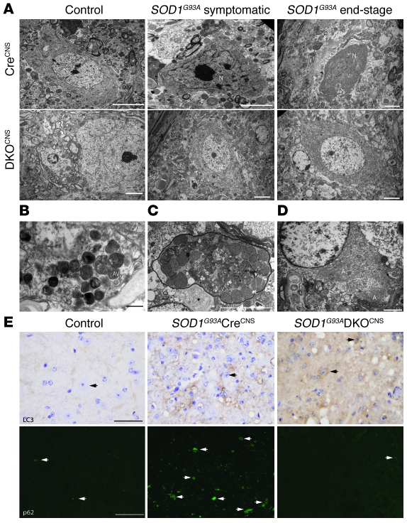
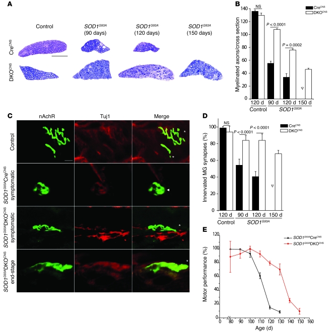
Apoptosis of motor neurons is a well-documented feature in amyotrophic lateral sclerosis (ALS) and related motor neuron diseases (MNDs). However, the role of apoptosis in the pathogenesis of these diseases remains unresolved. One possibility is that the affected motor neurons only succumb to apoptosis once they have exhausted functional capacity. If true, blocking apoptosis should confer no therapeutic benefit. To directly investigate this idea, we tested whether tissue-specific deletion in the mouse CNS of BCL2-associated X protein (BAX) and BCL2-homologous antagonist/killer (BAK), 2 proapoptotic BCL-2 family proteins that together represent an essential gateway to the mitochondrial apoptotic pathway, would protect against motor neuron degeneration. We found that neuronal deletion of Bax and Bak in a mouse model of familial ALS not only halted neuronal loss, but prevented axonal degeneration, symptom onset, weight loss, and paralysis and extended survival. These results show that motor neurons damaged in ALS activate the mitochondrial apoptotic pathway early in the disease process and that apoptotic signaling directly contributes to neuromuscular degeneration and neuronal dysfunction. Hence, inhibiting apoptosis upstream of mitochondrial permeabilization represents a possible therapeutic strategy for preserving functional motor neurons in ALS and other MNDs.


Figures

Figure 1
Figure 1. Deletion of BAX/BAK-dependent apoptosis delays symptom onset, prolongs survival, and preserves motor neurons in a mouse model of ALS.
(C and E) Control mice were harvested at 120 days of age. Symptom onset occurred at 90 days and 120 days for SOD1G93ACreCNS and SOD1G93ADKOCNS mice, respectively. End-stage occurred at 120 days and 150 days for SOD1G93ACreCNS and SOD1G93ADKOCNS mice, respectively. (A) Symptom onset of SOD1G93ACreCNS mice (111.3 ± 4.3 days) and SOD1G93ADKOCNS mice (135.6 ± 4.6 days). P < 0.0001, unpaired 2-tailed Student’s t test. n = 10. (B) Survival of SOD1G93ACreCNS mice (138.6 ± 3.8 days) and SOD1G93ADKOCNS mice (167.2 ± 8.7 days). P < 0.0001, log-rank test. n = 10. (C) Representative choline acetyltransferase staining (brown) of the anterior horn region of spinal cords from the indicated genotypes. Arrowheads indicate motor neurons. Scale bar: 200 μm. (D) Quantitation of anterior horn motor neurons from control and SOD1G93A mice using choline acetyltransferase staining. (E) Representative spinal cord anterior horn sections stained with antibody to caspase-3 (brown). Arrowheads indicate activated caspase-3 staining. Scale bar: 100 μm. (F) Numbers of apoptotic cells (positive for activated caspase-3) from control and SOD1G93A mice. The solid colors represent motor neurons, while the hatched pattern represents all other cell types. Quantification of data was analyzed via unpaired 2-tailed Student’s t test (for all quantitation data, n = 3). ψ indicates that all mice were deceased at the indicated time point.
Figure 2
Figure 2. SOD1G93ADKOCNS neurons lack morphological features of apoptosis but show evidence of autophagy.
(AD) Transmission electron microscope images of motor neurons from control and SOD1G93A mice. (A and E) Control mice were harvested at 120 days. Symptom onset [symptomatic] began at 90 days and 120 days for SOD1G93ACreCNS and SOD1G93ADKOCNS mice, respectively. End-stage began at 120 days and 150 days for SOD1G93ACreCNS and SOD1G93ADKOCNS mice, respectively. (BD) The SOD1G93ADKOCNS animal was harvested at end-stage (156 days). (A) Motor neurons from SOD1G93ACreCNS animals appear apoptotic, while motor neurons from SOD1G93ADKOCNS animals appear healthy. Scale bars: 5 μm. (B) SOD1G93ADKOCNS motor neurons display morphological features of autophagy. Scale bar: 0.5 μm. (C) SOD1G93ADKOCNS motor axons are dystrophic and contain lysosomes. Scale bar: 5 μm. (D) Increased intracellular aggregates in SOD1G93ADKOCNS motor neurons. Scale bar: 2 μm. (E) Representative LC3 (brown) and p62 (green) staining of the anterior horn region of spinal cords from control and SOD1G93A mice. Arrows indicate motor neurons (top row) and positive p62 staining (bottom row). Scale bar: 100 μm (top row); 50 μm (bottom row). A, aggregates; AV, autophagic vesicle; L, lysosome; M, mitochondria; N, nucleus.
Figure 3
Figure 3. Deletion of BAX/BAK preserves neuronal function.
(A and C) Control, symptomatic, and end-stage are as defined as in the legend for Figure 2. (A) Representative ventral root sections from control and SOD1G93A mice stained with toluidine blue. Scale bar: 200 μm. (B) Quantitation of myelinated ventral root axons from control and SOD1G93A mice. n = 3. (C) Representative neuromuscular junction images from control and SOD1G93A mice stained with FITC-conjugated α-bungarotoxin (green) and antibody to Tuj1 (red). Asterisks and arrowheads indicate fully innervated and partially innervated neuromuscular junctions, respectively. Notice the rounded, degenerated appearance of the SOD1G93ACreCNS symptomatic neuromuscular junction. nAchR, nicotinic acetylcholine receptor. Scale bar: 8 μm. (D) Percentage of innervated synapses in control and SOD1G93A mice as quantified from neuromuscular junction staining. n = 2. (E) Percentage of the longest rotarod performance by each mouse (n = 3 for each group). P < 0.05 via ANOVA. (B and E) Data were analyzed using unpaired 2-tailed Student’s t test. ψ indicates that all mice were deceased at the indicated time point.

References

    1. Bredesen DE. Programmed cell death mechanisms in neurological disease. Curr Mol Med. 2008;8(3):173–186. doi: 10.2174/156652408784221315. - DOI - PubMed
    1. Trushina E, McMurray CT. Oxidative stress and mitochondrial dysfunction in neurodegenerative diseases. NeuroScience. 2007;145(4):1233–1248. doi: 10.1016/j.neuroscience.2006.10.056. - DOI - PubMed
    1. Ellerby LM, et al. Kennedy’s disease: caspase cleavage of the androgen receptor is a crucial event in cytotoxicity. J Neurochem. 1999;72(1):185–195. doi: 10.1046/j.1471-4159.1999.0720185.x. - DOI - PubMed
    1. Sathasivam S, Shaw PJ. Apoptosis in amyotrophic lateral sclerosis--what is the evidence? Lancet Neurol. 2005;4(8):500–509. doi: 10.1016/S1474-4422(05)70142-3. - DOI - PubMed
    1. Tsai MS, Chiu YT, Wang SH, Hsieh-Li HM, Lian WC, Li H. Abolishing Bax-dependent apoptosis shows beneficial effects on spinal muscular atrophy model mice. Mol Ther. 2006;13(6):1149–1155. doi: 10.1016/j.ymthe.2006.02.008. - DOI - PubMed

Publication types

MeSH terms