. 2010 Nov;42(11):1015-20.
doi: 10.1038/ng.683. Epub 2010 Oct 3.

Mutations in WDR62, encoding a centrosome-associated protein, cause microcephaly with simplified gyri and abnormal cortical architecture

Affiliations

Mutations in WDR62, encoding a centrosome-associated protein, cause microcephaly with simplified gyri and abnormal cortical architecture

Timothy W Yu et al. Nat Genet. 2010 Nov.

Abstract

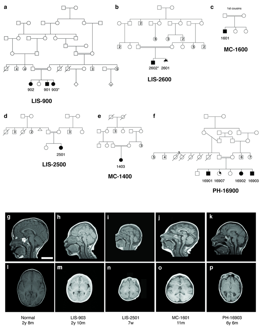
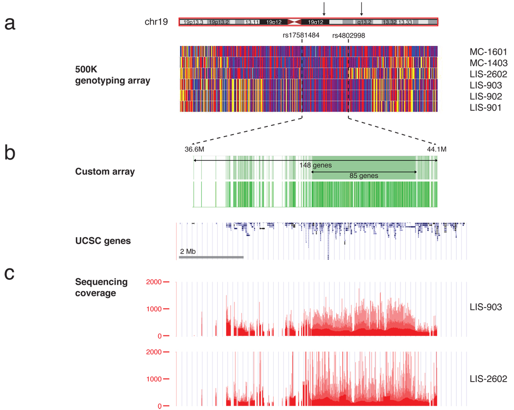
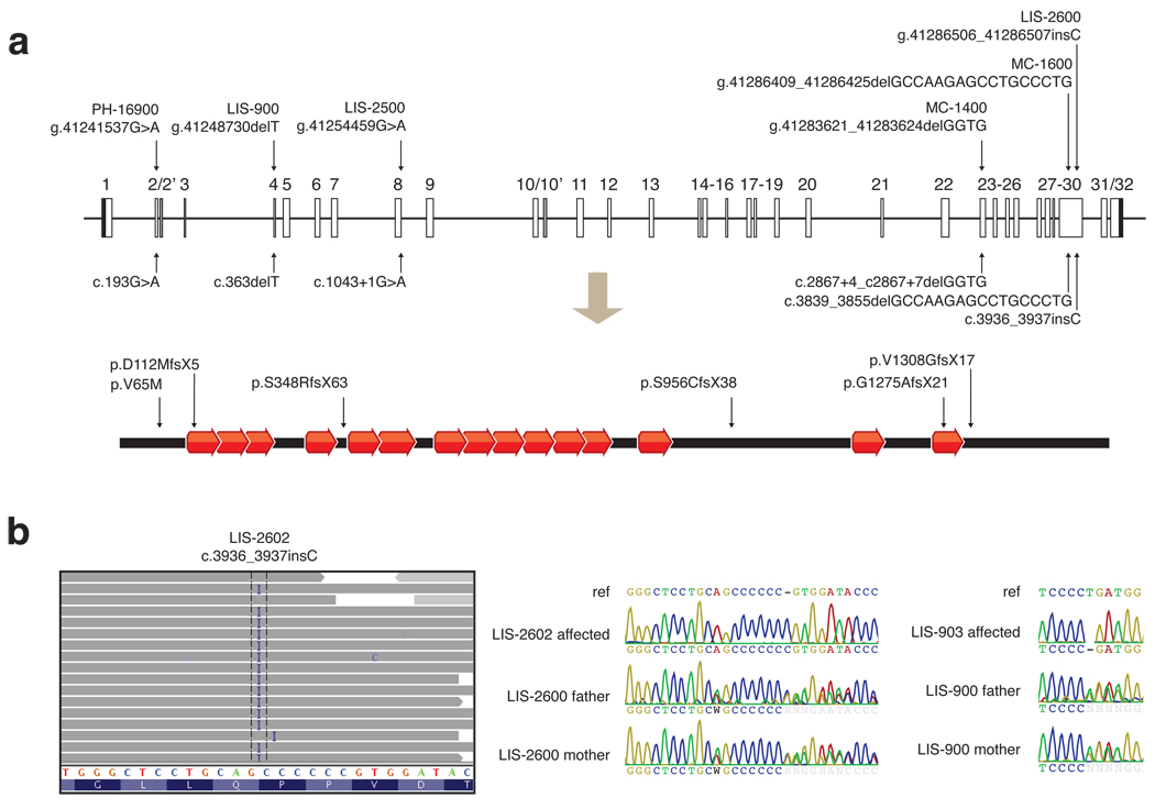
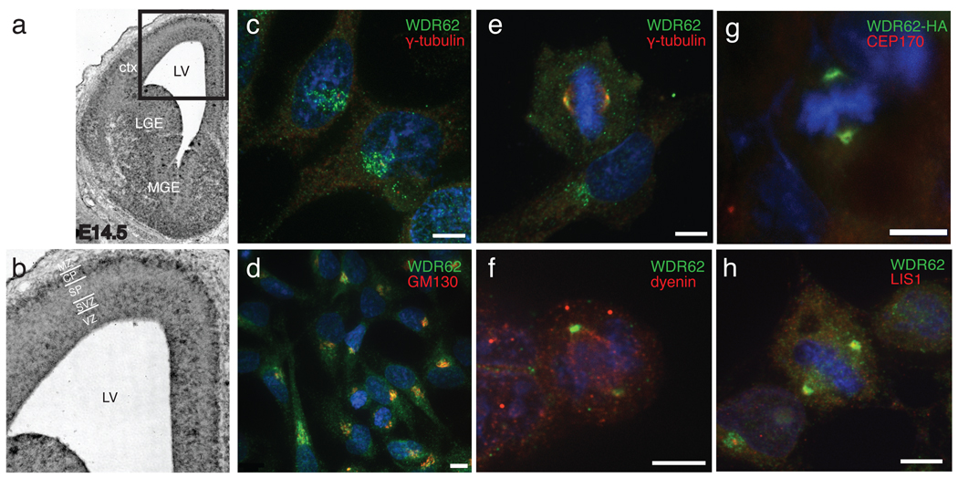
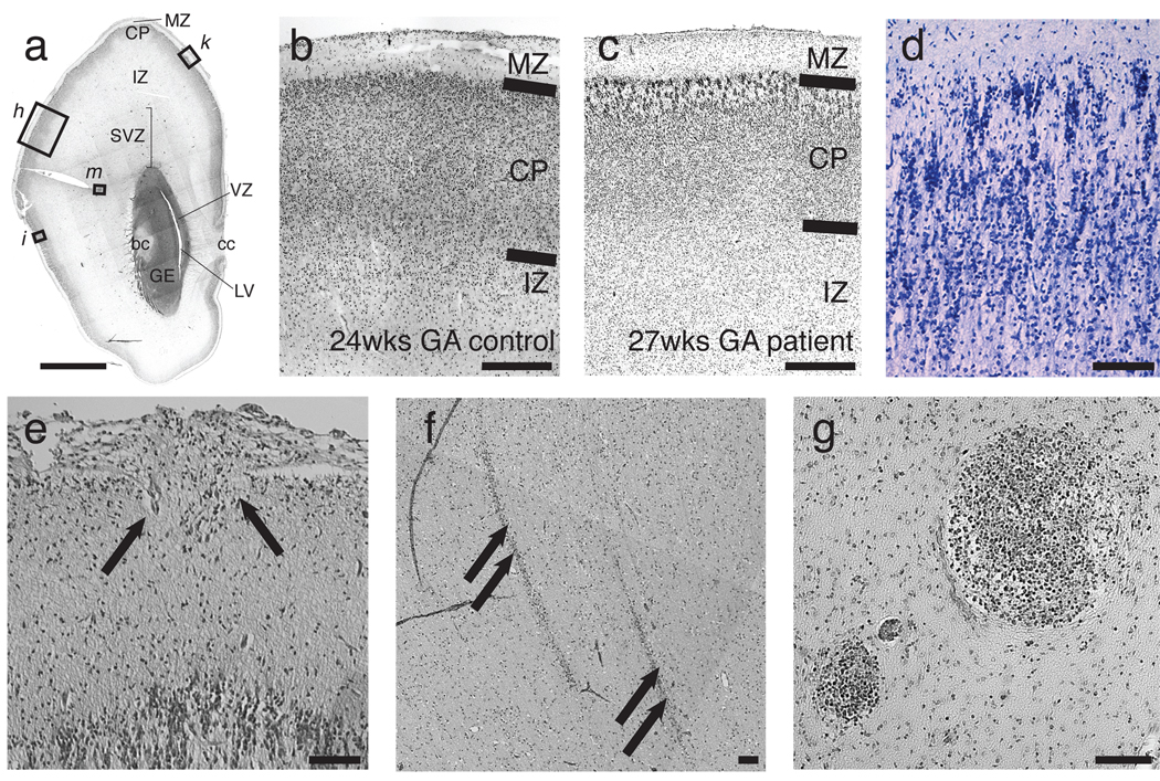
Genes associated with human microcephaly, a condition characterized by a small brain, include critical regulators of proliferation, cell fate and DNA repair. We describe a syndrome of congenital microcephaly and diverse defects in cerebral cortical architecture. Genome-wide linkage analysis in two families identified a 7.5-Mb locus on chromosome 19q13.12 containing 148 genes. Targeted high throughput sequence analysis of linked genes in each family yielded > 4,000 DNA variants and implicated a single gene, WDR62, as harboring potentially deleterious changes. We subsequently identified additional WDR62 mutations in four other families. Magnetic resonance imaging and postmortem brain analysis supports important roles for WDR62 in the proliferation and migration of neuronal precursors. WDR62 is a WD40 repeat-containing protein expressed in neuronal precursors as well as in postmitotic neurons in the developing brain and localizes to the spindle poles of dividing cells. The diverse phenotypes of WDR62 suggest it has central roles in many aspects of cerebral cortical development.


Figures

Figure 1
Figure 1
Pedigrees and radiographic findings in six consanguineous families with microcephaly and simplified gyri (MCSG). (a–f) Shaded symbols denote affected individuals. (a) LIS-900, a Mexican family with three affected children. (b) LIS-2600, a Turkish family with two affected children. (c) LIS-2500, (d) MC-1400, and (e) MC-1600, three Turkish families (not known to be related to LIS-2600 or each other). (f) PH-16900, a Saudi family with three affected children; a fourth child (partial shading) had mild speech delay and articulation and attention difficulties but no brain abnormalities. Whole blood DNA was obtained and analyzed from each nuclear family, with the exception of LIS-2601, who died as a fetus. Asterisks denote the two individuals chosen for targeted high-throughput sequencing. (g–p) MRI features of patients with MCSG, demonstrating the breadth of cortical phenotypes associated with WDR62. Mid-sagittal T1 sections (g–k) and axial T1 sections at the level of the insula (l–p) are shown for LIS-903 (h,m), LIS-2501 (i,n), MC-1601 (j,o), and PH-16903 (k, p) individuals, as well as a control individual (g, l) age-matched to LIS-903. Common findings include microcephaly (h–k, m–p), anomalies of the corpus callosum (small splenium in h and k, absent splenium in i, thick body in k), simplification of the normal gyral pattern (h, i, k, m, n, p) as well as more variable features such as mild asymmetries of cortical size (p), possible heterotopia and cortical clefts (not shown, see Supplementary Note, Clinical Data). Scale bar = 5 cm.
Figure 2
Figure 2
Mapping, capture, and sequencing of 148 genes in the MCSG locus. (a) Homozygosity analysis in four pedigrees (LIS-900, LIS-2600, MC-1400 and MC-1600) revealed a 7.5 Mb interval on chr19, bounded by SNP markers rs17581484 and rs4802998. Homozygous SNPs are shown in red or blue, heterozygous SNPs are shown in yellow, and SNPs for which no genotype could be assigned are shown in white. The homozygous region contained 148 annotated UCSC genes. (b) Custom Nimblegen microarrays were designed to target all coding and noncoding regions of the 85 genes in the center of the linkage peak and all exonic regions of the remaining 63 genes (upper green track; the location of probes on the array is shown on the lower track). These were used to capture genomic DNA and generate sequencing libraries from two individuals, LIS-903 and LIS-2602. (c) Libraries were sequenced on a Illumina GA II to an average depth of >200X and completeness of 88–90% (bases covered by >= 10 reads). Depth of sequence coverage over the region is shown.
Figure 3
Figure 3
Six WDR62 mutations reported in association with microcephaly with simplified gyri. (a) Alterations are shown in genomic, coding DNA, and protein contexts. The human WDR62 gene consists of 32 exons shown as boxes, and encodes a protein of 1518 amino acids containing 15 WD40 repeats. Black shaded boxes represent untranslated regions, open boxes represent coding regions, and gray shaded boxes represent alternatively spliced exons. Lines connecting boxes represent introns. The diagram is drawn to scale. Five of the six alleles (from families LIS-900, LIS-2500, LIS-2600, MC-1400, MC-1600, and LIS-2600) disrupt splice sites or cause frameshifts resulting in protein truncations and are likely nulls. The sixth allele, found in PH-16900, is a missense alteration of V65M, a conserved residue. (b) Illustration of the c.3936_3937insC mutation in LIS-2602 by high throughput sequencing, and representative Sanger traces confirming proper segregation. High throughput sequencing data is shown using the Integrated Genome Viewer. Aligned reads are shown as gray tags shaded by quality score, SNPs are identified by the letter code of the substituted base, and the position of the LIS-2602 single basepair insertion is denoted by the letter I. Representative Sanger traces confirm this change in the affected individual and show that both parents are heterozygous for the insertional event. Similarly, representative Sanger traces illustrate the c.363delT mutation in LIS-903 and the carrier status of both parents.
Figure 4
Figure 4
WDR62 expression in developing mouse brain and subcellular localization. (a) in situ hybridization of E14.5 mouse brain with antisense probe to mouse WDR62. Sense strand (not shown) showed no specific hybridization. (b) Higher power view. Strong WDR62 message is seen in the ventricular zone, subventricular zone, ventral portion of the intermediate zone, and ganglionic eminences, with some hybridization in the cortical plate as well. (c–h) Confocal microscopy demonstrating WDR62 subcellular localization. (c, d) endogenous WDR62 localization in interphase HeLa cells. (c) anti-WDR62 (green), also stained with anti γ-tubulin (red) and Hoechst for DNA (blue), showing perinuclear localization, surrounding but not overlapping the centrosome. (d) co-staining of interphase HeLa cells with anti-WDR62 and anti-GM130 shows localization of both to the Golgi apparatus near the Hoechst-positive nucleus. (e–g) WDR62 localization in HeLa and HEK cells during M phase. (e) Endogenous WDR62 localizes to the spindle poles in M phase HeLa cells, visualized by double labeling with γ-tubulin. (f) Co-staining with antisera to WDR62 and dynein in HEK cells shows WDR62 at the spindle poles and dynein throughout the spindle. (g) Transfection of C-terminal HA-tagged WDR62 in HEK cells confirms ring-like localization around the centrosome and overlaps with CEP170, another centrosomal protein. (h) Endogenous WDR62 immunoreactivity surrounds LIS1 but does not overlap fully.
Figure 5
Figure 5
Histopathologic analysis of a 27 week human fetus with MCSG. (a) H&E stained coronal section from the forebrain of a 27 week gestational age fetus with microcephaly with simplified gyri (LIS-2601). Section is at the rostral end of the caudate along the A–P axis. Locations of higher magnification views for additional panels are indicated. (b, c) H&E stained section demonstrating cortical layering. Compared to a (b) 24 week gestational age control fetus, (c) LIS-2601 exhibits an abnormally thin cortical plate (despite being three weeks older), suggestive of a proliferative defect and absence of normal appearing layer II/III cells underneath the molecular layer. The dimensions of the marginal zone are relatively preserved. (d) In addition to a smaller cortical plate, neurons in the cortical plate display abnormal persistence of a radial columnar pattern and disorganized clustering (Kluver-Barrera stain). (e) Occasionally, eruptions of neuroglial cells (arrows) through the pial surface into the subarachnoid space are seen, as are occasional (f) streaky heterotopia in the intermediate zone, and (g) clusters of small, darkly staining cells in the outer subventricular zone that resemble dividing cells. Abbreviations: IZ, intermediate zone; GE, ganglionic eminence; LV, lateral ventricle; bc, body of the caudate; cc, corpus callosum; ctx, cortex; LGE, lateral ganglionic eminence; MGE, medial ganglionic eminence; MZ, marginal zone; CP, cortical plate; SP, subplate; SVZ, subventricular zone; VZ, ventricular zone.

Comment in

References

    1. Sergi C, Zoubaa S, Schiesser M. Norman-Roberts syndrome: prenatal diagnosis and autopsy findings. Prenat Diagn. 2000;20:505–509. - PubMed
    1. Albert TJ, et al. Direct selection of human genomic loci by microarray hybridization. Nat Methods. 2007;4:903–905. - PubMed
    1. Li H, Durbin R. Fast and Accurate Short Read Alignment with Burrows-Wheeler Transform. Bioinformatics. 2009 - PMC - PubMed
    1. Sherry ST, et al. dbSNP: the NCBI database of genetic variation. Nucleic Acids Research. 2001;29:308–311. - PMC - PubMed
    1. Desmet F-O, et al. Human Splicing Finder: an online bioinformatics tool to predict splicing signals. Nucleic Acids Res. 2009;37:e67. - PMC - PubMed

Publication types

MeSH terms

Associated data