. 2010 Nov;42(11):1010-4.
doi: 10.1038/ng.682. Epub 2010 Oct 3.

WDR62 is associated with the spindle pole and is mutated in human microcephaly

Affiliations

WDR62 is associated with the spindle pole and is mutated in human microcephaly

Adeline K Nicholas et al. Nat Genet. 2010 Nov.

Abstract

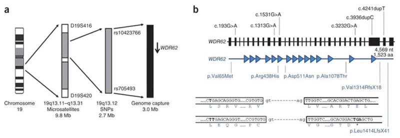
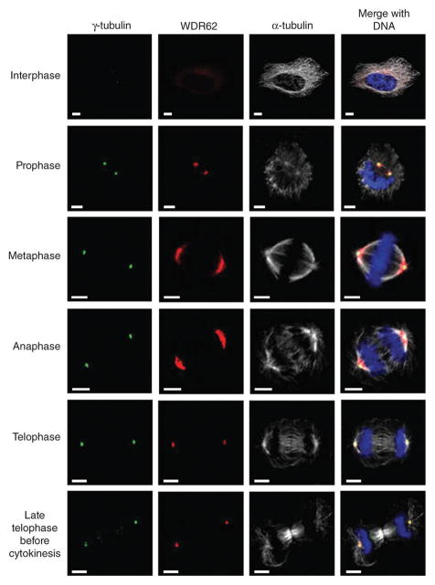
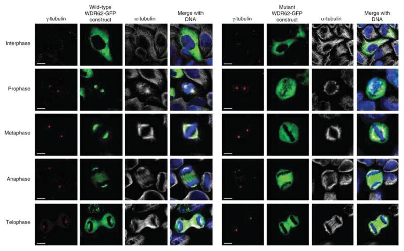
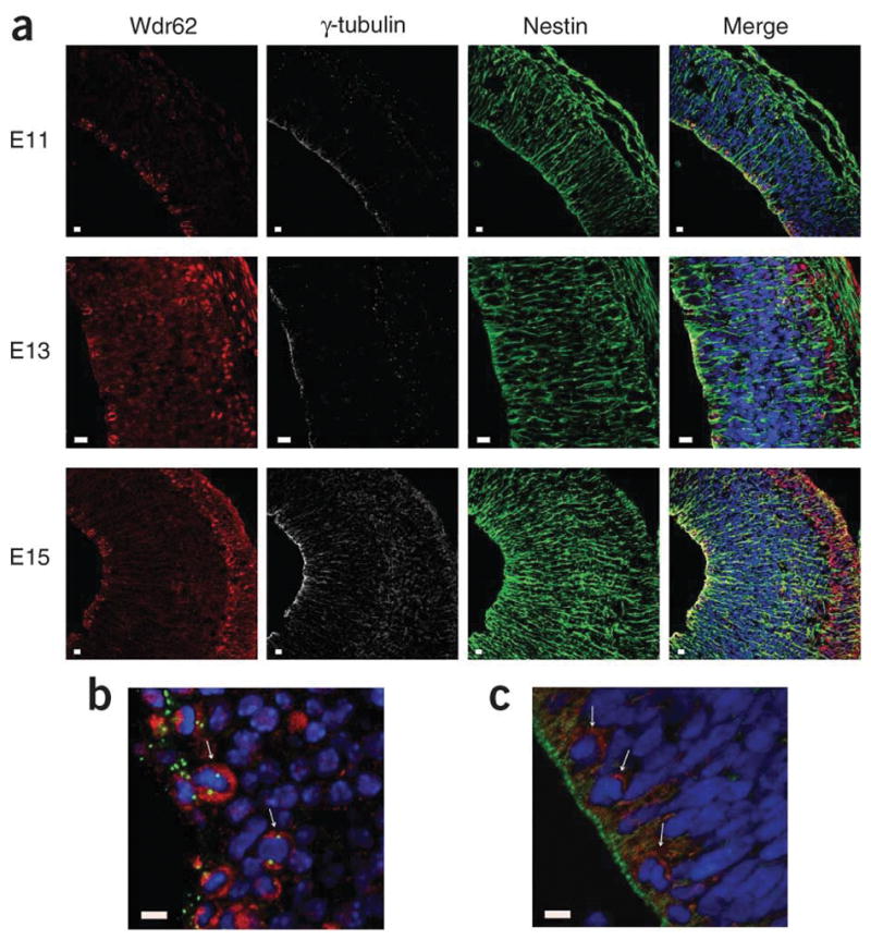
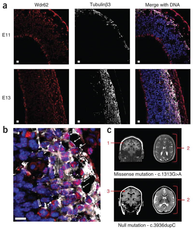
Autosomal recessive primary microcephaly (MCPH) is a disorder of neurodevelopment resulting in a small brain. We identified WDR62 as the second most common cause of MCPH after finding homozygous missense and frame-shifting mutations in seven MCPH families. In human cell lines, we found that WDR62 is a spindle pole protein, as are ASPM and STIL, the MCPH7 and MCHP7 proteins. Mutant WDR62 proteins failed to localize to the mitotic spindle pole. In human and mouse embryonic brain, we found that WDR62 expression was restricted to neural precursors undergoing mitosis. These data lend support to the hypothesis that the exquisite control of the cleavage furrow orientation in mammalian neural precursor cell mitosis, controlled in great part by the centrosomes and spindle poles, is critical both in causing MCPH when perturbed and, when modulated, generating the evolutionarily enlarged human brain.


Conflict of interest statement

COMPETING FINANCIAL INTERESTS

The authors declare no competing financial interests.

Figures

Figure 1
Figure 1
A summary of the linkage strategy used to define the MCPH2 region and mutations found in WDR62 in MCPH2 families. (a) From left to right, chromosome 19, shown as a G-banded cartoon; the initial linkage region defined by homozygous microsatellite markers; the final minimal linkage region defined by homozygous SNPs; the region subject to genome capture (slightly larger and overlapping the minimal linkage region); and WDR62 shown as an arrow pointing from 5′ _to 3′. Critical defining heterozygous markers are shown that bound each defined region. (b) WDR62 is shown from 5′ _to 3′, from left to right. Exons are shown to scale and introns are shown as an artificial fixed interval for clarity. The position of each homozygous mutation is shown. The lengths of the gene and protein are given. Below the gene, the WDR62 protein is shown with each WD repeat detected by PFAM, shown as a filled in triangle. The position that each DNA mutation affects the WDR62 protein is shown beneath, with indication of the resultant amino acid or peptide change. For the c.4241dupT mutation (resulting in the p.Leu1414LeufsX41 alteration), an explanatory cartoon is shown at the bottom of the figure. This mutation causes a frame shift in the penultimate exon of WDR62, which does not give rise to a new stop codon until the terminal exon. After the frame shift, 17 novel amino acids were found, and those amino acids at the start of the frame shift, at the splice sites and at the position of the new premature stop codon are shown.
Figure 2
Figure 2
Subcellular localization of WDR62 throughout the cell cycle. Confocal microscopy analysis of HeLa cells during each stage of the cell cycle. WDR62 staining is weak and cytoplasmic during interphase, with a concentration at a perinuclear position suggestive of the Golgi apparatus, and shows spindle pole localization during mitosis. Cells were stained with antibodies against human WDR62 (red), γ-tubulin (green) as a centrosome marker, α-tubulin (white) as a microtubule marker, and DNA (blue) stained with DAPI. Scale bar, 5 μm.
Figure 3
Figure 3
Overexpression of WDR62-GFP wild type and c.1313G>A ( p.Arg438His) mutant constructs in HeLa cells. The first 4.5 panel of images (on the left) shows the results for the wild-type WDR62-GFP construct, whereas the second 4 . 5 panel of images (on the right) shows the comparable results for the c.1313G>A (p.Arg438His) missense mutation in the WDR62-GFP construct. The wild-type WDR62-GFP protein localized to the spindle pole during mitosis (in 50 of 50 cells analyzed), paralleling the localization found for the endogenous wild type protein. Conversely, in cells expressing the mutant construct, there was no spindle pole accumulation of GFP during mitosis (in 50 of 50 cells analyzed). Cells were stained with γ-tubulin (red) as a centrosome marker and α-tubulin (white) identifying microtubules and the mitotic spindle. Both WDR62-GFP (green) proteins were directly visualized with DNA (blue) stained with DAPI. (See Supplementary Fig. 4 for the results for the c.4241dupT mutation). Scale bar, 5 μm.
Figure 4
Figure 4
Endogenous expression pattern of WDR62 in human and mouse embryonic brain. (a) Wdr62 expression in mouse cerebral cortex neuroepithelium from early to late neurogenesis: E11, E13 and E15. Wdr62 is seen to concentrate in the cytoplasm of mitotic apical and basal neural precursor cells. In the cortical plate containing newly born neurons, seen after E13, Wdr62 is also found in the nucleus of newly born neurons. (b) A magnified image of mouse E13 neuroepithelium clearly showing Wdr62 cytoplasmic aggregation in apical mitotic precursors. The arrows indicate examples of apical neural precursor cells undergoing mitosis with a pair of centrosomes on either side of a metaphase DNA plate. In the neuroepithelium, Wdr62 expression is only clearly seen in cells undergoing mitosis. (c) A magnified image of human CS22 cerebral cortex neuroepithelium showing WDR62 cytoplasmic aggregation in apical mitotic precursors. Arrows indicate cells in which WDR62 expression can be most clearly seen, which by their position and nuclear morphology are likely to be undergoing mitosis. Due to collection, fixation and subsequent paraffin embedding, immunohistochemistry is more difficult in human embryonic brain sections; however, the apically positioned centrosomes of the apical neural precursors are clearly visible. For each set of images, the result are shown for WDR62 and Wdr62 (red), with γ-tubulin (white) as a centrosome marker, Nestin (green) marking apical neural precursors in the cytoplasm and DNA (blue) stained with DAPI. Scale bars, 10 μm.
Figure 5
Figure 5
Wdr62 expression in newborn, newly arrived cortical neurons in the developing cerebral cortex and brain imaging from two individuals with WDR62 mutations. (a) Newborn neuron data. Mouse cerebral cortex neuroepithelium in early and later neurogenesis stages E11 and E13 stained with antibodies to Wdr62 (red) and costained against the neuronal marker protein Tubulinβ3 (white) with DNA (blue) stained with DAPI. The apical ventricular margin of the neuroepithelium is shown on the left of each image; on the right of each image is the outer, pial surface of the developing cerebral cortex. The developing cerebral cortex is bounded by Tubulinβ3 staining to the right of each image. Localization of Wdr62 to the developing cortical plate can be seen at the right of each image. An outermost Wdr62 staining is also seen, beyond the cerebral cortex, which represents staining of pial membrane cells. Scale bars, 10 μm. (b) An enlargement of an E13 merged image allowing easier visualization of neurons staining with Tubulinβ3 (white). The single arrows indicate neurons in which Wdr62 staining is nuclear. Also visible are cells, indicated by double arrows, undergoing mitosis in the subventricular zones with prominent Wdr62 staining in the cytoplasm, which are assumed to be basal or intermediate neural progenitors. (c) MRI images of individuals with MCPH2 from this study with homozygous WDR62 mutations; the c.1313G>A missense mutation is shown above and the protein truncating c.3936dupC mutation is shown below. The annotation in red indicates: 1, cortex normal thickness (3–4 mm) but having an indistinct border; 2, diffuse simplified gyral pattern with frontal cortex most severely affected (this feature is more marked for the null mutation); and 3, cortex appears mildly thickened (~5 mm).

Comment in

References

    1. Mochida GH, Walsh CA. Genetic basis of developmental malformations of the cerebral cortex. Arch Neurol. 2004;61:637–640. - PubMed
    1. Woods CG, Bond J, Enard W. Autosomal recessive primary microcephaly (MCPH): a review of clinical, molecular, and evolutionary findings. Am J Hum Genet. 2005;76:717–728. - PMC - PubMed
    1. Bond J, et al. ASPM is a major determinant of cerebral cortical size. Nat Genet. 2002;32:316–320. - PubMed
    1. Pfaff KL, et al. The zebra fish cassiopeia mutant reveals that SIL is required for mitotic spindle organization. Mol Cell Biol. 2007;27:5887–5897. - PMC - PubMed
    1. Bond J, Woods CG. Cytoskeletal genes regulating brain size. Curr Opin Cell Biol. 2006;18:95–101. - PubMed

Publication types