. 2010 Nov;11(11):868-75.
doi: 10.1038/embor.2010.134. Epub 2010 Oct 1.

The protein phosphatase 1 regulator PNUTS is a new component of the DNA damage response

Affiliations

The protein phosphatase 1 regulator PNUTS is a new component of the DNA damage response

Helga B Landsverk et al. EMBO Rep. 2010 Nov.

Abstract

The function of protein phosphatase 1 nuclear-targeting subunit (PNUTS)--one of the most abundant nuclear-targeting subunits of protein phosphatase 1 (PP1c)--remains largely uncharacterized. We show that PNUTS depletion by small interfering RNA activates a G2 checkpoint in unperturbed cells and prolongs G2 checkpoint and Chk1 activation after ionizing-radiation-induced DNA damage. Overexpression of PNUTS-enhanced green fluorescent protein (EGFP)--which is rapidly and transiently recruited at DNA damage sites--inhibits G2 arrest. Finally, γH2AX, p53-binding protein 1, replication protein A and Rad51 foci are present for a prolonged period and clonogenic survival is decreased in PNUTS-depleted cells after ionizing radiation treatment. We identify the PP1c regulatory subunit PNUTS as a new and integral component of the DNA damage response involved in DNA repair.


Conflict of interest statement

The authors declare that they have no conflict of interest.

Figures

Figure 1
Figure 1
Depletion of protein phosphatase 1 nuclear-targeting subunit induces a checkpoint-dependent delay at the G2–M transition. PNUTS-depleted (siPNUTS) and control HeLaH2B-EGFP cells transfected with a scrambled oligonucleotide (Scr) monitored through mitosis by live cell imaging. (A) Chart showing average time spent in indicated stages of mitosis. Difference in prophase, P<0.0001; differences in metaphase and telophase, P<0.05. (B) Representative siRNA-transfected cells in prophase. Numbers indicate time in minutes before nuclear envelope breakdown. (C) Frequency distribution of prophase duration for Scr and siPNUTS cells. For control cells, median, 18 min; 95% CI, 18–21 min. For PNUTS-depleted cells, median, 48; 95% CI, 42–56 min. (D) Mean condensation (±s.d.; log scale) kinetics for 14 Scr cells and 15 siPNUTS cells. (E) siRNA-transfected HeLa cells grown in the presence of 1 μg/ml nocodazole alone (Noco) or in the presence of 2 mM caffeine or 2.5 μM SB218078 for 1 h. Mitotic cells were labelled with pH3S10 antibodies. Difference between Scr and siPNUTS for caffeine, P<0.05 and for SB218078, P<0.005. (F) Average median pH3S10 levels from three independent experiments as in (E). CI, confidence interval; PNUTS, protein phosphatase 1 nuclear-targeting subunit; siRNA, small interfering RNA.
Figure 2
Figure 2
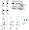
Protein phosphatase 1 nuclear-targeting subunit negatively affects G2 arrest after ionizing radiation treatment. (A) Flow cytometry analysis of siRNA-transfected HeLa cells untreated (Exp) or 24 h after irradiation with 5, 7.5 or 10 Gy IR. (B) Western blot using indicated antibodies of siRNA-transfected cells collected after 2 and 3 days or treated with 10 Gy IR at 2 days and collected after 2, 6 and 24 h. (C) Flow analysis of gated GFP-negative, PNUTS–EGFP- or PNUTS(W401A)–EGFP-expressing cells either untreated (Exp) or 20 h after 5 Gy IR in the absence or presence of 1 μg/ml nocodazole (noco) added 2 h after IR treatment. IR treatment was carried out 4 h after transfection. G2/M gating is indicated. (D) Average relative percentage of cells in G2 from (C) determined as the ratio of cells in G2–M after IR over cells in G2/M after IR and nocodazole treatment. Data from two independent experiments. Differences between PNUTS–EGFP and EGFP-negative and between PNUTS(W401A)–EGFP and EGFP-negative cells were at the P<0.01 level for both. EGFP, enhanced green fluorescent protein; IR, ionizing radiation; PNUTS, protein phosphatase 1 nuclear-targeting subunit; siRNA, small interfering RNA.
Figure 3
Figure 3
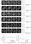
Protein phosphatase 1 nuclear-targeting subunit–enhanced green fluorescent protein is rapidly and transiently recruited to sites of double-strand breaks in the nucleus. (AE) A single spot of (A,B) nuclear PNUTS–EGFP, (C,D) PNUTS(W401A)–EGFP, (F) GFP–53BP1 or (E) NIPP1–EGFP was bleached with a 500-ms pulse at 405 nm (A,C,E,F) or 488 nm (B,D), and fluorescence intensity in the bleached area was monitored over time. Graphs on the right-hand side show data points for individual cells. Scale bar, 10 μm. (G,H) Superposition of recruitment dynamics of indicated constructs. Measured intensities were normalized to the values immediately before bleaching (set to 1) and plotted against time. Average results from several experiments as indicated in Table 2. 53BP1, p53-binding protein 1; EGFP, enhanced green fluorescent protein; NIPP1, nuclear inhibitor of PP1; PNUTS, protein phosphatase 1 nuclear-targeting subunit; wt, wild type.
Figure 4
Figure 4
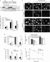
Protein phosphatase 1 nuclear-targeting subunit depletion prolongs the presence of DNA damage markers. (A) Western blot using indicated antibodies of siRNA-transfected cells collected after 2 and 3 days or treated with 10 Gy IR at 2 days and collected after 2, 6 and 24 h. (B) Average median γH2AX levels from flow cytometry analysis of γH2AX labelling compared with DNA content of siRNA-transfected cells 3 days after transfection or treated with 5 Gy IR and collected after 90 min or 24 h. (C) Immunofluorescence analysis of siRNA-transfected cells 3 days after transfection or collected at indicated times after 5 Gy IR using γH2AX antibodies. Scale bar, 20 μm. (D) Average percentage of cells with more than 10 γH2AX foci from (C). (E) Flow cytometry analysis 24 h after transfection of PNUTS–EGFP or PNUTS(W401A)–EGFP in cells left untreated (Exp), or treated with 5 Gy IR 4 h after transfection (IR). Average median γH2AX levels from gated EGFP-expressing or GFP-negative cells. (F) Percentage of cells with more than 10 53BP1, Rad51 or RPA foci 24 h after 5 Gy IR from immunofluorescence analysis as in (C). Average data from two (53BP1, RAD51) or three (RPA) experiments are shown. *P<0.05, **P<0.005. (G) Clonogenic survival assays showing survival fraction of Scr and siPNUTS cells as a function of radiation dose (Gy). Average data from three experiments. 53BP1, p53-binding protein; EGFP, enhanced green fluorescent protein; IR, ionizing radiation; PNUTS, protein phosphatase 1 nuclear-targeting subunit; RPA, replication protein A; Scr, scrambled oligonucleotide; siRNA, small interfering RNA.

References

    1. Allen PB, Kwon YG, Nairn AC, Greengard P (1998) Isolation and characterization of PNUTS, a putative protein phosphatase 1 nuclear targeting subunit. J Biol Chem 273: 4089–4095 - PubMed
    1. Ayoub N, Jeyasekharan AD, Bernal JA, Venkitaraman AR (2008) HP1-beta mobilization promotes chromatin changes that initiate the DNA damage response. Nature 453: 682–686 - PubMed
    1. Bekker-Jensen S, Lukas C, Kitagawa R, Melander F, Kastan MB, Bartek J, Lukas J (2006) Spatial organization of the mammalian genome surveillance machinery in response to DNA strand breaks. J Cell Biol 173: 195–206 - PMC - PubMed
    1. Bollen M, Gerlich DW, Lesage B (2009) Mitotic phosphatases: from entry guards to exit guides. Trends Cell Biol 19: 531–541 - PubMed
    1. De Leon G, Sherry TC, Krucher NA (2008) Reduced expression of PNUTS leads to activation of Rb-phosphatase- and caspase-mediated apoptosis. Cancer Biol Ther 7: 833–841 - PubMed

Publication types

MeSH terms