. 2010 Sep 15:2:26.
doi: 10.3389/fnene.2010.00026. eCollection 2010.

High resolution measurement of the glycolytic rate

Affiliations

High resolution measurement of the glycolytic rate

Carla X Bittner et al. Front Neuroenergetics. .

Abstract

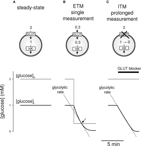
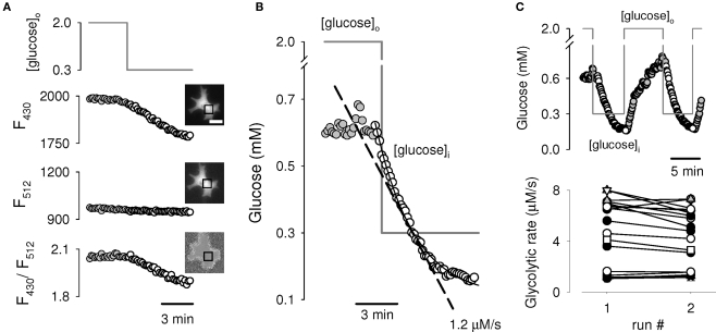
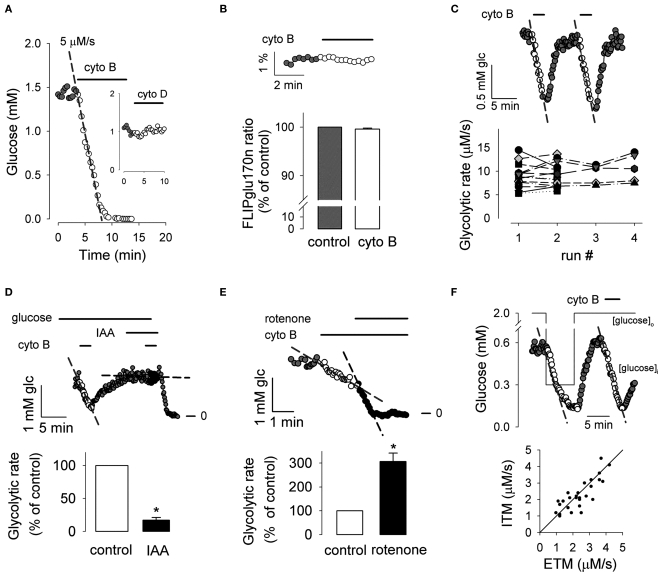
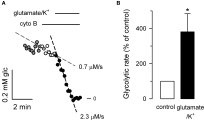
The glycolytic rate is sensitive to physiological activity, hormones, stress, aging, and malignant transformation. Standard techniques to measure the glycolytic rate are based on radioactive isotopes, are not able to resolve single cells and have poor temporal resolution, limitations that hamper the study of energy metabolism in the brain and other organs. A new method is described in this article, which makes use of a recently developed FRET glucose nanosensor to measure the rate of glycolysis in single cells with high temporal resolution. Used in cultured astrocytes, the method showed for the first time that glycolysis can be activated within seconds by a combination of glutamate and K(+), supporting a role for astrocytes in neurometabolic and neurovascular coupling in the brain. It was also possible to make a direct comparison of metabolism in neurons and astrocytes lying in close proximity, paving the way to a high-resolution characterization of brain energy metabolism. Single-cell glycolytic rates were also measured in fibroblasts, adipocytes, myoblasts, and tumor cells, showing higher rates for undifferentiated cells and significant metabolic heterogeneity within cell types. This method should facilitate the investigation of tissue metabolism at the single-cell level and is readily adaptable for high-throughput analysis.

Keywords: FRET nanosensor; cytochalasin B; glucose; glycolysis.


Figures

Figure 1
Figure 1
Two strategies to measure the rate of glycolysis. Glucose dynamics were modeled in cell endowed with the equilibrative glucose transporter GLUT1 and hexokinase (HK), using numerical simulation as described in Materials and Methods. (A). At 2 mM extracellular glucose, the intracellular concentration of glucose is kept constant at 1 mM by the dynamic balance between glucose influx and its intracellular phosphorylation by hexokinase. (B) For ETM, the steady-state is disrupted by a step decrease in extracellular glucose, in this case to 0.3 mM, which causes a slow decrease in intracellular glucose. When intracellular and extracellular glucose are equal (arrow), there is no net transfer of sugar across the plasma membrane, and therefore the rate of glucose decrease in that instant is identical to the rate of glycolysis, represented by the interrupted line. (C) For ITM, the steady-state is disrupted by blockage of the glucose transporter, leading to a progressive decline in intracellular glucose at the rate of glycolysis (interrupted line), providing an extended window of measurement.
Figure 2
Figure 2
Experimental demonstration of ETM in astrocytes. (A) A single astrocyte expressing the FRET glucose nanosensor FLII12Pglu600μΔ6 was switched from 2 mM to 0.3 mM glucose. Data are background-corrected average fluorescence intensities in the specified region of interest (square) for Citrine emission as excited by FRET (430 nm; Top panel), as excited directly (512 nm; middle panel) and the ratio between both emissions (lower panel). Bar represents 20 μm. (B) Ratios from Figure 2A, were converted to intracellular glucose concentrations using the calibration parameters (Materials and Methods). A monoexponential function was fitted to the data during the decrease in glucose concentration (continuous line), yielding a glycolytic rate of 1.2 μM/s as computed from the slope at 0.3 mM glucose (interrupted line). (C) Reproducibility of the assay. The glycolytic rate was measured twice in the same cell (top trace). The bottom graph summarizes data from 20 cells in five independent experiments in which the protocol was applied twice. Each cell is represented by a different symbol.
Figure 3
Figure 3
Experimental demonstration of ITM in astrocytes. (A) Exposure of a nanosensor-expressing astrocyte to 20 μM cytochalasin B resulted in a linear decrease in glucose concentration at a rate of 5 μM/s (interrupted line). The inset shows that the same concentration of cytochalasin D had no effect on glucose concentration. (B) Lack of effect of cytochalasin B on the ultra-high affinity form of the sensor (FLIPglu170n) saturated with 4 mM galactose (n = 8 cells in three experiments). (C) Reproducibility of the assay. The glycolytic rate was measured twice in the same cell (top trace). The graph summarizes sixteen independent experiments in which the protocol was applied two to four times in the same cell, giving similar rates. Each cell is represented by a different symbol. (D) Rates were measured before (white symbols) and during (black symbols) inhibition of glycolysis with 500 μM IAA (n = 15 cells in three experiments; *p < 0.05 with respect to control rate). (E) The inhibitor of oxidative phosphorylation rotenone (2 μM) was applied during glycolytic rate determination (n = 12 cells in three experiments; *p < 0.05 with respect to control rate). (F) Glycolytic rates were measured with ETM and ITM in succession as illustrated in the top panel. The bottom panel summarizes the data for 28 cells in seven experiments, with a solid line indicating equal rates for both methods.
Figure 4
Figure 4
Temporal resolution: fast and strong activation of astrocytic glycolysis by neuronal signals. (A) Application of ITM to a single astrocyte (with 20 μM cytochalasin B) revealed a basal glycolytic rate of 0.7 μM/s (interrupted line over white symbols), which jumped to 2.3 μM/s after addition of 50 μM glutamate and 12 mM K+ (interrupted line over black symbols). (B) Summary of four similar experiments (n = 17 cells; *p < 0.05 with respect to control rate).
Figure 5
Figure 5
Effect of glutamate/K+ on astrocytic cell volume. (A) Fluorescence was measured in a calcein-loaded cell during addition of 50 μM glutamate/12 mM K+, followed by a solution in which NaCl had been reduced to make the solution 30% hypotonic (hypo). Relative calcein concentration was calculated from calcein fluorescence using the response to hypotonicity as a calibration factor. (B) The initial time course of the response to glutamate/K+ shown in A (calcein) is plotted together with an example of relative decrease in glucose concentration elicited by K+ (glucose, same data as shown in Figure 4A). (C) Initial rates of decrease are given for a series of experiments with glutamate/K+ for glucose (n = 25 cells in five experiments) and calcein (n = 20 cells in three experiments), *p < 0.05.
Figure 6
Figure 6
Spatial resolution: simultaneous measurement of glycolytic rate in neighboring astrocytes and neurons. Two examples of simultaneous measurement of glycolytic rate in a neuron (n) and an adjacent astrocyte (a) in a dispersed culture are shown. Whereas for example (A), the astrocyte is faster than the neuron, in (B) the opposite is true. The other astrocyte pictured in b also presented a low metabolic rate. Bars represent 25 μm. Image contrast was enhanced to make neuronal processes more visible. (C) Hippocampal slices were infected as described in Materials and Methods. Confocal images show colocalization of the glucose nanosensor (top) and the astrocytic marker GFAP (bottom). Bar represents 10 μm. (D) Slices were superfused with 2 mM glucose and ITM was applied, giving a rate of 6.3 μM/s. (E) Distribution histogram of the rates obtained for 68 cells in 25 slices.
Figure 7
Figure 7
Heterogeneity of glycolytic rates in various cell types. Confocal imaging of the glucose sensor expressed in astrocytes (A), neurons (B), 3T3-L1 fibroblasts (C), 3T3-L1 adipocytes (D), C2C12 myoblasts (E), and HeLa cells (F), displaying its expected cytosolic distribution, with exclusion of nuclei and organelles. Graphs summarize glycolytic rates obtained with ITM, with averages represented by a horizontal line (n > 14 cells in at least four experiments in each cell type).
Figure 8
Figure 8
Lack of clear correlation between level of sensor expression and glycolytic rate. The glycolytic rate was measured in cultured astrocytes using ITM and the result is plotted against the fluorescence intensity of the sensor as excited at 512 nm, divided by camera exposure time in milliseconds. Excitation intensity and camera gain were identical in all experiments, so this normalized intensity serves as a parameter of sensor abundance. Each kind of symbol represents cells from a single experiment (n = 80 cells from seven experiments).

References

    1. Barros L. F. (1999). Measurement of sugar transport in single living cells. Pflugers Arch. 437, 763–770 10.1007/s004240050843 - DOI - PubMed
    1. Barros L. F. (2010). Towards single-cell real-time imaging of energy metabolism in the brain. Front. Neuroenerg. 2:4 doi:10.3389/fnene.2010.00004. PMID: 20577639 10.3389/fnene.2010.00004 - DOI - PMC - PubMed
    1. Barros L. F., Bittner C. X., Loaiza A., Porras O. H. (2007). A quantitative overview of glucose dynamics in the gliovascular unit. Glia 55, 1222–1237 - PubMed
    1. Barros L. F., Courjaret R., Jakoby P., Loaiza A., Lohr C., Deitmer J. W. (2009). Preferential transport and metabolism of glucose in Bergmann glia over Purkinje cells: a multiphoton study of cerebellar slices. Glia 57, 962–970 - PubMed
    1. Barros L. F., Deitmer J. W. (2010). Glucose and lactate supply to the synapse. Brain Res. Rev. 63, 149–159 10.1016/j.brainresrev.2009.10.002 - DOI - PubMed

LinkOut - more resources