. 2011 Mar;16(2):173-9.
doi: 10.1007/s12192-010-0230-x. Epub 2010 Oct 2.

Interactions between the actin filament capping and severing protein gelsolin and the molecular chaperone CCT: evidence for nonclassical substrate interactions

Affiliations

Interactions between the actin filament capping and severing protein gelsolin and the molecular chaperone CCT: evidence for nonclassical substrate interactions

Karen I Brackley et al. Cell Stress Chaperones. 2011 Mar.

Abstract

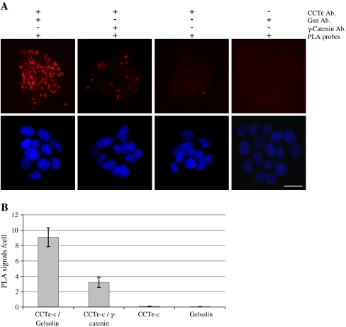
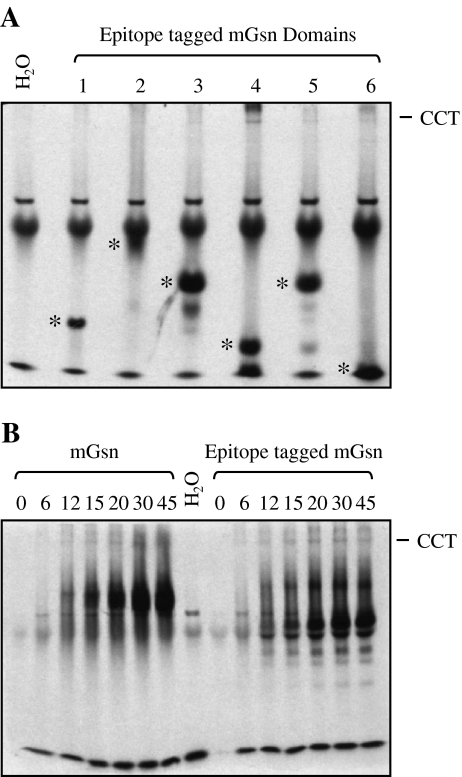
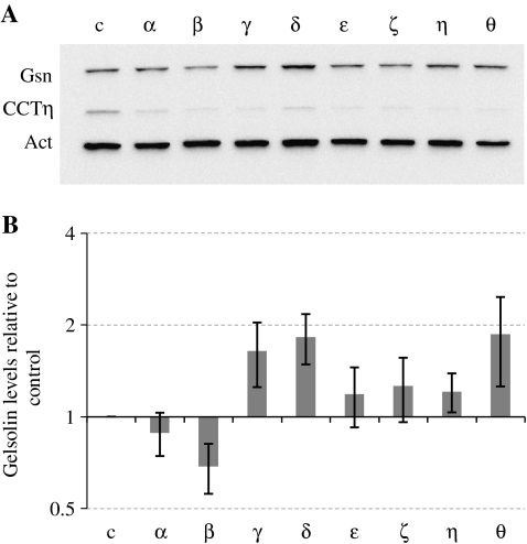
CCT is a member of the chaperonin family of molecular chaperones and consists of eight distinct subunit species which occupy fixed positions within the chaperonin rings. The activity of CCT is closely linked to the integrity of the cytoskeleton as newly synthesized actin and tubulin monomers are dependent upon CCT to reach their native conformations. Furthermore, an additional role for CCT involving interactions with assembling/assembled microfilaments and microtubules is emerging. CCT is also known to interact with other proteins, only some of which will be genuine folding substrates. Here, we identify the actin filament remodeling protein gelsolin as a CCT-binding partner, and although it does not behave as a classical folding substrate, gelsolin binds to CCT with a degree of specificity. In cultured cells, the levels of CCT monomers affect levels of gelsolin, suggesting an additional link between CCT and the actin cytoskeleton that is mediated via the actin filament severing and capping protein gelsolin.


Figures

Fig. 1
Fig. 1
Detection of potential CCT–gelsolin interactions in T47D cells using a proximity ligation assay. Proximity ligation assays were performed using primary antibody pairs against CCTε/gelsolin and CCTε/γ-catenin, both with anti-rabbit plus and anti-mouse minus PLA probes. Controls with one primary antibody and both PLA probes are indicated (a). The scale bar represents 20 μm. PLA signals/cell were counted from five fields of view (b). Error bars represent the standard error where n = 5
Fig. 2
Fig. 2
Cytoplasmic gelsolin binds the chaperonin CCT, but not as a classical folding substrate. Gelsolin was translated in the presence of 35S methionine at 30°C in a rabbit reticulocyte lysate in vitro translation system. Samples (2 μl) were separated by native PAGE, the CCT oligomer is indicated, and a negative control with water instead of DNA was included in all experiments (a, c, and d). Samples were collected at the time points shown (in minutes) during translation of mouse cytoplasmic gelsolin and separated by nondenaturing electrophoresis and visualized by autoradiography (a). A 45-min translation of human cytoplasmic gelsolin was performed in (b) with and without a further incubation with an anti-gelsolin antibody for 20 min on ice. The positions of unshifted (u) and shifted (s) gelsolin are indicated. 2 μl of 45-min time in vitro translations of human gelsolin and β-actin were separated by SDS PAGE (c). Time courses of human cytoplasmic gelsolin (with subsequent antibody incubation as in b) and β-actin in vitro translations were compared on the same native gel (d), where an arrowhead and bracket denote gelsolin and native actin products, respectively. An asterisk indicates actin bound to endogenous CAP (McCormack et al. 2001). The counts on CCT were calculated as a percentage of total counts per lane by phosphor imaging using a Molecular Imager FX (BioRad) (e) ▲ represents gelsolin and ■ β-actin
Fig. 3
Fig. 3
Domain-specific binding of gelsolin to CCT. Individual epitope (2 × LM-myc) tagged domains of cytoplasmic gelsolin were translated for 45 min in the presence of l-[35S] methionine, separated by native PAGE (a) and visualized by autoradiography. Asterisks denote individual domain products and the position of the CCT oligomer is indicated. In vitro translations of full-length cytoplasmic gelsolin with and without the epitope tag were compared by resolving time course samples by native page followed by autoradiography (b)
Fig. 4
Fig. 4
Altering levels of CCT monomers affects gelsolin expression. BE cells were transfected with either a control nontargeting duplex (c) or siRNA duplexes to each of the CCT subunits (α-θ) and harvested four days after transfection. Western blot analysis (a) was performed probing for gelsolin (Gsn), CCTη, and actin (Act). Quantification of the gelsolin signals in each transfection was performed from three individual experiments, using the actin signal to standardize for gel loading. Gelsolin levels in transfected samples were compared to those of the control and the standard error indicated (b)

References

    1. Altschuler GM, Willison KR. Development of free-energy-based models for chaperonin containing TCP-1 mediated folding of actin. J R Soc Interface. 2008;5:1391–1408. doi: 10.1098/rsif.2008.0185. - DOI - PMC - PubMed
    1. Azuma TK, Koths K, Flanagan L, Kwiatkowski D. Gelsolin in complex with phosphatidylinositol4, 5-bisphosphate inhibits caspase-3 and -9 to retard apoptotic progression. J Biol Chem. 2000;275:3761–3766. doi: 10.1074/jbc.275.6.3761. - DOI - PubMed
    1. Brackley KI, Grantham J. Activities of the chaperonin containing TCP-1 (CCT): implications for cell cycle progression and cytoskeletal organization. Cell Stress Chaperones. 2009;14:23–31. doi: 10.1007/s12192-008-0057-x. - DOI - PMC - PubMed
    1. Brackley KI, Grantham J. Subunits of the chaperonin CCT interact with F-actin and influence cell shape and cytoskeletal assembly. Exp Cell Res. 2010;316:543–553. doi: 10.1016/j.yexcr.2009.11.003. - DOI - PubMed
    1. Burtnick LD, Koepf EK, Grimes J, Jones EY, Stuart DI, McLaughlin PJ, Robinson RC. The crystal structure of plasma gelsolin: implications for actin severing, capping and nucleation. Cell. 1997;90:661–670. doi: 10.1016/S0092-8674(00)80527-9. - DOI - PubMed

Publication types