

doi: 10.1002/wsbm.96.
Branch formation during organ development
Affiliations
- PMID: 20890968
- PMCID: PMC2951286
- DOI: 10.1002/wsbm.96
Item in Clipboard
Branch formation during organ development
Wiley Interdiscip Rev Syst Biol Med.
2010 Nov-Dec.
Abstract
Invertebrates and vertebrates use branching morphogenesis to build epithelial trees to maximize the surface area of organs within a given volume. Several molecular regulators of branching have recently been discovered, a number of which are conserved across different organs and species. Signals that control branching at the cellular and tissue levels are also starting to emerge, and are rapidly unveiling the physical nature of branch development. Here we discuss the molecular, cellular, and physical processes that govern branch formation, and highlight the major outstanding questions in the field.
© 2010 John Wiley & Sons, Inc.
Figures
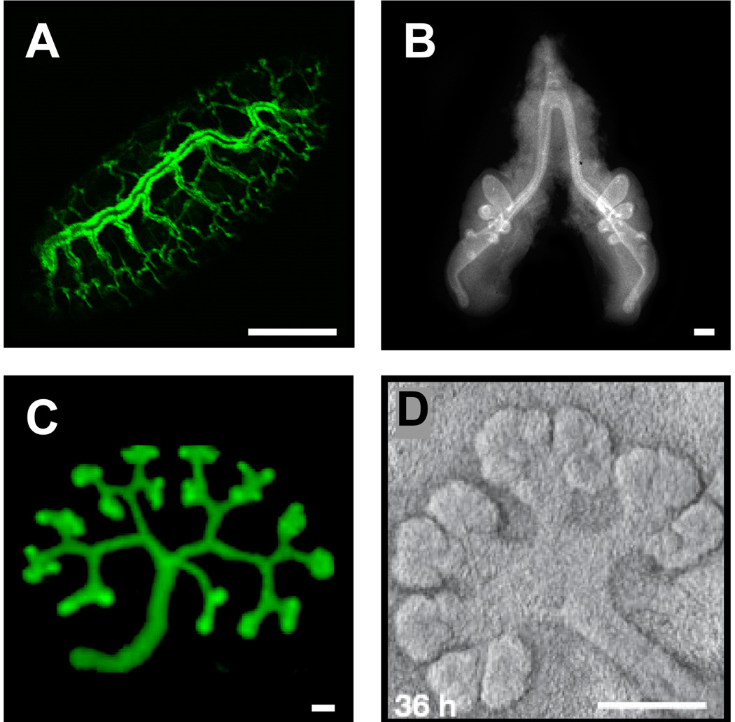
Organs constructed by branch formation. (A) Drosophila trachea, (B) chicken lung, and mouse (C) kidney (reprinted from with permission from Elsevier, © 2009) and (D) salivary gland (reprinted with permission from Macmillan Publishers Ltd: Nature 423: 876–881, © 200629). Scale bars, 100 µm.
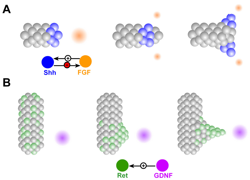
Branch formation by chemoattraction. (A) FGF source (orange) guides branch extension by enhancing motility of tip cells. FGF induces expression of Shh (blue), which in turn suppresses and splits the FGF source. The split source of FGF gives rise to branch bifurcation. (B) Cells expressing high levels of Ret (green), persistently migrate toward the source of GDNF (purple), forming a patch of high Ret activity, which ultimately forms the ureteric bud.
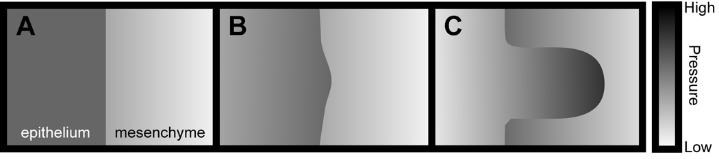
Branch formation by viscous fingering. (A) The pressure in the epithelium is initially uniform, whereas that of the mesenchyme decreases away from the epithelium. (B) A small bulge in the epithelium protrudes and encounters a sharper pressure drop, which drives further protrusion. (C) As the bud grows, it displaces the mesenchyme toward the stalk regions, reducing the pressure drop there and lowering the driving force for motion.

Mechanical control of branching. The high mechanical fields at the tips enhance branching by physically propelling the epithelium forward and by activating proteases and signaling pathways that enhance cellular motility and invasiveness.
References
Publication types
MeSH terms
Substances
Grants and funding
LinkOut - more resources
Full Text Sources
Molecular Biology Databases
