Review
. 2011 Aug 1;15(3):831-44.
doi: 10.1089/ars.2010.3412. Epub 2011 Mar 31.

Peroxiredoxin 6: a bifunctional enzyme with glutathione peroxidase and phospholipase A₂ activities

Affiliations
Review

Peroxiredoxin 6: a bifunctional enzyme with glutathione peroxidase and phospholipase A₂ activities

Aron B Fisher. Antioxid Redox Signal. .

Abstract

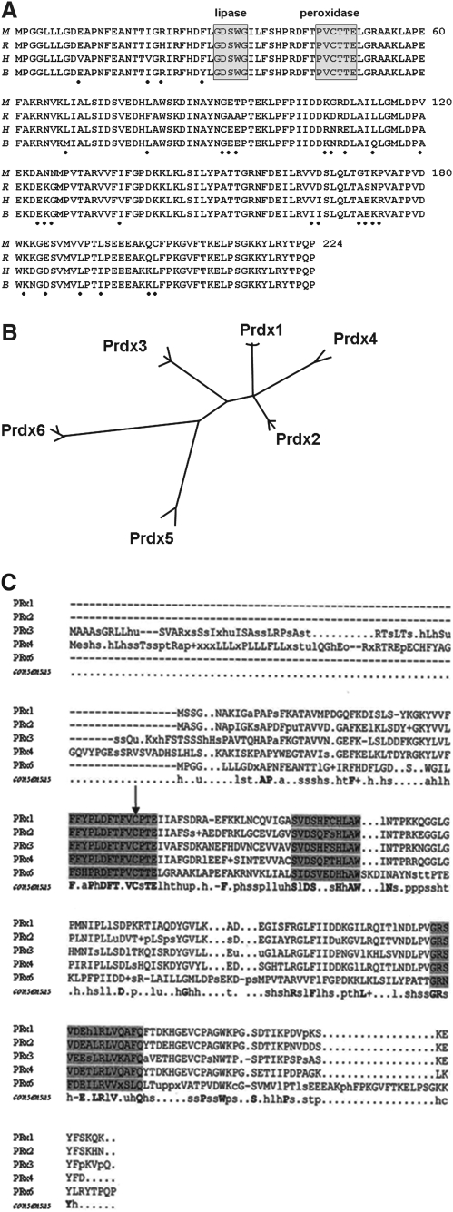
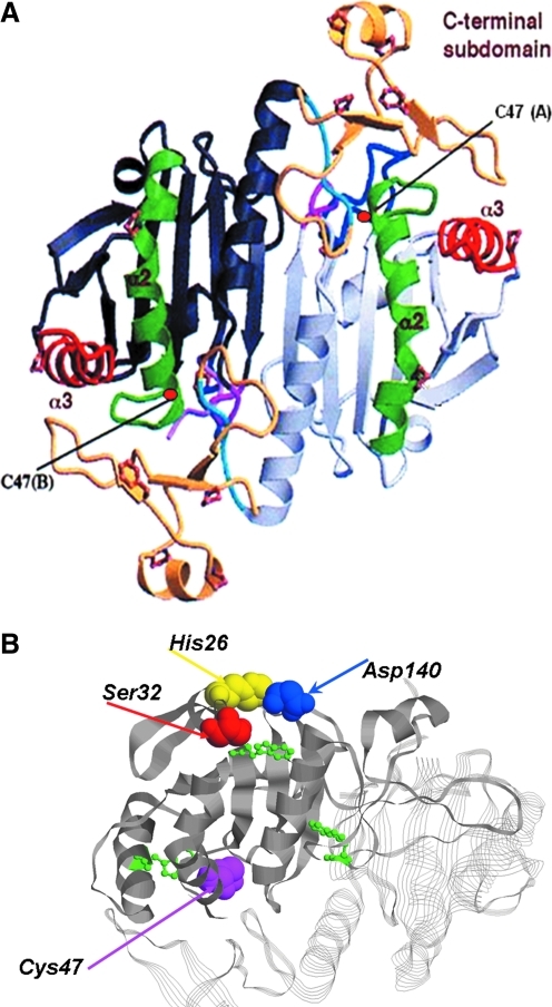
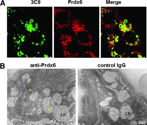
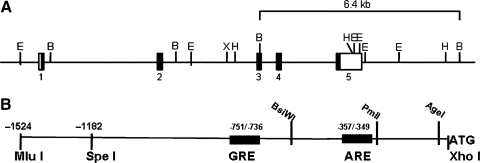
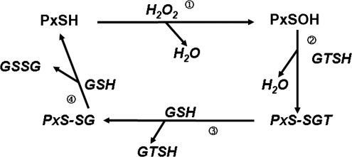
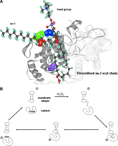
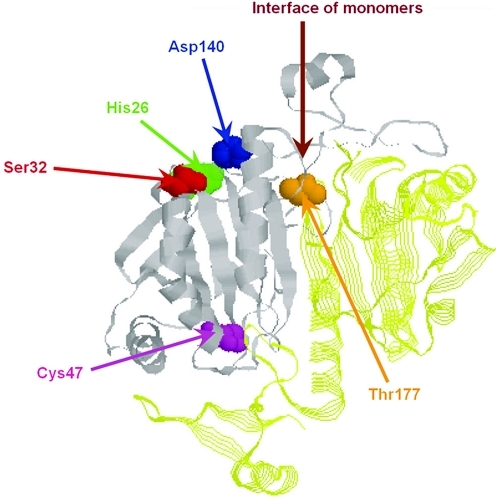
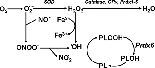
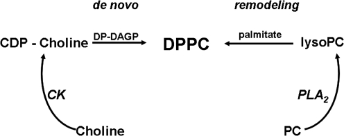
Peroxiredoxin 6 (Prdx6) is the prototype and the only mammalian 1-Cys member of the Prdx family. Major differences from 2-Cys Prdxs include the use of glutathione (GSH) instead of thioredoxin as the physiological reductant, heterodimerization with πGSH S-transferase as part of the catalytic cycle, and the ability either to reduce the oxidized sn-2 fatty acyl group of phospholipids (peroxidase activity) or to hydrolyze the sn-2 ester (alkyl) bond of phospholipids (phospholipase A(2) [PLA(2)] activity). The bifunctional protein has separate active sites for peroxidase (C47, R132, H39) and PLA(2) (S32, D140, H26) activities. These activities are dependent on binding of the protein to phospholipids at acidic pH and to oxidized phospholipids at cytosolic pH. Prdx6 can be phosphorylated by MAP kinases at T177, which markedly increases its PLA(2) activity and broadens its pH-activity spectrum. Prdx6 is primarily cytosolic but also is targeted to acidic organelles (lysosomes, lamellar bodies) by a specific targeting sequence (amino acids 31-40). Oxidant stress and keratinocyte growth factor are potent regulators of Prdx6 gene expression. Prdx6 has important roles in both antioxidant defense based on its ability to reduce peroxidized membrane phospholipids and in phospholipid homeostasis based on its ability to generate lysophospholipid substrate for the remodeling pathway of phospholipid synthesis.


Figures

FIG. 1.
FIG. 1.
The primary structure of peroxiredoxin 6 (Prdx6). (A) Deduced Prdx6 amino acid sequence for the mouse (M), rat (R), human (H), and bovine (B) proteins. The dots beneath the sequence indicate amino acids that are not fully conserved among the four species. The lipase and peroxidase motifs are indicated. Modified from Kim et al. (37) and Fisher et al. (21). (B) Phylogenetic tree for mammalian Prdxs constructed using ClustalW (1.7). Modified from Knoops et al. (41). (C) Consensus sequences aligned using ClustalW (1.7). The consensus sequences for the human, mouse, and rat homologs for Prdxs 1, 2, 3, 4, and 6 were generated using the consensus C program. The catalytic Cys residue is indicated by the arrow. The shading indicates regions of relatively high homology. Modified from Phelan (73).
FIG. 2.
FIG. 2.
Tertiary structure of Prdx6. (A) Crystal structure of mutated (C91S) human Prdx6 that was oxidized by air exposure. The protein crystallized as a homodimer. Notable features are the thioredoxin fold consisting of a four-stranded β sheet with two flanking α helices. The c-terminal domain comprises amino acid residues 175–224 and consists of 3 β strands and an α helix attached to the larger internal domain by a short loop. The catalytic C47 in each monomer is indicated. Modified from Choi et al. (7). (B) Ribbon diagram showing the relative position of the active site for peroxidase activity (C47) and the catalytic triad for phospholipase A2 (PLA2) activity (S32, H26, D140). The SDH catalytic triad is on the protein surface, whereas C47 is at the base of a narrow pocket. Also indicated (in green) are the positions of the three Trp residues that have been used to analyze substrate binding (54, 55). (To see this illustration in color the reader is referred to the web version of this article at www.liebertonline.com/ars).
FIG. 3.
FIG. 3.
Subcellular localization of Prdx6 in rat lung alveolar epithelial type II cells. (A) Immunofluorescence using antibodies to 3C9, a marker for lung lamellar body membranes, and Prdx6. The merged image shows colocalization for the two proteins, indicating the presence of Prdx6 in lung lamellar bodies. (B) Ultrastructural localization of Prdx6 using electron microscopic immunohistochemistry with 20 nm gold-coupled antibody. Arrows indicate gold grains of Prdx6 in lamellar bodies (LB) with a lesser concentration of grains in other organelles. The right panel is a control in the absence of primary antibody. Modified from Wu et al. (103).
FIG. 4.
FIG. 4.
Regulation of Prdx6 expression. (A) The structure of the Prdx6 gene indicating five exons and intervening introns. The black boxes represent the protein coding regions in the exons and the open boxes represent noncoding regions. The sequence from exon 1 to exon 5 represents 10.3 kb. Restriction sites are indicated: B, BAMH1; E, EcoR1; H, Hind3; X, Xho1. (B) Translational regulatory elements and restriction recognition sites upstream of the Prdx6 translational start site (ATG). The figure indicates the position of an antioxidant (electrophilic) response element (ARE) (8) and glutocorticoid response element (GRE) (unpublished results). The numbers indicated are relative to the site for start of transcription. Modified from Mo et al. (60).
FIG. 5.
FIG. 5.
The catalytic cycle for Prdx6 and role of GSH transferase (GT). Prdx6 (PxSH) is shown indicating the active-site sulfhydryl on C47. πGSH S-transferase (GTSH) also is shown with its active sulfhydryl group indicated (by coincidence, also at C47). In its antioxidant role, Prdx6 interacts with an oxidant (H2O2) to generate the sulfenic acid (reaction 1), which then interacts with the SH of πGST to generate the Prdx6:πGST heterodimer (reaction 2). The GSH bound to the πGST glutathionylates Prdx6, liberating πGST (reaction 3). Finally, a second GSH reduces the –SSG bond and regenerates the active (reduced) enzyme (reaction 4). Modified from Ralat et al. (81).
FIG. 6.
FIG. 6.
Binding of phospholipids to Prdx6. (A) Theoretical analysis of the binding of an oxidized phospholipid to Prdx6. The phospholipid head group binds at the protein surface in the vicinity of the PLA2 catalytic triad (SDH), whereas the oxidized sn-2 oxidized fatty acyl group inserts into the hydrophobic pocket where the oxidized double bond is in proximity to the catalytic Cys47. (B) Scheme for reduction of oxidized membrane phospholipids by cytosolic Prdx6. The resting state shows phospholipids of the membrane bilayer and Prdx6 contained in the cytosol (panel 1). Oxidative stress (H2O) generates a phospholipid hydroperoxide, increasing its aqueous solubility resulting in flotation of the peroxidized sn-2 acyl chain (panel 2). Cytosolic Prdx6 binds to the oxidized phospholipid (panel 3) resulting in reduction of the phospholipid hydroperoxide (panel 4) and dissociation of Prdx6 from the membrane (panel 5). Reduction of Prdx6 with GSH restores the resting state. At this time, it is not clear whether Prdx6 is reduced while attached to the membrane or in the cytosol subsequent to its dissociation. Modified from Manevich et al. (55). (To see this illustration in color the reader is referred to the web version of this article at www.liebertonline.com/ars).
FIG. 7.
FIG. 7.
Site for MAP kinase-mediated phosphorylation of Prdx6. The phosphorylation site is T177. This site, which is distant from the PLA2 catalytic triad and the peroxidatic Cys47, is located within the protein globule relatively close to the interface of Prdx6 monomers. The buried nature of the site suggests that change in configuration is required for phosphorylation. Reprinted from Wu et al. (102). (To see this illustration in color the reader is referred to the web version of this article at www.liebertonline.com/ars).
FIG. 8.
FIG. 8.
Scheme for intracellular generation of reactive oxygen species (ROS) and the role of Prdx6. O2•− is dismutated to H2O2, which can generate OH in the presence of Fe2+. OH is a powerful oxidant that can peroxidize cell membrane phospholipids (PL). Thus, cell resistance to oxidant stress and repair depends in large part on removal of H2O2 and reduction of phospholipid hydroperoxides (there is no specific scavenger for OH). H2O2 can be removed by multiple enzymes, including catalase, GSH peroxidases, and all Prdxs. Phospholipid hydroperoxides (PLOOH) are reduced in lung cells by Prdx6; GPx4 represents an alternate enzyme for reduction in some cells (not shown). The product of Prdx6 activity, the hydroxy phospholipid (PLOH), is further reduced by unspecified enzymes to regenerate the native phospholipid. Modified from Manevich and Fisher (53).
FIG. 9.
FIG. 9.
Pathways for synthesis of dipalmitoylphosphatidylcholine (DPPC) by lung alveolar epithelial cells. The de novo pathway proceeds by the phosphorylation of choline (choline kinase [CK]) and then generation of cytidine 5-diphosphocholine (CDP-choline), which combines with 1,2-palmitoyl diacylglycerol phosphate (DP-DAGP) to form DPPC. The remodeling pathway requires a PLA2 acting on phosphatidylcholine (PC) to generate a lysoPC, which then forms DPPC by reacylation in the sn-2 position with palmitate. Prdx6 has been shown to be the major PLA2 involved in the remodeling pathway for surfactant biosynthesis in lung alveolar epithelial cells.

References

    1. Akiba S. Dodia C. Chen X. Fisher AB. Characterization of acidic Ca(2+)-independent phospholipase A2 of bovine lung. Comp Biochem Physiol B Biochem Mol Biol. 1998;120:393–404. - PubMed
    1. Baek IJ. Seo DS. Yon JM. Lee SR. Jin Y. Nahm SS. Jeong JH. Choo YK. Kang JK. Lee BJ. Yun YW. Nam SY. Tissue expression and cellular localization of phospholipid hydroperoxide glutathione peroxidase (PHGPx) mRNA in male mice. J Mol Histol. 2007;38:237–244. - PubMed
    1. Baek YM. Hwang HJ. Kim SW. Hwang HS. Lee SH. Kim JA. Yun JW. A comparative proteomic analysis for capsaicin-induced apoptosis between human hepatocarcinoma (HepG2) and human neuroblastoma (SK-N-SH) cells. Proteomics. 2008;8:4748–4767. - PubMed
    1. Chae HZ. Robison K. Poole LB. Church G. Storz G. Rhee SG. Cloning and sequencing of thiol-specific antioxidant from mammalian brain: alkyl hydroperoxide reductase and thiol-specific antioxidant define a large family of antioxidant enzymes. Proc Natl Acad Sci U S A. 1994;91:7017–7021. - PMC - PubMed
    1. Chang XZ. Li DQ. Hou YF. Wu J. Lu JS. Di GH. Jin W. Ou ZL. Shen ZZ. Shao ZM. Identification of the functional role of peroxiredoxin 6 in the progression of breast cancer. Breast Cancer Res. 2007;9:R76. - PMC - PubMed

Publication types

MeSH terms

LinkOut - more resources