. 2011 Mar;17(5-6):631-44.
doi: 10.1089/ten.tea.2010.0298. Epub 2010 Nov 17.

Engineered pullulan-collagen composite dermal hydrogels improve early cutaneous wound healing

Affiliations

Engineered pullulan-collagen composite dermal hydrogels improve early cutaneous wound healing

Victor W Wong et al. Tissue Eng Part A. 2011 Mar.

Erratum in

  • Tissue Eng Part A. 2012 Mar;18(5-6):676. Neofyotou, Evgenios [corrected to Neofytou, Evgenios]

Abstract

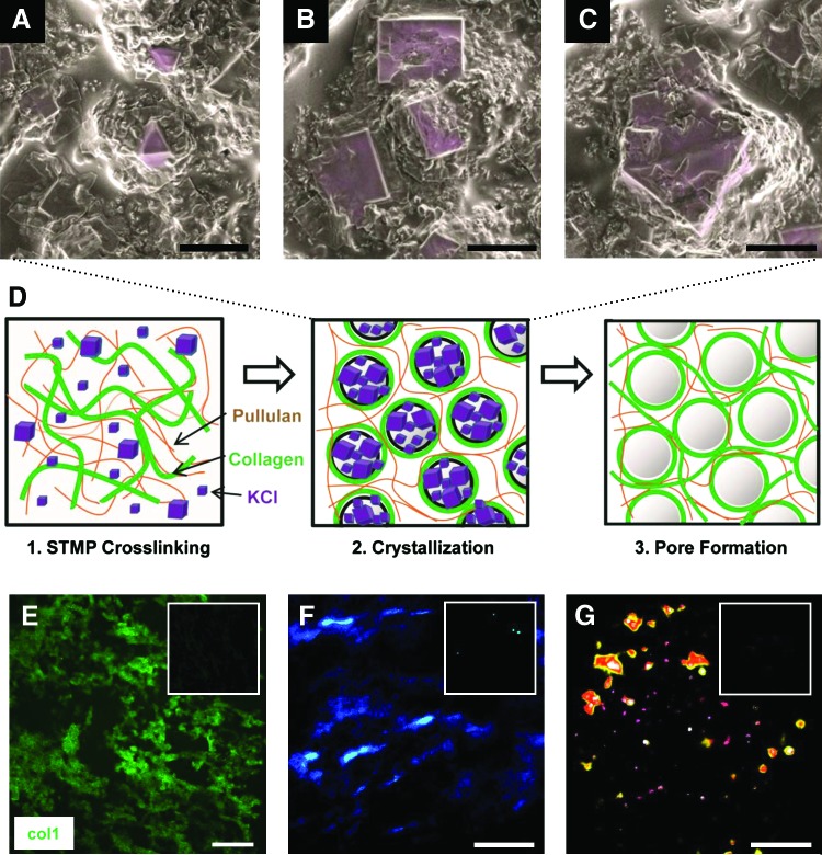
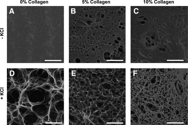
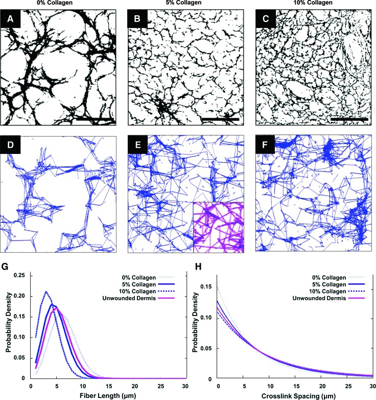
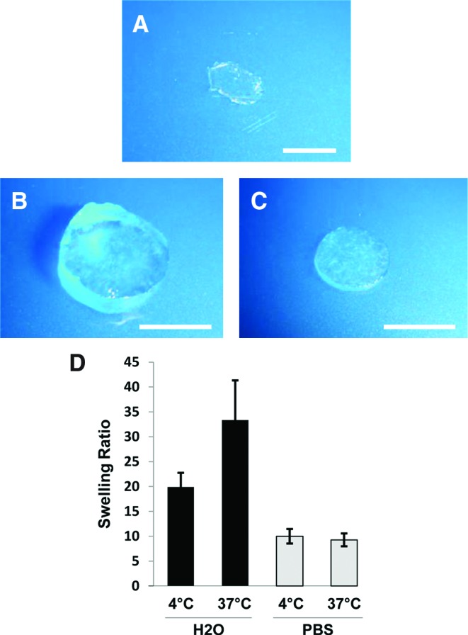
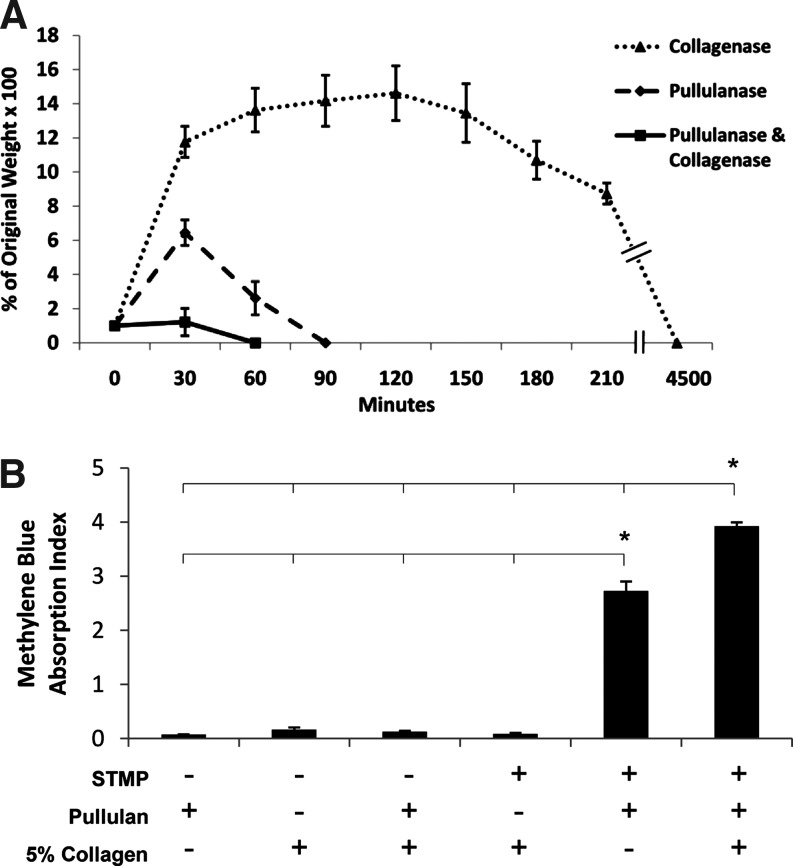
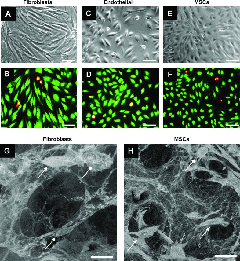
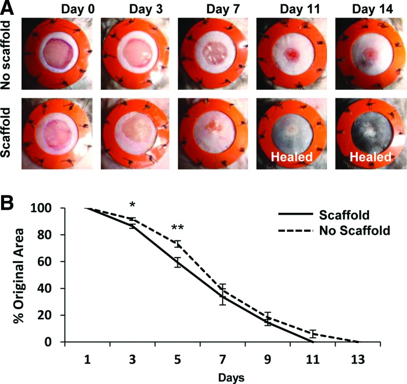
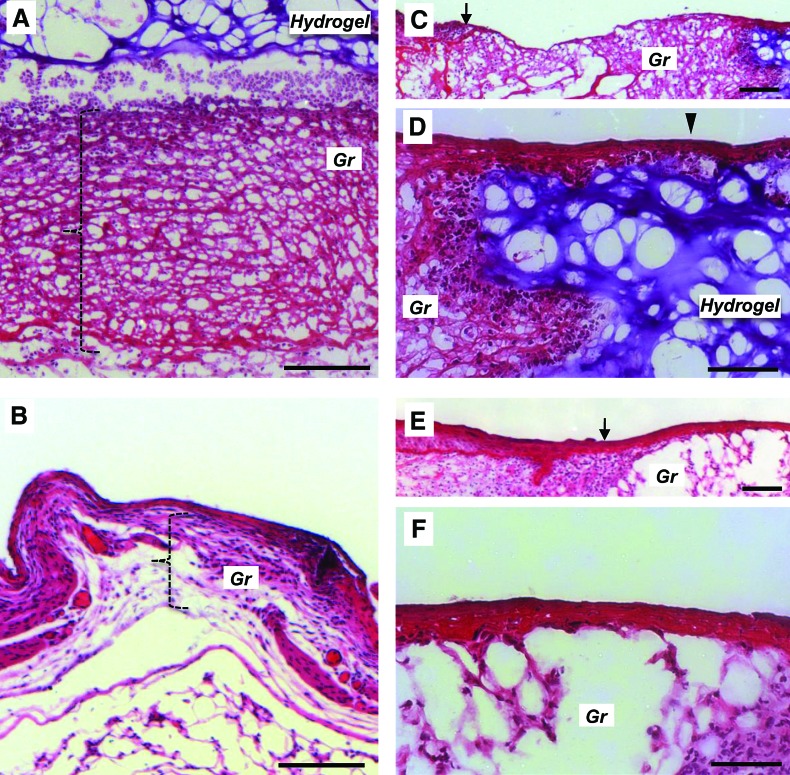
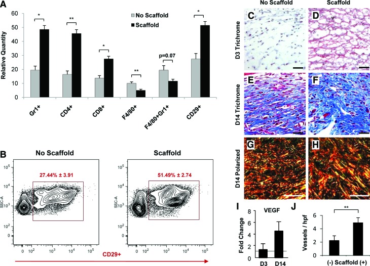
New strategies for skin regeneration are needed to address the significant medical burden caused by cutaneous wounds and disease. In this study, pullulan-collagen composite hydrogel matrices were fabricated using a salt-induced phase inversion technique, resulting in a structured yet soft scaffold for skin engineering. Salt crystallization induced interconnected pore formation, and modification of collagen concentration permitted regulation of scaffold pore size. Hydrogel architecture recapitulated the reticular distribution of human dermal matrix while maintaining flexible properties essential for skin applications. In vitro, collagen hydrogel scaffolds retained their open porous architecture and viably sustained human fibroblasts and murine mesenchymal stem cells and endothelial cells. In vivo, hydrogel-treated murine excisional wounds demonstrated improved wound closure, which was associated with increased recruitment of stromal cells and formation of vascularized granulation tissue. In conclusion, salt-induced phase inversion techniques can be used to create modifiable pullulan-collagen composite dermal scaffolds that augment early wound healing. These novel biomatrices can potentially serve as a structured delivery template for cells and biomolecules in regenerative skin applications.


Figures

FIG. 1.
FIG. 1.
Potassium chloride (KCl) crystallization and salt-induced phase inversion. After the dehydration processing step, scanning electron micrographs of 5% collagen–pullulan hydrogels demonstrated various KCl crystal sizes (A–C, pseudocolored purple) that correlated with hydrogel pore sizes produced after salt dissolution. We hypothesize that as water is rapidly removed from the hydrogel, localized supersaturation of KCl results in the formation of crystals around which polymers become organized (D, middle). As KCl is dissolved from the hydrogel, porous macrovoids remain, resulting in a reticular pullulan–collagen composite scaffold (D, right). The movement and organization of the polymers is further enhanced by mechanical mixing during the fabrication process (D, left). Collagen 1 immunofluorescence demonstrates reticular and homogeneous distribution of collagen throughout the hydrogel (E, green = collagen 1). Negative control shown in upper right inset (E). Polarized light analysis of hydrogels (F) and picrosirius red-stained hydrogels (G) further confirms the homogeneous distribution of collagen around porous macrovoids throughout the hydrogel. The 0% collagen hydrogels shown in upper right inset of (F) and (G) demonstrate the absence of collagen signal. Scale bar for (A–C) is 10 μm. Scale bar for (E–G) is 100 μm. Color images available online at www.liebertonline.com/ten.
FIG. 2.
FIG. 2.
Hydrogel pore formation. Scanning electron microscopy imaging revealed that pullulan hydrogels fabricated without KCl demonstrated poor porosity, despite increases in collagen content (A–C). With the addition of KCl, consistent interconnected porous domains were created (D–F). Alterations in collagen concentration significantly modified pore size. Scale bar is 100 μm.
FIG. 3.
FIG. 3.
Network extraction analysis. A network extraction algorithm was used to analyze the microstructure of pullulan-based hydrogels. Two-dimensional topographical data were extracted from representative scanning electron microscopy images for 0%, 5%, and 10% collagen–pullulan hydrogels (A–F). The same algorithm was applied to acellular human dermal matrix (E, inset). Quantitative analysis of fiber length (G) and crosslink spacing (H) distribution was performed, and 5% collagen hydrogel scaffolds (solid blue line) best approximated the microarchitecture of unwounded human dermis (purple line). Scale bar is 100 μm. Color images available online at www.liebertonline.com/ten.
FIG. 4.
FIG. 4.
Collagen–pullulan hydrogel general physical properties. Digital photographs of dry (A) and H2O- (B) and phosphate-buffered saline (PBS)-incubated (C) 5% collagen–pullulan hydrogels after overnight incubation. Swelling ratios for 5% collagen–pullulan hydrogels were calculated after incubation in both dH2O and PBS (D). n = 6 for each condition. Error bars are ± standard error of the mean (SEM). Scale bar is 5 mm. Color images available online at www.liebertonline.com/ten.
FIG. 5.
FIG. 5.
Five percent collagen–pullulan hydrogel degradation and crosslinking. Hydrogels were incubated with pullulanase/collagenase (solid line), pullulanase only (dashed line), or collagenase only (dotted line) (A). Five percent collagen without pullulan demonstrated minimal sodium trimetaphosphate (STMP) crosslinking based on methylene blue binding but appeared to synergistically augment the organization and crosslinking of pullulan hydrogels (B). Polymer mixtures without STMP exhibited negligible nonspecific binding of methylene blue to either collagen or pullulan. n = 6 for each condition. Error bars are ± SEM. *p < 0.05.
FIG. 6.
FIG. 6.
In vitro cellular incorporation. Hydrogels were biocompatible with fibroblasts (A, B), endothelial cells (C, D), and bone marrow-derived mesenchymal stem cells (MSCs) (E, F) for up to 7 days. Live cells are stained green, whereas dead cells are stained red/yellow (B, D, F). Scanning electron micrographs showed that both fibroblasts (arrows, G) and mesenchymal stem cells (arrows, H) were viably incorporated into 5% collagen–pullulan hydrogels. Additionally, the porous reticular network of the hydrogel scaffold is maintained in both images (G, H). Scale bar is 50 μm in (A–F). Scale bar is 25 μm in (G) and (H). Color images available online at www.liebertonline.com/ten.
FIG. 7.
FIG. 7.
Humanized excisional wound model. Digital photographs of stented excisional wounds treated with no scaffold (top row) or 5% collagen–pullulan scaffolds (bottom row) from days 0 to 14 postinjury (A). The silicone rings prevent wound contraction and ensure that healing proceeds via granulation tissue formation and re-epithelialization, which is more similar to human healing. Five percent hydrogel scaffold-treated wounds demonstrated significantly improved wound closure at days 3 and 5 postinjury (B). n = 10 wounds for each condition. Errors bars are ± SEM (*p < 0.01 and **p < 0.005). Color images available online at www.liebertonline.com/ten.
FIG. 8.
FIG. 8.
Wound histology. Hematoxylin and eosin staining of day 3 hydrogel-treated excisional wounds (top series) compared with nontreated wounds (bottom series). Hydrogel-treated wounds demonstrated marked granulation (Gr) matrix formation (A, bracket) compared with thinner matrix in nontreated wounds (B). A wide bed of granulation matrix is seen between the wound margin (C, arrow) and the hydrogel (purple staining). Layered keratinocytes are seen migrating across the wound bed over the hydrogel (D, arrowhead). This is in contrast to nontreated excisional wounds, which demonstrate thinner granulation matrix (B, bracket) and sparse granulation tissue at the margin of the immature wound bed (E, arrow, and F). Scale bar is 100 μm in (A) and (B) and 50 μm in (C–F). Color images available online at www.liebertonline.com/ten.
FIG. 9.
FIG. 9.
Analysis of wound healing parameters. Flow cytometric analysis of wound cell populations at day 3 postinjury demonstrated increased recruitment of neutrophils and T cells and decreased recruitment of macrophage populations with hydrogel scaffold treatment (A). Importantly, there was increased recruitment of stromal fibroblast-like CD29+ cells that are associated with granulation tissue formation and improved wound healing (A). Representative flow cytometric graphs of CD29+ cell populations in control and hydrogel scaffold-treated wounds at day 3 postinjury (B). n = 4 for each condition for flow cytometric analysis. Scaffold-treated wounds demonstrated improved granulation tissue formation based on trichrome and polarized light analysis compared with untreated controls (C–H). Note the greater prevalence of mature collagen (blue on trichrome staining, red/orange on polarized light analysis) with hydrogel treatment at day 14 postinjury. Hydrogel treatment was also associated with greater gene expression of vascular endothelial growth factor (VEGF) and a significantly greater quantity of microvessels at day 14 postinjury (I, J). Scale bar is 20 μm in (C–H). Errors bars are ± SEM (*p < 0.01 and **p < 0.05). Color images available online at www.liebertonline.com/ten.

References

    1. Priya S.G. Jungvid H. Kumar A. Skin tissue engineering for tissue repair and regeneration. Tissue Eng B Rev. 2008;14:105. - PubMed
    1. Hodde J.P. Johnson C.E. Extracellular matrix as a strategy for treating chronic wounds. Am J Clin Dermatol. 2007;8:61. - PubMed
    1. Bello Y.M. Falabella A.F. Eaglstein W.H. Tissue-engineered skin. Current status in wound healing. Am J Clin Dermatol. 2001;2:305. - PubMed
    1. Wainwright D.J. Use of an acellular allograft dermal matrix (AlloDerm) in the management of full-thickness burns. Burns. 1995;21:243. - PubMed
    1. Gentzkow G.D. Gidner A. Davis M. Kealey G.P. Mozingo D.W. Hansbrough J.F. Clinical trials of a biosynthetic temporary skin replacement, dermagraft-transitional covering, compared with cryopreserved human cadaver skin for temporary coverage of excised burn wounds. J Burn Care Rehabil. 1997;18:43. - PubMed

Publication types

MeSH terms

LinkOut - more resources