. 2011 Jan 15;81(2):211-21.
doi: 10.1016/j.bcp.2010.09.024. Epub 2010 Oct 12.

Thioredoxin reductase 1 knockdown enhances selenazolidine cytotoxicity in human lung cancer cells via mitochondrial dysfunction

Affiliations

Thioredoxin reductase 1 knockdown enhances selenazolidine cytotoxicity in human lung cancer cells via mitochondrial dysfunction

Robyn L Poerschke et al. Biochem Pharmacol. .

Abstract

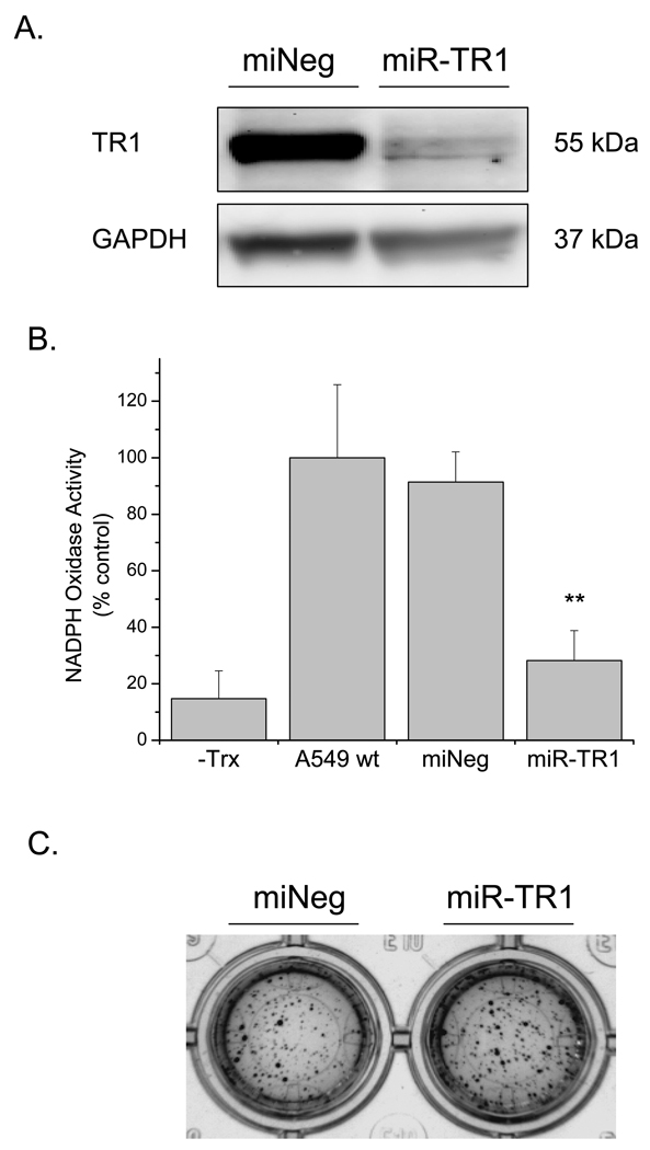
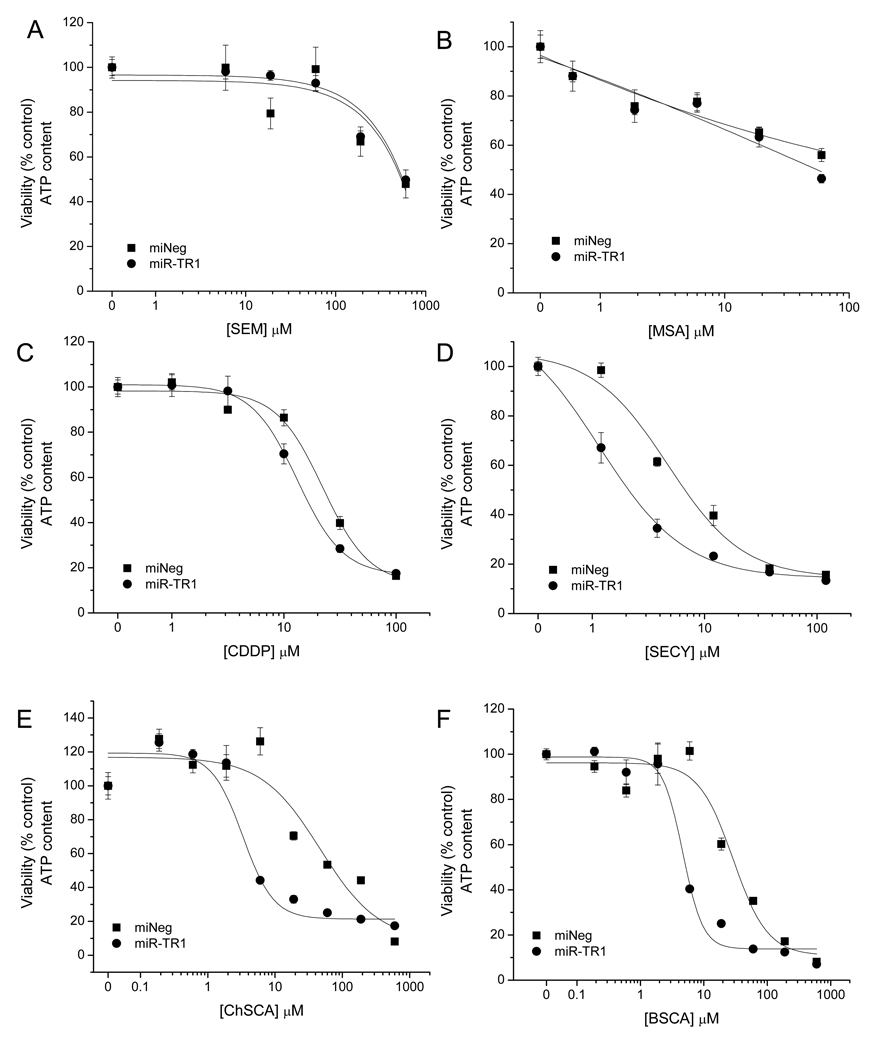
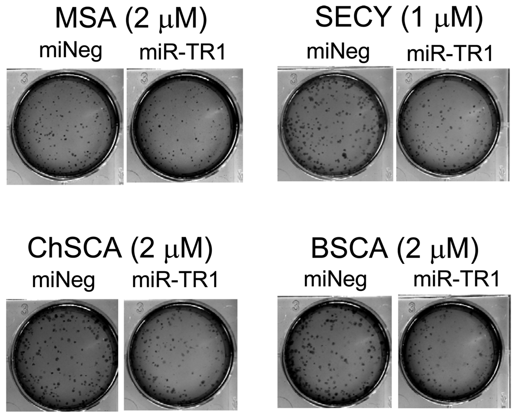
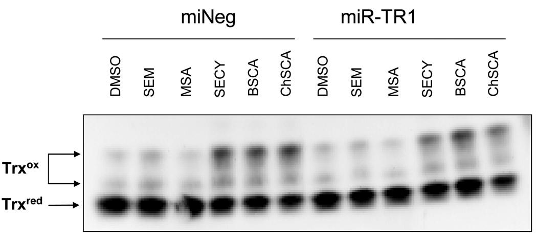
Thioredoxin reductase (TR1) is a selenoprotein that is involved in cellular redox status control and deoxyribonucleotide biosynthesis. Many cancers, including lung, overexpress TR1, making it a potential cancer therapy target. Previous work has shown that TR1 knockdown enhances the sensitivity of cancer cells to anticancer treatments, as well as certain selenocompounds. However, it is unknown if TR1 knockdown produces similar effect on the sensitivity of human lung cancer cells. To further elucidate the role of TR1 in the mechanism of selenocompounds in lung cancer, a lentiviral microRNA delivery system to knockdown TR1 expression in A549 human lung adenocarcinoma cells was utilized. Cell viability was assessed after 48 hr treatment with the selenocysteine prodrug selenazolidines 2-butylselenazolidine-4(R)-carboxylic acid (BSCA) and 2-cyclohexylselenazolidine-4-(R)-carboxylic acid (ChSCA), selenocystine (SECY), methylseleninic acid (MSA), 1,4-phenylenebis(methylene)selenocyanate (p-XSC), and selenomethionine (SEM). TR1 knockdown increased the cytotoxicity of BSCA, ChSCA, and SECY but did not sensitize cells to MSA, SEM, or p-XSC. GSH and TR1 depletion together decreased cell viability, while no change was observed with GSH depletion alone. Reactive oxygen species generation was induced only in TR1 knockdown cells treated with the selenazolidines or SECY. These three compounds also decreased total intracellular glutathione levels and oxidized thioredoxin, but in a TR1 independent manner. TR1 knockdown increased selenazolidine and SECY-induced mitochondrial membrane depolarization, as well as DNA strand breaks and AIF translocation from the mitochondria. These results indicate the ability of TR1 to modulate the cytotoxic effects of BSCA, ChSCA and SECY in human lung cancer cells through mitochondrial dysfunction.


Figures

Figure 1
Figure 1. Knockdown of TR1 in A549 miR-TR1 cells by miRNA
A549 cells were transduced with lenitviral miRNA constructs to produce miNeg and miR-TR1 cell lines (see Materials and Methods). Cells were treated with 1 µg/mL tetracycline for 72 hrs to induce miRNA expression. A) Determination of TR1 protein expression by Western blotting. TR1 is the top band of the doublet. GAPDH was probed as a loading control using 1:500 anti-GAPDH polyclonal primary antibody. A similar decrease in TR1 protein expression was observed in A549 miR-TR1 cells up to 72 hrs after removal of 1 µg/mL tetracycline. The blot shown is representative of two individual experiments. B) Measurement of TR activity via NADPH oxidation. Data are presented as % A549 wt activity. n=2 for all groups. ‘*’ indicates p<0.05 versus miNeg. NADPH oxidative activity in A549 cell lysates without added Trx (-Trx) in the reaction mixture, basal activity, activity in miNeg cells, and in the miR-TR1 cells. C) Anchorage independent growth assay. Cells were allowed to grow in 2% growth factor reduced Matrigel (BD Biosciences; Bedford, MA) and advanced DMEM supplemented with 1 µg/mL tet for 7 days, with media and tet refreshed every three days. Colonies were stained with MTT overnight and visualized on the Kodak ImageStation. No difference in colony growth was observed between the two cell lines. The data shown are representative of six replicates.
Figure 2
Figure 2. Effects of TR1 knockdown on drug cytotoxicity in A549 cells
Viability of A549 miNeg (▪) and miR-TR1 (•) cells was assessed by measuring cellular ATP content after 48 hr drug treatment at the indicated concentrations. Viability values are expressed as % relative to vehicle controls, which were set to 100%. n=4 for all data points. No sensitization was observed in cells treated with A) SEM or B) MSA. Sensitization was observed in cells treated with C) CDDP (miNeg IC50: 22 ± 4 µM; miR-TR1 IC50: 13 ± 0.5), D) SECY (miNeg IC50: 5 ± 1.4 µM; miR-TR1 IC50: 1.2 ± 0.3), E) ChSCA (miNeg IC50: 48 ± 8 µM; miR-TR1 IC50: 3 ± 1.1), and F) BSCA (miNeg IC50: 28 ± 8 µM; miR-TR1 IC50: 5 ± 0.7).
Figure 3
Figure 3. TR1 knockdown did not increase sensitization in clonogenic survival in A549 cells
Representative images of 7-day selenocompound treatment of A549 cells stained by the addition of MTT to the media and then visualized. Measurements of formazan-converted MTT indicate that statistically significant differences were not obtained between the miNeg and miR-TR1 cells.
Figure 4
Figure 4. TR1 expression and selenocompound cytotoxicity in H1666 human NSCLC cells
A) Determination of TR1 protein expression by Western blotting. 10 µg protein was loaded for each sample. H1666 cells have ∼85% less TR1 protein expression than A549 cells as determined by densitometry. B) H1666 cell viability following 48 hr selenocompound treatments. Concentrations ranged from 0-200 µM (ChSCA) or 0-120 µM (SECY). Viability was assessed by ATP content. Values are expressed as % relative to vehicle controls, which were set to 100%, and n=6 for all data points. Both SECY and ChSCA have similar cytotoxicity (SECY IC50: 8.5 ± 0.6; ChSCA IC50: 8.6 ± 1.7 µM).
Figure 5
Figure 5. Effects of TR1 and GSH attenuation on A549 cell viability
miNeg and miR-TR1 cells were treated with 20 µM BSO or PBS vehicle for 48 or 72 hrs. Cell viability was determined by measuring cellular ATP content. BSO-treated viability values are expressed as % matched vehicle controls. n=8 for all groups. ‘***’ indicates p<0.001 versus -BSO (control); ‘###’ indicates p<0.001 versus 48 hr miR-TR1 +BSO.
Figure 6
Figure 6. Assessment of cellular redox status parameters
Treatments were for 48 hrs at the following concentrations: SEM, 20 µM; MSA, 1 µM; SECY, BSCA, and ChSCA, 5 µM; CDDP, 20 µM. A) DCF fluorescence concentration. B) Measurement of total intracellular GSH. GSH concentration values were normalized to cell viability fluorescence values and are expressed as % miNeg DMSO control. n=3 for all DCF assay treatment groups; n=4 for GSH assay treatment groups. ‘**’ indicates p<0.01 versus matched DMSO vehicle control; ‘***’ indicates p<0.001 versus matched DMSO vehicle control; ‘###’ indicates p<0.001 versus matched miNeg treatment.
Figure 7
Figure 7. Selenocompounds induce expression of Trx in an oxidized state
Iodoacetic acid/iodoacetamine-labeled samples were collected following 48 hr treatments at the following concentrations: SEM, 20 µM; MSA, 1 µM; SECY, BSCA, and ChSCA, 5 µM. Equal amounts of protein were analyzed by urea-PAGE and transferred to PVDF membranes that were probed with 1:200 anti-Trx primary antibody. Bands correspond to reduced Trx (bottom band) or Trx in various oxidation states (upper two bands). SECY, BSCA, and ChSCA increased the expression of Trx in an oxidized state in both A549 miNeg (left six lanes) and miR-TR1 (right six lanes) cells. The blot shown is representative of at least two independent experiments.
Figure 8
Figure 8. TR1 knockdown increases selenocompound-mediated mitochondrial membrane depolarization
Determination of mitochondrial membrane potential by JC-1 after 48 hr treatment. Compounds were used at the following concentrations: SEM, 20 µM; MSA, 1 µM; SECY, BSCA, and ChSCA, 5 µM. Data are expressed as % total population, with the 525 nm fluorescence population (JC-1 green) represented as depolarized mitochondria and the 575 nm fluorescence population (JC-1 red) represented as polarized mitochondria. Carbonyl cyanide 3-chlorophenylhydrazone (CCCP; 25 µM), a known mitochondrial membrane disrupter,served as positive control. ‘*’ indicates p<0.05 versus matched DMSO vehicle control; ‘***’ indicates p<0.001 versus matched DMSO vehicle control; ‘##’ indicates p<0.01 versus matched miNeg treatment; ‘###’ indicates p<0.001 versus matched miNeg treatment.
Figure 9
Figure 9. SECY and the selenazolidines induce cell death through a caspase-independent mechanism
For all data shown, cells were treated at the following concentrations for 48 hrs: SEM, 20 µM; MSA, 1 µM; SECY, BSCA, and ChSCA, 5 µM A) Measure of DNA strand breaks by the TUNEL assay. 10 000 events were measured for each sample. Data shown are for BrdU positive, PI negative cell populations and are presented as % total population. ‘***’ indicates p<0.001 versus matched DMSO vehicle control. All samples were analyzed at least in duplicate. B) Caspase 3/7 activity. Data are presented as expressed as % matched DMSO vehicle control. No significance difference between treatment groups was found. C) AIF protein expression in mitochondrial fractions. 1 µg mitochondrial protein was loaded for each sample. GAPDH was probed as a protein loading control. α-tubulin protein was not detected (data not shown), indicating that the mitochondrial fractions were free of cytosolic contamination. The blot shown is representative of two individual experiments.

References

    1. Yoo MH, Xu XM, Carlson BA, Patterson AD, Gladyshev VN, Hatfield DL. Targeting thioredoxin reductase 1 reduction in cancer cells inhibits self-sufficient growth and DNA replication. PLoS One. 2007;2(10):e1112. - PMC - PubMed
    1. Sun QA, Wu Y, Zappacosta F, Jeang KT, Lee BJ, Hatfield DL, Gladyshev VN. Redox regulation of cell signaling by selenocysteine in mammalian thioredoxin reductases. J Biol Chem. 1999;274(35):24522–24530. - PubMed
    1. Schenk H, Klein M, Erdbrugger W, Droge W, Schulze-Osthoff K. Distinct effects of thioredoxin and antioxidants on the activation of transcription factors NF-kappa B and AP-1. Proc Natl Acad Sci U S A. 1994;91(5):1672–1676. - PMC - PubMed
    1. Soini Y, Kahlos K, Napankangas U, Kaarteenaho-Wiik R, Saily M, Koistinen P, Paaakko P, Holmgren A, Kinnula VL. Widespread expression of thioredoxin and thioredoxin reductase in non-small cell lung carcinoma. Clin Cancer Res. 2001;7(6):1750–1757. - PubMed
    1. Kakolyris S, Giatromanolaki A, Koukourakis M, Powis G, Souglakos J, Sivridis E, Georgoulias V, Gatter KC, Harris AL. Thioredoxin expression is associated with lymph node status and prognosis in early operable non-small cell lung cancer. Clin Cancer Res. 2001;7(10):3087–3091. - PubMed

Publication types

MeSH terms