. 2010 Oct 6;68(1):19-31.
doi: 10.1016/j.neuron.2010.08.023.

Regulation of tau pathology by the microglial fractalkine receptor

Affiliations

Regulation of tau pathology by the microglial fractalkine receptor

Kiran Bhaskar et al. Neuron. .

Abstract

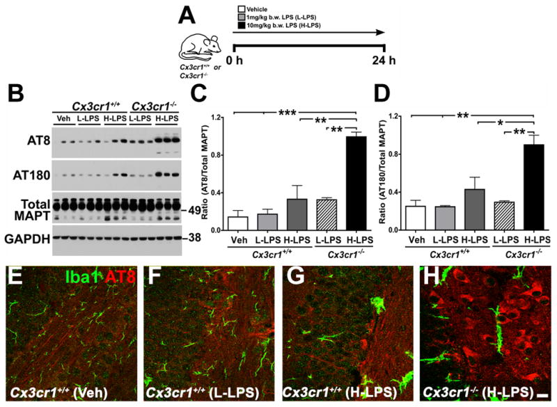
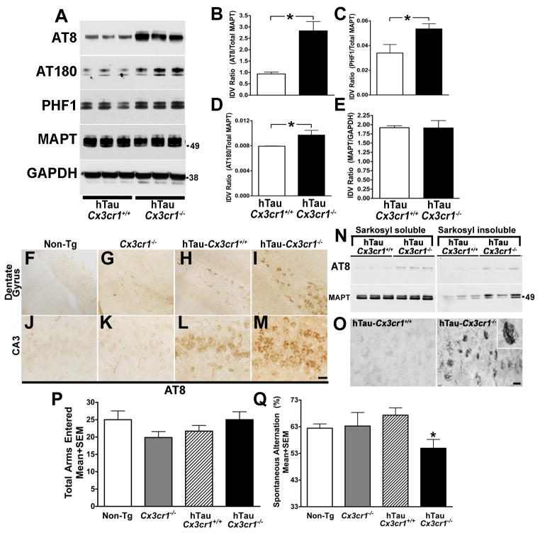
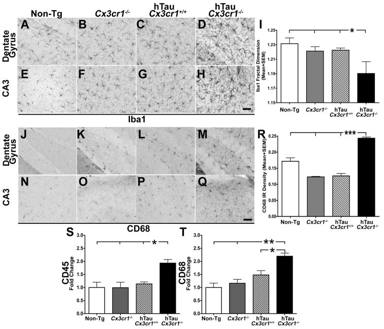
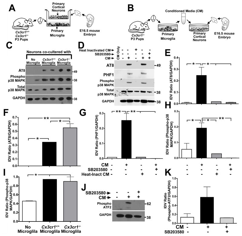
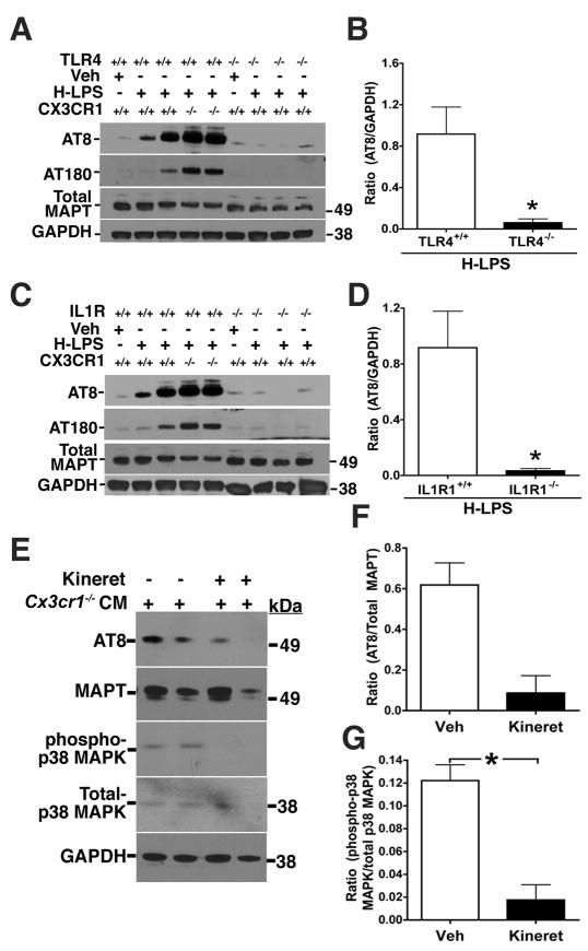
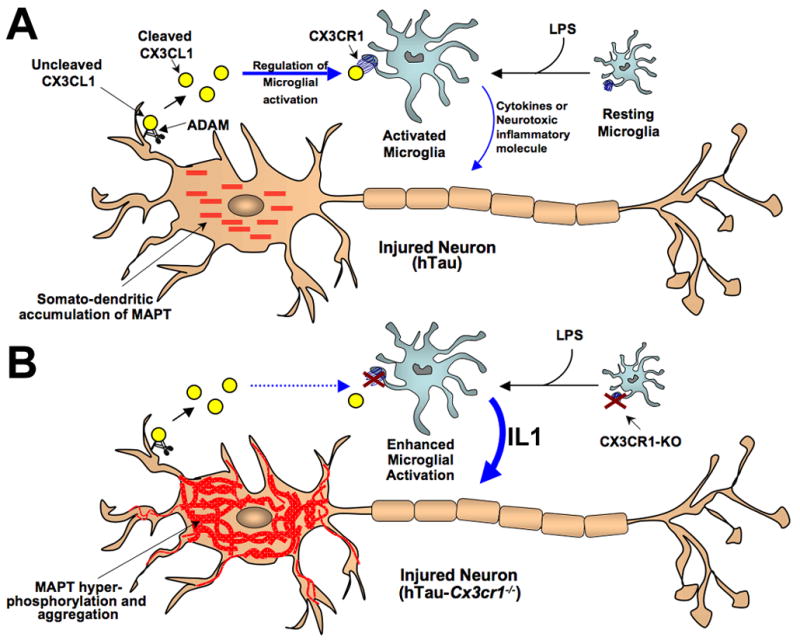
Aggregates of the hyperphosphorylated microtubule-associated protein tau (MAPT) are an invariant neuropathological feature of tauopathies. Here, we show that microglial neuroinflammation promotes MAPT phosphorylation and aggregation. First, lipopolysaccharide-induced microglial activation promotes hyperphosphorylation of endogenous mouse MAPT in nontransgenic mice that is further enhanced in mice lacking the microglial-specific fractalkine receptor (CX3CR1) and is dependent upon functional toll-like receptor 4 and interleukin-1 (IL-1) receptors. Second, humanized MAPT transgenic mice lacking CX3CR1 exhibited enhanced MAPT phosphorylation and aggregation as well as behavioral impairments that correlated with increased levels of active p38 MAPK. Third, in vitro experiments demonstrate that microglial activation elevates the level of active p38 MAPK and enhances MAPT hyperphosphorylation within neurons that can be blocked by administration of an interleukin-1 receptor antagonist and a specific p38 MAPK inhibitor. Taken together, our results suggest that CX3CR1 and IL-1/p38 MAPK may serve as novel therapeutic targets for human tauopathies.


Figures

Figure 1
Figure 1. Enhancement of microglial activation via injection of LPS or genetic deletion of Cx3cr1 promotes MAPT hyperphosphorylation
(A) Schematic diagram showing that two-month-old Cx3cr1+/+ or Cx3cr1−/− mice were injected with a single dose of L-LPS (1.0 mg/kg b.w; i.p.) or H-LPS (10.0 mg/kg b.w; i.p) or Vehicle (Veh), sacrificed after 24 h and brains processed either for biochemical or immunohistochemical analysis. (B) Western immunoblot of hippocampal extracts shows LPS induces dose-dependent increases in AT8 (ser202) and AT180 (thr231) site phosphorylation of endogenous mouse MAPT in Cx3cr1+/+ mice, which was further elevated in Cx3cr1−/− mice injected with L-LPS or H-LPS. Total MAPT and glyceraldehyde 3-phosphate dehydrogenase (GAPDH) levels were similar across all samples. (C–D) Quantification of Western blots revealed a statistically significant (n=4 for Cx3cr1+/+ with Veh; n=5 for Cx3cr1+/+ with L-LPS; n=4 for Cx3cr1−/− with L-LPS; n=3 for Cx3cr1+/+ with H-LPS and n=3 for Cx3cr1−/− with H-LPS; mean±s.e.m of integrated density value – IDV ratio; *p<0.05; **p<0.01; ***p<0.001; one-way ANOVA with Tukey multiple comparison posthoc test) increase in the AT8/Total MAPT and AT180/Total MAPT with H-LPS treatment of wild-type mice and an even greater increase in LPS injected Cx3cr1−/− mice. (E–H) Double immunofluorescence revealing Iba1+ morphological activation of microglia (green) and AT8 + neurons (red) in wild-type mice injected with LPS that was further enhanced in LPS injected Cx3cr1−/− mice. Scale bar 10 μm. See also Figure S1.
Figure 2
Figure 2. CX3CR1 deficiency induces MAPT hyperphosphorylation and aggregation as well as p38 MAPK activation in six-month-old hTau mice
(A) Western blot analysis of hippocampal lysates revealed an increase in MAPT phosphorylation at AT8, AT180 and PHF1 in hTau-Cx3cr1−/− mice, but no alterations in total MAPT levels (antibody Tau5); GAPDH was loading control. (B–E) Quantification of western blots for AT8, PHF1, AT180 and total MAPT revealed a statistically significant (n=5 for hTau-Cx3cr1+/+; n=5 for hTau-Cx3cr1−/−; mean±s.e.m of IDV; *p<0.05; unpaired t test) increase in AT8 (B), AT180 (D) and PHF1 (C), but not detergent-soluble total MAPT (E) in hTau-Cx3cr1−/− mice compared to hTau-Cx3cr1+/+ mice. (F–M) Immunohistochemistry revealed an increase in the number of AT8 positive neurons in the dentate gyrus (DG) (I) and CA3 (M) in hTau-Cx3cr1−/− mice compared to hTau-Cx3cr1+/+ mice (H, L), Cx3cr1−/− (G, K) or non-transgenic (Non-Tg) controls (F, J), Scale bar 20 μm. (N) Sarkosyl insoluble and AT8 + MAPT was higher in the hippocampi of hTau-Cx3cr1−/− mice than hTau-Cx3cr1+/+ mice. (O) Numerous Gallyas silver positive CA3 neurons were detected in the hippocampus of hTau-Cx3cr1−/− mice but not hTau-Cx3cr1+/+ mice. Scale bar 10 μm. (P–Q) Six-month-old Cx3cr1−/−, hTau-Cx3cr1+/+ and hTau-Cx3cr1−/− mice (n=8 per age group) were subjected to Y-maze test. While hTau-Cx3cr1−/− mice appeared similar to Cx3cr1−/− and hTau-Cx3cr1+/+ mice motor abilities, including the number of arms entered in the Y-maze (P), hTau-Cx3cr1−/− mice exhibited a significant deficit (*p<0.05; one-way ANOVA with Dunnett’s multiple comparison test at 99% confidence interval) in the percent spontaneous alternation when compared to control group (Q), indicating deficiencies in spatial working memory. See also Figure S2.
Figure 3
Figure 3. CX3CR1 deficiency results in enhanced microglial activation hTau mice
(A–H) Iba1+ microglia in the DG (A–D) and CA3 (E–H) of six-month-old hTau-Cx3cr1−/− mice displayed activated phenotype compared to hTau-Cx3cr1+/+, Cx3cr1−/− and non-transgenic (Non-Tg) controls. (I) Morphometric analysis of Iba1+ microglia from hTau-Cx3cr1−/− mice displayed significantly (n=3 animals per group; mean±s.e.m; *p<0.05 for hTau-Cx3cr1−/− compared to control groups; one-way ANOVA followed by Dunnett’s posthoc test) lower fractal dimension value compared to hTau-Cx3cr1+/+, Cx3cr1−/− and non-tg controls. (J–Q) Increase in the number of CD68 (phagocytic marker) positive cells in the DG (J–M) and CA3 (N–Q) of hTau-Cx3cr1−/− mice compared to hTau-Cx3cr1+/+, Cx3cr1−/− or non-tg controls. (R) Quantification of the CD68+ density revealed a significant increase (n=3 mice per group; mean ± s.e.m; ***p<0.0001; one-way ANOVA followed by Tukey posthoc test) in hTau-Cx3cr1+/+ mice when compared to hTau-Cx3cr1+/+, Cx3cr1−/− or non-tg controls. Scale bar 10 μm. (S–T) Quantitative real-time PCR analysis for CD45 and CD68 transcripts revealed a significant increase (normalized to non-transgenics; n=3 mice per group; mean±s.d; *p<0.05, **p<0.01; one-way ANOVA followed by Tukey posthoc test) in both CD45 and CD68 in the hemibrains of six-month-old hTau-Cx3cr1−/− mice compared to age-matched controls. See also Figure S3.
Figure 4
Figure 4. Microglial-derived soluble factors induce MAPT phosphorylation in neurons via p38 MAPK pathway
(A–B) Schematics displaying details of microglia–neuron co-culture experiment (A) and microglia conditioned media (CM) treatment on primary neurons (B). Non-transgenic controls (Cx3cr1+/+) or Cx3cr1−/− microglia were co-cultured with 21 DIV primary cortical neurons derived from non-transgenic (Cx3cr1+/+) embryos for 24 h and the neuronal lysates were processed for biochemical analysis. For the CM experiments, 21 DIV Cx3cr1+/+ neurons were treated with 25% ( + ) Cx3cr1−/− microglial CM for 24 h prior to biochemical analysis of neuronal lysates. CM from empty wells ( − ) served as a negative control. (C, F, I) Neurons co-cultured with Cx3cr1+/+ microglia displayed significant induction of AT8 site MAPT phosphorylation (C, F) and p38 MAPK activation (C, I) compared to neurons co-cultured with no-microglia. AT8 site phosphorylation was further enhanced when the neurons were co-cultured with Cx3cr1−/− microglia (n=3 independent cultures; mean±s.e.m of IDV ratios; *p<0.05, **p<0.01; one-way ANOVA followed by Tukey posthoc test). (D, E, G, H) Cx3cr1−/− microglial CM significantly induced neuronal AT8 and PHF1 site MAPT phosphorylation and p38 MAPK (phospho-thr180/tyr182) activation. Heat inactivation of microglial CM (microwaved until boiling and cooled) prior to neuronal treatment blocked neuronal MAPT phosphorylation and p38 MAPK activation. Pre-treatment (30 min) of primary neurons with SB203580, a p38 MAPK inhibitor, also significantly blocked the effects of microglial CM (n=3 independent cultures; mean±s.e.m of IDV ratios; *p<0.05, **p<0.01; one-way ANOVA-Dunnett’s posthoc test for AT8 and Tukey posthoc test for PHF1 and phospho-p38 MAPK). (J, K) Levels of activated-ATF2 (phospho-thr71) were elevated in microglia CM treated neuronal lysates and was blocked by SB203580 pre-treatment. All experiments were performed in duplicates in three independent cultures.
Figure 5
Figure 5. IL1R1 and TLR4 deficiency blocks LPS-induced MAPT phosphorylation
Two-month-old Cx3cr1+/+, Cx3cr1−/−, Tlr4−/− (A, B) and Il1r1−/− (C, D) mice were injected with either a single dose of vehicle or high dose LPS (10 mg/kg). (A, C) Western immunoblot of hippocampal extracts shows LPS induces dose-dependent increases in AT8 and AT180 site phosphorylation of endogenous mouse MAPT in Cx3cr1+/+ mice, that was further elevated in Cx3cr1−/− mice, but blocked in Tlr4−/− (A) and Il1r1−/− (C) mice to levels similar to that observed in vehicle injected Cx3cr1+/+ mice. Total MAPT and GAPDH levels were similar across all samples. (B, D) Quantification of Western blots revealed a statistically significant decrease in the ratios of AT8/GAPDH and AT180/GAPDH (data not shown) in the IL1R1−/− (n=3) and TLR4−/− (n=3) mice injected with H-LPS when compared to H-LPS injected treated Cx3cr1+/+ mice (n=4; mean ± s.e.m of integrated density value – IDV ratio; * p<0.05; unpaired t test). (E–G) IL1 Receptor Antagonist (IL1-RA/Kineret) Attenuates Microglial-Induced Neuronal MAPT Phosphorylation. (E) 21 DIV primary cortical neurons were pretreated (15 min) with vehicle (−) or 50 ng/ml IL1-RA (Kineret) prior to incubation with conditioned media (CM) from Cx3cr1−/− microglia. 24 h after the incubation, the neuronal lysates were probed for AT8, total MAPT (Tau5), phospho-p38 MAPK, total p38 MAPK and GAPDH. Note reduction in AT8 and phospho-p38 MAPK immunoreactive bands in IL1-RA treated neurons. (F and G) Quantification of the AT8/Total MAPT and phospho-p38 MAPK/total p38 MAPK ratios revealed more than four-fold reduction in the AT8 site phosphorylation (F) and statistically significant (*p<0.05; mean±s.e.m; n=3; unpaired t test) reduction in phospho-p38 MAPK levels (G) as a result of Kineret pretreatment. All experiments were performed in duplicates in three independent cultures.
Figure 6
Figure 6. Model of fractalkine (CX3CL1/CX3CR1) signaling in neurodegenerative tauopathies
(A) In the CNS, the chemokine fractalkine (CX3CL1) is expressed by neurons and cleaved by the A Disintegrin And Metalloproteases (ADAM10 and ADAM17) to release cleaved soluble CX3CL1, which binds to the G-coupled transmembrane receptor (CX3CR1) present on microglia and regulates microglial activity. Lipopolysaccharide (LPS) induces microglial activation, while neuronal stress (such as overexpression and somato-dendritic accumulation of human MAPT in case of hTau) results in enhanced release of CX3CL1. An interaction between fractalkine (CX3CL1) and its receptor (CX3CR1) downregulates microglial activity and dampens the toxic effects of activated microglia. (B) Lack of CX3CR1 (Cx3cr1−/−) in microglia disrupts CX3CL1-CX3CR1 communication (dashed blue arrow), dysregulates microglial activity induced by LPS or neuronal stress (human MAPT overexpression in hTau mice) and results in enhanced release of soluble factors (including IL1) from activated microglia, can induce active neuronal p38 MAPK and lead to enhanced phosphorylation and aggregation of MAPT. Notably, this molecular mechanism would lead to a feed-forward induction of MAPT phosphorylation/aggregation and microglial activation.

References

    1. Andorfer C, Acker CM, Kress Y, Hof PR, Duff K, Davies P. Cell-cycle reentry and cell death in transgenic mice expressing nonmutant human tau isoforms. J Neurosci. 2005;25:5446–5454. - PMC - PubMed
    1. Andorfer C, Kress Y, Espinoza M, de Silva R, Tucker KL, Barde YA, Duff K, Davies P. Hyperphosphorylation and aggregation of tau in mice expressing normal human tau isoforms. J Neurochem. 2003;86:582–590. - PubMed
    1. Bhaskar K, Miller M, Chludzinski A, Herrup K, Zagorski M, Lamb BT. The PI3K-Akt-mTOR pathway regulates Abeta oligomer induced neuronal cell cycle events. Mol Neurodegener. 2009;4:14. - PMC - PubMed
    1. Braak H, Braak E. Demonstration of amyloid deposits and neurofibrillary changes in whole brain sections. Brain Pathol. 1991;1:213–216. - PubMed
    1. Cardona AE, Pioro EP, Sasse ME, Kostenko V, Cardona SM, Dijkstra IM, Huang D, Kidd G, Dombrowski S, Dutta R, et al. Control of microglial neurotoxicity by the fractalkine receptor. Nat Neurosci. 2006;9:917–924. - PubMed

Publication types

MeSH terms