. 2010 Oct 4;191(1):61-74.
doi: 10.1083/jcb.200912046.

Sds22 regulates aurora B activity and microtubule-kinetochore interactions at mitosis

Affiliations

Sds22 regulates aurora B activity and microtubule-kinetochore interactions at mitosis

Markus Posch et al. J Cell Biol. .

Abstract

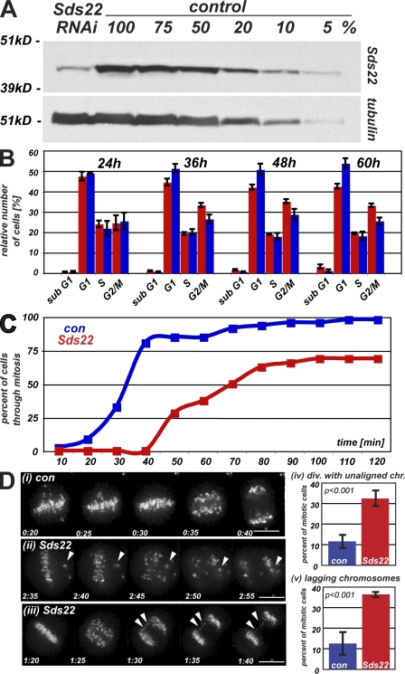
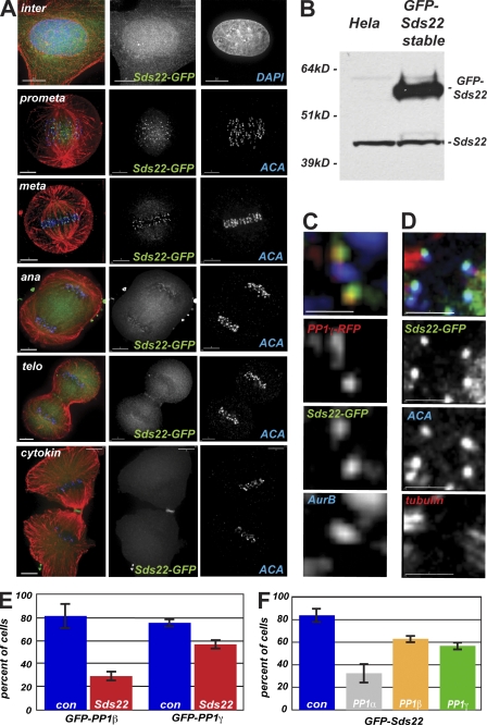
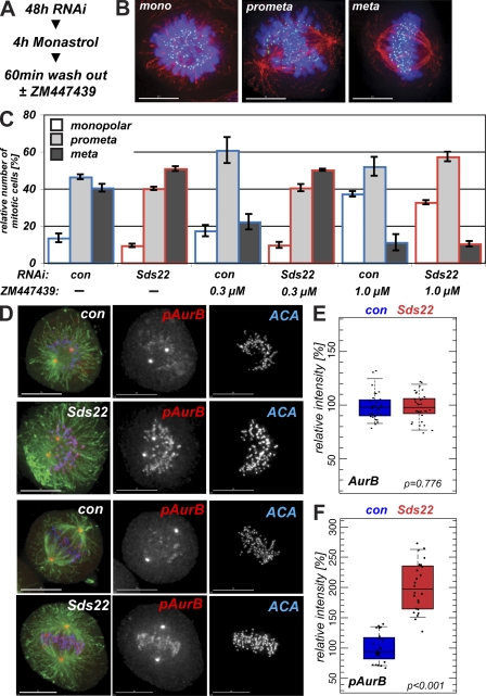
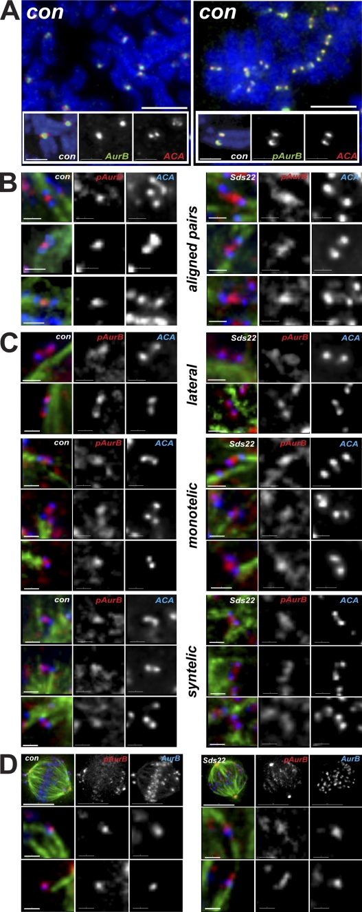
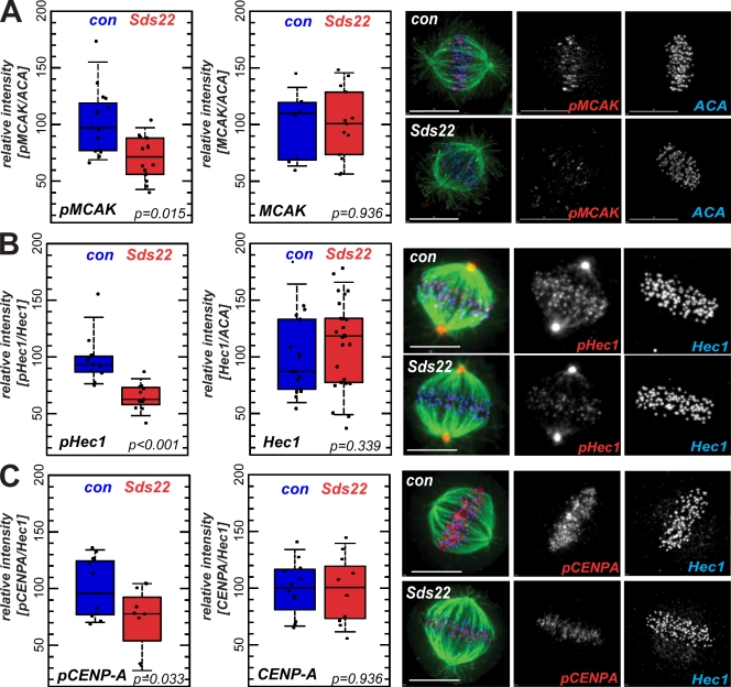
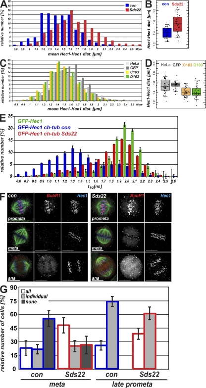
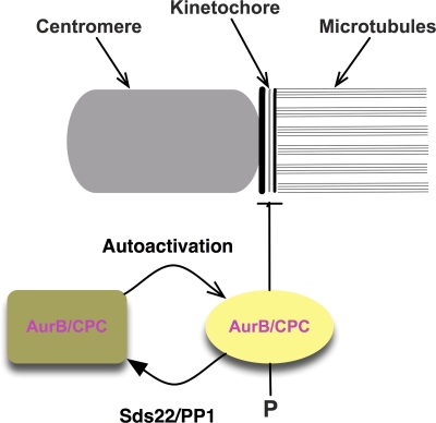
We have studied Sds22, a conserved regulator of protein phosphatase 1 (PP1) activity, and determined its role in modulating the activity of aurora B kinase and kinetochore-microtubule interactions. Sds22 is required for proper progression through mitosis and localization of PP1 to mitotic kinetochores. Depletion of Sds22 increases aurora B T-loop phosphorylation and the rate of recovery from monastrol arrest. Phospho-aurora B accumulates at kinetochores in Sds22-depleted cells juxtaposed to critical kinetochore substrates. Sds22 modulates sister kinetochore distance and the interaction between Hec1 and the microtubule lattice and, thus, the activation of the spindle assembly checkpoint. These results demonstrate that Sds22 specifically defines PP1 function and localization in mitosis. Sds22 regulates PP1 targeting to the kinetochore, accumulation of phospho-aurora B, and force generation at the kinetochore-microtubule interface.


Figures

Figure 1.
Figure 1.
Depletion of Sds22 causes defects in mitotic progression. (A) Depletion of Sds22 by siRNAi. 70 µg extracts of logarithmically growing HeLa cells 48 h after transfection with Sds22-specific and control siRNAi were probed with Sds22-specific polyclonal antibody by Western blotting and reprobed with anti-tubulin as a loading control. (B) Cell cycle profile of HeLa cells after Sds22 RNAi. Subpopulation of HeLa in specific cell cycle stages as determined by DNA content analysis (FACS of propidium iodide–stained cells). Cells were transfected with control (blue) or Sds22-specific (red) siRNAi and analyzed at the time points indicated (hours). Results are from four separate experiments. (C) Timing of mitosis in Sds22-depleted and control cells. HeLa (Kyoto) cells stably expressing EGFP–CENP-A were imaged at 5- or 10-min intervals starting 48 h after transfection with Sds22-specific and control RNAi. Time elapsed from NEB to the onset of anaphase was plotted against cumulative percentage of cells having completed mitosis. Data were generated from three independent experiments. Data from two experiments recorded at 5-min intervals were binned to 10 min and combined with one experiment recorded at 10-min intervals. Total number of cells: control, 314; Sds22 RNAi, 293. (D) Mitotic defects in Sds22-depleted cells. Still images of C are shown with time after NEB indicated (hours:minutes). (i) Control cell. (ii and iii) Sds22-depleted cell. Defects are indicated by arrowheads. (iv) Mean percentage of mitotic cells dividing with unaligned chromosomes in C. (v) Mean percentage of mitotic cells dividing with lagging kinetochores in the midzone in C. Error bars indicate SEM. Bars, 10 µm.
Figure 2.
Figure 2.
Sds22 functions at kinetochores. (A) Intracellular localization of Sds22-GFP. Immunofluorescence of HeLa cells fixed 48 h after transient transfection with GFP-Sds22 plasmid. Overlay shows DAPI or anti–human centromere antibody (ACA; blue), Sds22-GFP (green), and microtubules (red). (B) Stable expression of Sds22-GFP in HeLa cells. Western blot analysis of extracts from untransfected HeLa cells and cell clone D103 stably expressing GFP-Sds22 probed with polyclonal Sds22-specific antibody. (C) Relative localization of Sds22 and PP1 at the kinetochore. Immunofluorescence of HeLa cells transiently transfected with mRFP–PP1-γ and GFP-Sds22 fixed 48 h after transient transfection. Overlay shows mRFP1–PP1-γ (red), Sds22-GFP (green), and aurora B (blue). (D) Relative localization of Sds22 and PP1 at the kinetochore. Immunofluorescence of HeLa cells transiently transfected with GFP-Sds22 plasmid fixed 48 h after transient transfection. Overlay shows Sds22-GFP (green), ACA (blue), and anti-tubulin (red). (E) Dependency of PP1 kinetochore localization on Sds22. Kinetochore localization GFP–PP1-β and GFP–PP1-γ were scored in mitotic spreads of HeLa cells 48 h after transfection with Sds22-specific and control siRNA. Data are from three separate experiments testing localization of either GFP–PP1-β or GFP–PP1-γ. Total number of cells for GFP–PP1-β: control, 47; Sds22 RNAi, 61. Total number of cells for GFP–PP1-γ: control, 162; Sds22 RNAi, 134. P < 0.001. P-values were calculated by Fisher’s exact test. (F) Dependency of Sds22-GFP kinetochore localization on PP1 isoforms. Relative number of cells showing EGFP-Sds22 at kinetochores in mitotic spreads of a stable HeLa cell line (clone D103) 48 h after transfection with control or PP1 isoform–specific RNAi. Data are from five independent experiments. Total number of cells and p-values: control, 93; PP1-α, 40 (P < 0.001); PP1-β, 84 (P = 0.024); PP1-γ, 81 (P = 0.001). Error bars indicate SEM. Bars: (A, top row) 10 µm; (A, rows 2–6) 5 µm; (C and D) 2 µm.
Figure 3.
Figure 3.
Sds22 depletion affects aurora B activity. (A) Experimental protocol. (B) Monopolar, prometaphase, and metaphase data from monastrol recovery. Examples of cells at the three points of recovery counted in C. Overlay shows DAPI (blue), ACA (green), and microtubules (red). (C) Effect of Sds22 depletion on the recovery from monastrol arrest. 48 h after transfection of HeLa cells with control or Sds22-specific RNAi, duplexes were treated for 4 h with monastrol and released into fresh medium or medium containing 0.3 or 1 µM ZM. After 1 h, cells were fixed, stained (A), and mitotic cells were scored for their progression through mitosis. Total number of cells: 0 µM control, 347; 0 µM Sds22, 1,056; 0.3 µM control, 1,336; 0.3 µM Sds22, 1,230; 1.0 µM control, 1,261; 1.0 µM Sds22, 1,360. (D) Aurora B T232 phosphorylation depends on Sds22. HeLa cells were fixed and immunostained with anti–phospho-T232 antibody 48 h after transfection with control (con) or Sds22-specific duplexes. Overlay shows tubulin (green), anti–phospho-T232 aurora B (red), and ACA (blue). The spindle pole staining is not sensitive to inhibition with 1 µM ZM and is likely spurious and the result of cross-reaction with phospho–aurora A (Fuller et al., 2008). (E and F) Effect of Sds22 depletion on total aurora B and T232 aurora B phosphorylation in metaphase. Quantification of metaphase cells fixed and stained with aurora B (E) and anti-phospho–aurora B T232 (F) antibodies in fixed HeLa cells 48 h after transfection with control or Sds22-specific duplexes (see Materials and methods). Intensities were normalized relative to the general kinetochore marker ACA. Box plots show mean (middle line), top and bottom quartiles of the data as top and bottom of the box, and whiskers as the extent of 90% of the data. P-values were calculated using a Kolmogorov-Smirnov test. (E) Total number of cells: control RNAi, 30; Sds22 RNAi, 34. (F) Total number of cells: control RNAi, 24; Sds22 RNAi, 24. Error bars show SEM from three independent experiments. Bars, 10 µm.
Figure 4.
Figure 4.
Localization of phospho–T232 aurora B is determined by Sds22. (A) Localization of aurora B and phospho-T232 aurora B in chromosome spreads after arrest with nocodazole. Phospho-T232 aurora B is concentrated at kinetochores, whereas bulk aurora B remains concentrated at centromeres. Overlay shows DAPI (blue), aurora B or anti–phospho-T232 aurora B (green), and ACA (red). (B) Localization of phospho-T232 aurora B in bioriented chromosomes in fixed intact cells. Sds22 depletion causes phospho-T232 aurora B to spread toward kinetochores. Overlay shows ACA (blue), tubulin (green), and anti–phospho-T232 aurora B (red). (C) Localization of phospho-T232 aurora B in laterally and monooriented chromosomes in fixed intact cells. Phospho-T232 aurora B appears on kinetochores in control and Sds22-depleted cells. Overlay shows ACA (blue), tubulin (green), and anti–phospho-T232 aurora B (red). (D) Colocalization of phospho-T232 aurora B and total aurora B in fixed intact cells. Sds22 depletion causes phospho-T232 aurora B to spread toward kinetochores. Overlay shows phospho-T232 aurora B (red), aurora B (blue), and tubulin (green). Bars: (A, top) 5 µm; (A, insets) 2 µm; (B, C, and D [bottom]) 1 µm; (D, top) 10 µm.
Figure 5.
Figure 5.
Effect of Sds22 depletion on substrate phosphorylation and centromere binding of aurora B. (A–C) Representative images and quantification of metaphase cells fixed and stained with anti–phospho-Ser92 MCAK and total MCAK (A), anti–phospho-Hec1 and total Hec1 (B), and anti-phospho–CENP-A and total CENP-A (C) antibodies in fixed HeLa cells 48 h after transfection with control (con) or Sds22-specific duplexes (see Materials and methods). Intensities were normalized relative to general kinetochore markers (Hec1 and ACA as indicated). Box plots and statistical analysis are as in Fig. 3 (E and F). Bars, 10 µm.
Figure 6.
Figure 6.
Effects of Sds22 on sister kinetochore dis-tances and interactions between microtubules and kinetochores. (A–D) Histograms showing distribution of interkinetochore distances (A and C) and interkinetochore distances per cell (B and D). (A and B) Sister kinetochore distances after Sds22 depletion. Hec1–Hec1 distances of sister pairs were measured in HeLa cells fixed 48 h after transfection with control siRNA or Sds22-specific siRNA (C and D) Hec1–Hec1 distances of sister pairs in untransfected HeLa cells or stable cell lines expressing GFP alone or GFP-Sds22 (C103 and D103). Data from two independent experiments are shown. Total number of cells: control siRNA, 28; Sds22 RNAi, 33; normal HeLa, 37; HeLa GFP, 30; C103, 42; D103, 28. (B) Kolmogorov-Smirnov test of significance: control siRNA and Sds22-depleted cells, P < 6 × 10⁻⁵. (D) Kolmogorov-Smirnov test of significance: normal HeLa and HeLa GFP, P < 0.05; HeLa GFP and C103, P < 6 × 10⁻⁶; HeLa GFP and D103, P < 1 × 10⁻⁵; C103 and D103, P < 0.9. (E) Effect of Sds22 on the microtubule–kinetochore attachment. Fluorescence lifetime measurement of FRET between EGFP-Hec1 and mCherry-tubulin is shown. Fluorescence lifetime of EGFP was determined in HeLa cells 48 h after transfection with either EGFP-Hec1 (green bars) or cotransfection with EGFP-Hec1 and mCherry-tubulin together with either control (blue bars) or Sds22-specific (red bars) RNAi duplexes. (F) Recruitment of BubR1 in Sds22-depleted cells. HeLa cells were fixed and immunostained 48 h after transfection with control or Sds22-specific RNAi duplexes. Bars, 10 µm. (G) Effect of Sds22 depletion in localization of BubR1. HeLa cells in F that showed a defined plate with aligned chromosomes were subdivided into cells where all chromosomes had clearly aligned to the metaphase plate as opposed to cells with one or more unaligned chromosomes. Both categories were scored on whether all, individual, or no kinetochores at all stained positive for BubR1. Data from four independent experiments are shown. Total number of cells: control RNAi, 153; Sds22 RNAi, 99. P-values between control and Sds22 RNAi were calculated by χ2 test: aligned kinetochores, P = 0.0004; unaligned kinetochores, P = 0.1172. Error bars indicate mean ± SEM.
Figure 7.
Figure 7.
A model for regulation of aurora B by Sds22 and PP1. The drawing shows a schematic model of the activation of aurora B kinase activity and its regulation by Sds22/PP1. Aurora B activates through autophosphorylation of phospho-T232 at the both the centromere and kinetochore. Phospho–aurora B can destabilize interactions between kinetochores and microtubules through phosphorylation of unknown targets. Sds22/PP1 dephosphorylates phospho–aurora B and, thus, indirectly functions to stabilize kinetochore–microtubule interactions. Centromere-associated aurora B may inhibit Sds22/PP1 when distance between centromere and kinetochore are low, e.g., in syntelic or merotelic attachments.

Similar articles

Cited by

References

    1. Adams R.R., Maiato H., Earnshaw W.C., Carmena M. 2001. Essential roles of Drosophila inner centromere protein (Incenp) and aurora B in histone H3 phosphorylation, metaphase chromosome alignment, kinetochore disjunction, and chromosome segregation. J. Cell Biol. 153:865–880 10.1083/jcb.153.4.865 - DOI - PMC - PubMed
    1. Andrews P.D., Ovechkina Y., Morrice N., Wagenbach M., Duncan K., Wordeman L., Swedlow J.R. 2004. Aurora B regulates MCAK at the mitotic centromere. Dev. Cell. 6:253–268 10.1016/S1534-5807(04)00025-5 - DOI - PubMed
    1. Ceulemans H., Vulsteke V., De Maeyer M., Tatchell K., Stalmans W., Bollen M. 2002. Binding of the concave surface of the Sds22 superhelix to the alpha 4/alpha 5/alpha 6-triangle of protein phosphatase-1. J. Biol. Chem. 277:47331–47337 10.1074/jbc.M206838200 - DOI - PubMed
    1. Cheeseman I.M., Desai A. 2008. Molecular architecture of the kinetochore-microtubule interface. Nat. Rev. Mol. Cell Biol. 9:33–46 10.1038/nrm2310 - DOI - PubMed
    1. Cheeseman I.M., Anderson S., Jwa M., Green E.M., Kang J., Yates J.R., III, Chan C.S., Drubin D.G., Barnes G. 2002. Phospho-regulation of kinetochore-microtubule attachments by the Aurora kinase Ipl1p. Cell. 111:163–172 10.1016/S0092-8674(02)00973-X - DOI - PubMed

Publication types