. 2010 Oct 4;191(1):75-86.
doi: 10.1083/jcb.201004030.

The differential interaction of snRNPs with pre-mRNA reveals splicing kinetics in living cells

Affiliations

The differential interaction of snRNPs with pre-mRNA reveals splicing kinetics in living cells

Martina Huranová et al. J Cell Biol. .

Abstract

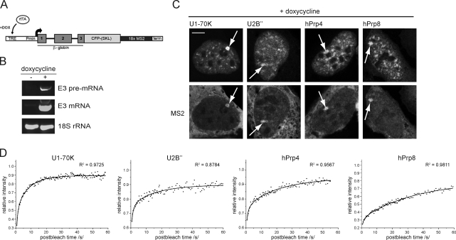
Precursor messenger RNA (pre-mRNA) splicing is catalyzed by the spliceosome, a large ribonucleoprotein (RNP) complex composed of five small nuclear RNP particles (snRNPs) and additional proteins. Using live cell imaging of GFP-tagged snRNP components expressed at endogenous levels, we examined how the spliceosome assembles in vivo. A comprehensive analysis of snRNP dynamics in the cell nucleus enabled us to determine snRNP diffusion throughout the nucleoplasm as well as the interaction rates of individual snRNPs with pre-mRNA. Core components of the spliceosome, U2 and U5 snRNPs, associated with pre-mRNA for 15-30 s, indicating that splicing is accomplished within this time period. Additionally, binding of U1 and U4/U6 snRNPs with pre-mRNA occurred within seconds, indicating that the interaction of individual snRNPs with pre-mRNA is distinct. These results are consistent with the predictions of the step-wise model of spliceosome assembly and provide an estimate on the rate of splicing in human cells.


Figures

Figure 1.
Figure 1.
GFP-tagged snRNP proteins incorporate into snRNPs. (A) Images of cells expressing GFP-tagged snRNP proteins. GFP-tagged proteins were localized to the nucleoplasm and enriched in splicing factor compartments and Cajal bodies (arrows). Bar, 5 µm. (B) snRNPs were immunoprecipitated using anti-GFP or anti-Sm antibodies, and coprecipitated RNAs were visualized. The positions of snRNAs and rRNAs are depicted. Note that a small amount of U6 and U5 snRNAs coprecipitate with U2 snRNP (U2A′ line) and U2 snRNA associated with U5 snRNP (hPrp8 and Snu114 lines). Vertical black lines indicate that intervening lanes have been spliced out. (C) Proliferation of control cells (hPrp31-GFP) and cells expressing mouse Snu114-GFP was assayed by DDAO staining after knockdown of endogenous hSnu114. Reduction of hSnu114 levels (see Western blot [WB]) resulted in proliferation defects that were partially rescued by expression of RNAi-resistant mouse Snu114-GFP. NC, negative control.
Figure 2.
Figure 2.
FCS measures the in vivo mobility of snRNPs. (A) FCS measurements were performed in the nucleoplasm of cells either stably expressing U1-70K–GFP or transiently expressing GFP. The autocorrelation function of free GFP is fitted with a one-component anomalous diffusion model. The autocorrelation function of U1-70K is fitted with a two-component diffusion model. (B) The autocorrelation function of U1-70KΔ1–197 is fitted with a two-component diffusion model. Deletion of the RNA-binding domain of U1-70K results in a fourfold increase in diffusion of the slow component. (C) DRB treatment (5 h) inhibits RNA polymerase II and results in the enlargement and rounding up of splicing factor compartments. DRB treatment had only a minimal effect on snRNP diffusion, with both fast and slow components still present. (A–C) Crosses indicate spots in the nucleoplasm where FCS measurements were performed, and weighted residuals are shown to assess the fit quality. Bar, 5 µm.
Figure 3.
Figure 3.
snRNPs interact independently with pre-mRNA, as analyzed by FRAP. (A) FRAP measurements were performed in the nucleoplasm, as depicted by circles. Bar, 5 µm. (B–D) U1-70K (B), U2A′ (C), and hPrp8 (D) FRAP curves representing a mean of 10–15 measurements before and after DRB treatment are shown. FRAP curves were fitted either with a pure diffusion model (DRB treatment) or the full model (no treatment). Calculated diffusion coefficients and dissociation constants koff are shown in Table II. R2 values evaluating fit quality are shown next to the curves. (E) Coimmunoprecipitation of snRNAs with anti-GFP antibodies from U1-70K-GFP, U2A′-GFP, hPrp4-GFP, and hPrp8-GFP cell lines before and after DRB or α-amanitin treatment. Transcriptional inhibition had no effect on the precipitation of U1 and U2 snRNAs but eliminated U6 snRNA association with the U2 snRNP. Transcriptional inhibition reduced the formation of U4/U6 and U4/U6•U5 snRNPs, as shown by the decrease of U4 and U6 snRNAs levels in hPrp4 (asterisks) and hPrp8 precipitates. NC, negative control. (F) Schematic representation of snRNP interaction times with pre-mRNA. We assume that the U4/U6 proteins hPrp4 and hPrp31 leave with the U4 snRNA, whereas U6 snRNA stays associated with the activated spliceosome.
Figure 4.
Figure 4.
The prolonged interaction of U1 and U2 snRNPs with pre-mRNA after splicing inhibition. (A) After isoginkgetin treatment, GFP-tagged snRNP proteins relocalized to enlarged splicing factor compartments. FRAP measurements were performed in the nucleoplasm, as depicted by circles. Bar, 5 µm. (B) Splicing inhibition increased the immobile fractions and reduced the mobility of U1-70K and U2A′ (Table II). The mean of 10–12 measurements is shown. (C) Coimmunoprecipitation of U1 snRNA with U2A′-GFP after isoginkgetin (isog) treatment. (D) Splicing inhibition had minimal impact on U4/U6 snRNP mobility (Table II). (E) Splicing inhibition resulted in increased U5 snRNP mobility that can be described by a pure diffusion model. A portion of U5 snRNPs was bound in an immobile fraction. (F) Schematic representation of snRNP interaction times with pre-mRNA after splicing inhibition. (G) Coimmunoprecipitation of snRNAs from hPrp4-GFP and hPrp8-GFP cell lines with anti-GFP antibodies before and after isoginkgetin treatment. Splicing inhibition impairs U4/U6 di-snRNP and U4/U6•U5 tri-snRNP formation as shown by the reduced coprecipitation of U4 and U6 snRNAs with hPrp4 and hPrp8 proteins. The vertical black line indicates that intervening lanes have been spliced out. (H) The efficiency of GFP-tagged protein immunoprecipitation was verified by Western blot analysis. NC, negative control.
Figure 5.
Figure 5.
snRNP interaction with E3 pre-mRNA. (A) Schematic representation of the E3 gene stably integrated into the genome of the U2-OS Tet-On cell line. Expression of the E3 transgene is driven by a minimal cytomegalovirus promoter (Pmin) under the control of the tetracycline response element (TRE) and is induced by the presence of doxycycline (DOX) by the reverse transactivator, rtTA. The transgene transcript contains 18× MS2-binding sites, and the encoded protein (human β-globin) is fused to CFP-SKL. (B) E3 transgene expression before and after induction. (C) Doxycycline-treated E3 cells expressing different GFP-tagged snRNP proteins (top) and MS2-mRED protein (bottom). Note the localization of snRNP proteins at the site of active transgene transcription, as depicted by MS2-mRED accumulation (arrows). Bar, 5 µm. (D) FRAP analysis of snRNP dynamics at the active transcription site of the E3 transgene (Table II). A mean of 10–12 measurements is shown.

Similar articles

Cited by

References

    1. Ali G.S., Prasad K.V., Hanumappa M., Reddy A.S. 2008. Analyses of in vivo interaction and mobility of two spliceosomal proteins using FRAP and BiFC. PLoS One. 3:e1953 10.1371/journal.pone.0001953 - DOI - PMC - PubMed
    1. Audibert A., Weil D., Dautry F. 2002. In vivo kinetics of mRNA splicing and transport in mammalian cells. Mol. Cell. Biol. 22:6706–6718 10.1128/MCB.22.19.6706-6718.2002 - DOI - PMC - PubMed
    1. Azubel M., Habib N., Sperling R., Sperling J. 2006. Native spliceosomes assemble with pre-mRNA to form supraspliceosomes. J. Mol. Biol. 356:955–966 10.1016/j.jmb.2005.11.078 - DOI - PubMed
    1. Behzadnia N., Hartmuth K., Will C.L., Lührmann R. 2006. Functional spliceosomal A complexes can be assembled in vitro in the absence of a penta-snRNP. RNA. 12:1738–1746 10.1261/rna.120606 - DOI - PMC - PubMed
    1. Behzadnia N., Golas M.M., Hartmuth K., Sander B., Kastner B., Deckert J., Dube P., Will C.L., Urlaub H., Stark H., Lührmann R. 2007. Composition and three-dimensional EM structure of double affinity-purified, human prespliceosomal A complexes. EMBO J. 26:1737–1748 10.1038/sj.emboj.7601631 - DOI - PMC - PubMed

Publication types