. 2010 Oct 4;191(1):187-97.
doi: 10.1083/jcb.201002089.

Otoferlin is a calcium sensor that directly regulates SNARE-mediated membrane fusion

Affiliations

Otoferlin is a calcium sensor that directly regulates SNARE-mediated membrane fusion

Colin P Johnson et al. J Cell Biol. .

Abstract

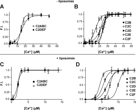
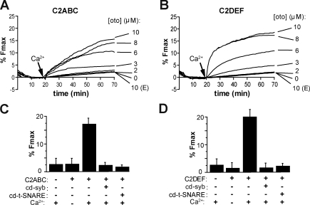
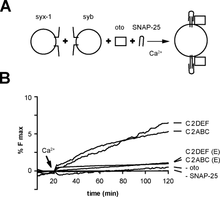
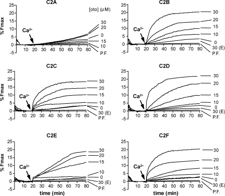
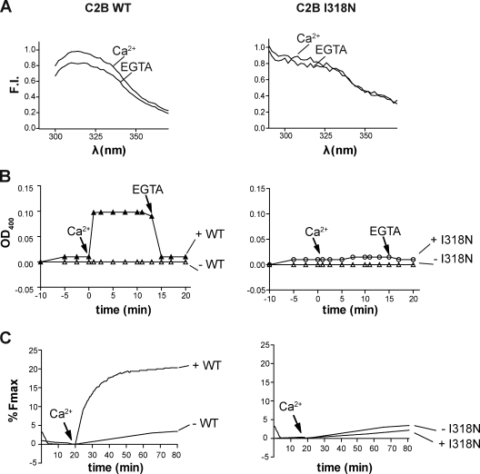
Otoferlin is a large multi-C2 domain protein proposed to act as a calcium sensor that regulates synaptic vesicle exocytosis in cochlear hair cells. Although mutations in otoferlin have been associated with deafness, its contribution to neurotransmitter release is unresolved. Using recombinant proteins, we demonstrate that five of the six C2 domains of otoferlin sense calcium with apparent dissociation constants that ranged from 13-25 µM; in the presence of membranes, these apparent affinities increase by up to sevenfold. Using a reconstituted membrane fusion assay, we found that five of the six C2 domains of otoferlin stimulate membrane fusion in a calcium-dependent manner. We also demonstrate that a calcium binding-deficient form of the C2C domain is incapable of stimulating membrane fusion, further underscoring the importance of calcium for the protein's function. These results demonstrate for the first time that otoferlin is a calcium sensor that can directly regulate soluble N-ethyl-maleimide sensitive fusion protein attachment protein receptor-mediated membrane fusion reactions.


Figures

Figure 1.
Figure 1.
Schematic diagram of full-length otoferlin and the otoferlin fragments used in this study. (A) Otoferlin is composed of six tandem C2 domains followed by a C-terminal transmembrane domain (TMD). Recombinant proteins composed of either isolated C2 domains or three–C2 domain fragments were generated according to the amino acid designations listed on the right. (B) Conservation of putative calcium-binding ligands of the C2 domains of otoferlin (oto). Synaptotagmin I (syt) C2A and C2B are listed for comparison. The numbers correspond to the order of the five aspartate residues that function as ligands in the C2A (D172, 178, 230, 232, and 238) and C2B domains (D303, 309, 363, 365, 371) of synaptotagmin I (Sutton et al., 1995; Shao et al., 1998; Fernandez et al., 2001).
Figure 2.
Figure 2.
Normalized fluorescence emission spectra of native aromatic side chains of otoferlin in the presence of 0.1 mM EGTA or 1 mM calcium. 1 µM purified otoferlin fragments were excited at 280 nm and the emission spectra collected from 290 to 380 nm. Representative spectra for each domain under EGTA or calcium conditions are shown. The traces are representative data from four independent trials. FI, fluorescence intensity.
Figure 3.
Figure 3.
Calcium-dependent changes in otoferlin fluorescence. (A and B) Normalized plots of the emission intensities of 1 µM of the three–C2 domain fragments C2ABC and C2DEF (A) and 1 µM of the isolated C2 domains C2B, C2C, C2D, C2E, and C2F (B) as a function of increasing calcium concentration in the absence of liposomes. (C and D) Calcium titrations of C2ABC, C2DEF (C), and each isolated C2 domain of otoferlin (D) in the presence of liposomes composed of 15% PS, 55% PC, and 30% PE. Error bars indicate mean ± SD (n = 4).
Figure 4.
Figure 4.
Coimmunoprecipitation of t-SNARE heterodimers with the isolated C2 domains of otoferlin. (A) The assays were performed using 20 µM of each C2 domain using an anti-syntaxin monoclonal antibody. T denotes the total C2 domain loaded into the sample, and Ca and E denote the presence of 1 mM calcium or 0.1 mM EGTA, respectively. H-chain denotes the heavy chain of the anti-syntaxin antibody (HPC-1). (B) Quantitation of the coimmunoprecipitation data using densitometry. (C) Representative gel in which each isolated C2 domain was assayed for its ability to cofloat with vesicles in an Accudenz gradient in the presence of 0.1 mM EGTA or 1 mM free calcium. (D) Calcium-triggered C2 domain–induced liposome aggregation as measured by OD400 is shown. Turbidity of samples containing liposomes was monitored in either 1 mM free calcium or 0.1 mM EGTA. Error bars indicate mean ± SD (n = 3).
Figure 5.
Figure 5.
Otoferlin stimulates SNARE-mediated proteoliposome fusion in a calcium-dependent manner. Fusion experiments were performed using donor v-SNARE proteoliposomes and t-SNARE acceptor proteoliposomes with varying concentrations of recombinant otoferlin fragments. The components were incubated together for 20 min at 37°C in the presence of 0.1 mM EGTA. At t = 20 min, calcium was added to a final concentration of 1 mM. (A and B) Both C2ABC (A) and C2DEF (B) stimulated SNARE-mediated proteoliposome fusion in response to calcium. (A and B) E indicates that the experiment was performed under EGTA conditions. (C and D) Mean value for the extent of fusion expressed as a percentage of maximum fluorescence intensity after addition of detergent for 10 µM C2ABC (C) and C2DEF (D) under the indicated conditions. Fusion was attenuated by addition of cd-syb or cd–t-SNARE. Error bars represent SD (n = 3).
Figure 6.
Figure 6.
Calcium/otoferlin enables t-SNAREs, which have not been preassembled into heterodimers, to drive membrane fusion. (A) Fusion assays were conducted using reconstituted syntaxin 1 and synaptobrevin 2 vesicles in the presence of 15 µM soluble SNAP-25 and 4 µM otoferlin. (B) Either half of otoferlin, C2ABC, or C2DEF was sufficient to accelerate membrane fusion using soluble SNAP-25. In the absence of calcium or otoferlin, fusion was not observed. E indicates that the experiment was performed under EGTA conditions.
Figure 7.
Figure 7.
The isolated C2 domains of otoferlin stimulate membrane fusion. Titrations of each isolated C2 domain, from 0 to 30 µM, demonstrate the ability of the otoferlin C2 domains C2B, C2C, C2D, C2E, and C2F to stimulate fusion between v- and t-SNARE proteoliposomes. The isolated C2 domains failed to stimulate membrane fusion between protein-free (PF) liposomes. E indicates that the experiment was performed under EGTA conditions.
Figure 8.
Figure 8.
A pathological isoleucine to asparagine missense mutation disrupts the structure and function of the C2B domain of otoferlin. (A) The fluorescence spectra of endogenous aromatic residues of the isolated WT (left) and I318N (right) mutant C2B domain differ in their spectral profiles. Although a notable difference in the fluorescence intensity (FI) of WT C2B is observed in 0.1 mM EGTA versus 1 mM calcium conditions, the spectra of the I318N mutant shows no discernable change in fluorescence in response to calcium. (B) The membrane aggregation activity of C2B is disrupted by the I318N mutation. After injection of protein at t = −5 min, calcium was added at t = 0 min to a final concentration of 1 mM. OD400 measurements for the WT C2B (left) and I318N (right) reveal that the point mutation abrogates calcium-triggered C2B-mediated liposome aggregation. The OD400 values after addition of calcium were 0.11 ± 0.02 for WT and 0.02 ± 0.01 for the mutant C2B (n = 3). (C) Reconstituted fusion assays performed in the presence of WT or I318N mutant C2B. After the addition of 1 mM free calcium, WT C2B accelerated SNARE-mediated fusion, whereas the I318N failed to enhance the rate or extent of fusion.

References

    1. Bai J., Wang C.T., Richards D.A., Jackson M.B., Chapman E.R. 2004. Fusion pore dynamics are regulated by synaptotagmin*t-SNARE interactions. Neuron. 41:929–942 10.1016/S0896-6273(04)00117-5 - DOI - PubMed
    1. Bhalla A., Chicka M.C., Tucker W.C., Chapman E.R. 2006. Ca(2+)-synaptotagmin directly regulates t-SNARE function during reconstituted membrane fusion. Nat. Struct. Mol. Biol. 13:323–330 10.1038/nsmb1076 - DOI - PubMed
    1. Brose N., Petrenko A.G., Südhof T.C., Jahn R. 1992. Synaptotagmin: a calcium sensor on the synaptic vesicle surface. Science. 256:1021–1025 10.1126/science.1589771 - DOI - PubMed
    1. Chapman E.R. 2008. How does synaptotagmin trigger neurotransmitter release? Annu. Rev. Biochem. 77:615–641 10.1146/annurev.biochem.77.062005.101135 - DOI - PubMed
    1. Chapman E.R., Hanson P.I., An S., Jahn R. 1995. Ca2+ regulates the interaction between synaptotagmin and syntaxin 1. J. Biol. Chem. 270:23667–23671 10.1074/jbc.270.40.23667 - DOI - PubMed

Publication types