. 2010 Oct 4;191(1):211-23.
doi: 10.1083/jcb.201006039.

Protein turnover of the Wallenda/DLK kinase regulates a retrograde response to axonal injury

Affiliations

Protein turnover of the Wallenda/DLK kinase regulates a retrograde response to axonal injury

Xin Xiong et al. J Cell Biol. .

Abstract

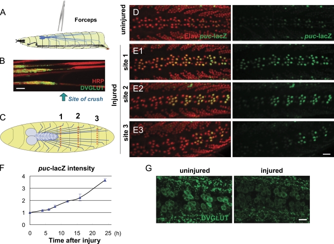
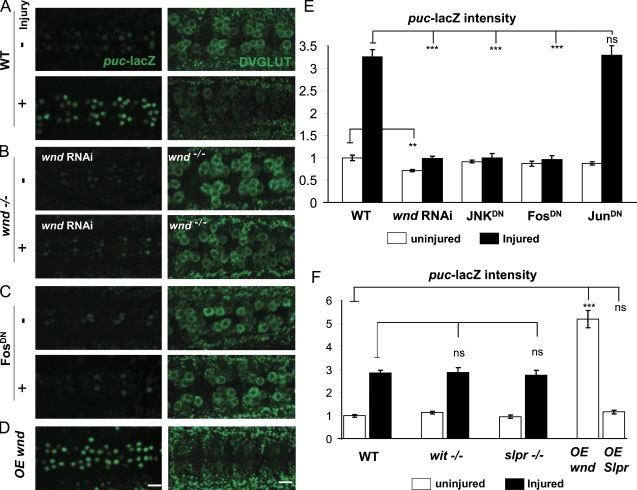
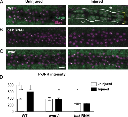
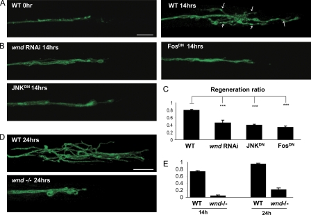
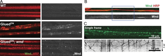
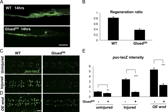
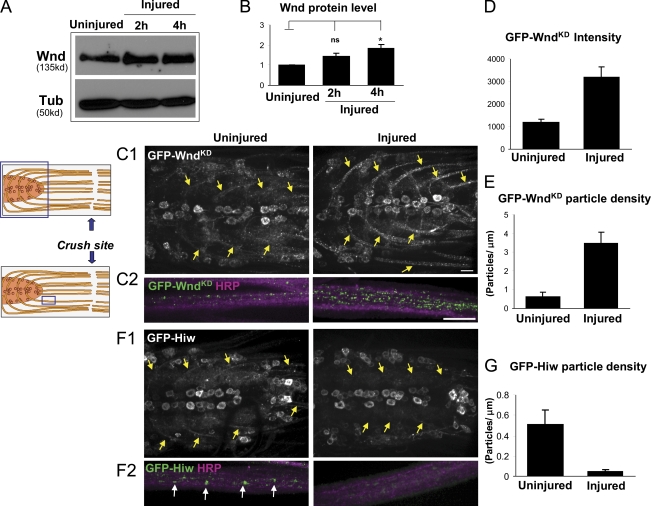
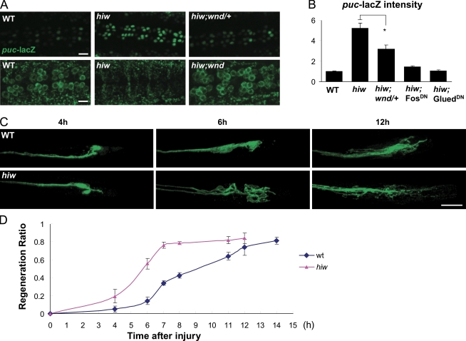
Regenerative responses to axonal injury involve changes in gene expression; however, little is known about how such changes can be induced from a distant site of injury. In this study, we describe a nerve crush assay in Drosophila melanogaster to study injury signaling and regeneration mechanisms. We find that Wallenda (Wnd), a conserved mitogen-activated protein kinase (MAPK) kinase kinase homologous to dual leucine zipper kinase, functions as an upstream mediator of a cell-autonomous injury signaling cascade that involves the c-Jun NH(2)-terminal kinase MAPK and Fos transcription factor. Wnd is physically transported in axons, and axonal transport is required for the injury signaling mechanism. Wnd is regulated by a conserved E3 ubiquitin ligase, named Highwire (Hiw) in Drosophila. Injury induces a rapid increase in Wnd protein concomitantly with a decrease in Hiw protein. In hiw mutants, injury signaling is constitutively active, and neurons initiate a faster regenerative response. Our data suggest that the regulation of Wnd protein turnover by Hiw can function as a damage surveillance mechanism for responding to axonal injury.


Figures

Figure 1.
Figure 1.
Axon injury induces transcriptional changes in the Motoneuron cell body. (A) Schematic of the nerve crush assay. The segmental nerves within a third instar larva are crushed by pinching the ventral cuticle with forceps. (B) Injured segmental nerves 24 h after nerve crush. Synaptic vesicle precursors detected by staining for DVGLUT staining (green) accumulate at the proximal side of the crush site (arrow). (C) Cartoon of neuron cell bodies (blue dots) and segmental nerves (blue lines). Different crush sites (dashed red lines) injure a predictable number of motoneurons. Crush site 1 injures more cells than crush sites 2 and 3. (D) In uninjured animals, puc-lacZ expression is barely detectable. A nuclear localization signal on lacZ (green) localizes the reporter to the nucleus, and neuronal nuclei are detected by staining for the ElaV (red) marker. (E1–3) Injury induces puc-lacZ expression. 24 h (at 25°C) after injury at sites 1–3 induces expression of puc-lacZ in a defined subset of motoneurons as predicted by the anatomy cartooned in C. (F) Time course quantitation of puc-lacZ. The mean intensity of puc-lacZ is measured as described in Materials and methods for the dorsal midline neurons. 24 h after injury, puc-lacZ intensity is increased 3.5-fold compared with uninjured animals (n > 15). (G) Axon injury leads to a decrease of DVGLUT protein in motoneuron cell bodies. Error bars indicate mean ± SEM. Bars, 25 µm.
Figure 2.
Figure 2.
The nuclear and cell body response to axonal injury specifically requires Wnd and downstream signaling components. (A–D) puc-lacZ expression (left) and staining for DVGLUT (right) in VNCs uninjured and 24 h after injury. (A) The nerve crush injury induces an increase in puc-lacZ expression (left) and decrease in staining for DVGLUT (right) in motoneuron cell bodies. (B) The response to injury requires Wnd function. No obvious change in puc-lacZ (left) and DVGLUT (right) is observed after injury when Wnd is disrupted. (C) The response to injury is inhibited by FosDN. (D) Overexpression (OE) of wnd in neurons is sufficient to activate the injury response, including induction of puc-lacZ and reduction in DVGLUT staining. (E and F) Quantification of the puc-lacZ expression level before (white bars) or 24 h after (black bars) injury in different genotypes. The BG380-Gal4 driver is used to drive expression of all UAS lines (wnd RNAi, JNKDN, FosDN, JunDN, and Slpr). P > 0.05 was not significant. **, P < 0.001; ***, P < 0.0001. Error bars indicate mean ± SEM. Bars, 25 µm.
Figure 3.
Figure 3.
Injury induces p-JNK accumulation in the cell bodies and nuclei of injured neurons. (A–C) VNCs are costained for p-JNK (green) and the nuclear marker Elav (magenta). In both uninjured and injured animals, p-JNK stains the neuropil (A, asterisks and dotted lines), which can be seen surrounding the cell bodies (A, yellow bracket). (A) p-JNK appears in motoneuron cell bodies within 12 h after injury. Bar, 25 µm. (B) This p-JNK staining is abolished by expression of bsk-RNAi (Dietzl et al., 2007) in neurons. (C) Mutations in wnd (wnd1/wnd3) inhibit the cell body accumulation of p-JNK after injury. The bright dots in the nuclei are fixation artifacts. (D) Quantification of p-JNK intensity in the cell body before (white bars) and 12 h after (black bars) injury. *, P < 0.05; **, P < 0.001; ***, P < 0.0001. Error bars indicate mean ± SEM.
Figure 4.
Figure 4.
Wnd/JNK/Fos injury signaling is required for axonal sprouting after injury. (A–E) Axons are labeled by driving expression of UAS-mCD8-GFP by m12-Gal4 (A–C) and RRa(eve)-Gal4 (D and E). (A) In WT animals, the proximal stump forms extensive new branches (arrows) by 14 h after injury. (B) Sprouting after injury is inhibited by expression of wnd-RNAi, JNKDN, or FosDN in the GFP-labeled neurons. (C) Quantification of regeneration ratio 14 h after injury. The fraction of injured axons that exhibited sprouting was measured for at least 100 axons per genotype while blinded to the genotype. (D) At 24 h after injury, branches from the proximal stump are even longer in the WT background; however, this sprouting remains strongly inhibited in the wnd1/wnd2 mutant background. Bar, 25 µm. (E) Quantification of the regeneration ratio for WT and wnd1/2 at 14 and 24 h after injury, conducted similarly as in C. ***, P < 0.0001. Error bars indicate mean ± SEM.
Figure 5.
Figure 5.
Wnd protein is transported in axons. (A) Wnd protein accumulates in segmental nerves when axonal transport is disrupted. Segmental nerves from third instar larvae are stained for axonal membrane (anti-HRP) in red and Wnd (anti-Wnd) in green. (right) Wnd staining alone is shown. (B) Wnd protein (top, green; bottom, alone) accumulates at the ligation (injury) site (blue rectangle). (C) Live imaging indicates that Wnd particles rapidly translocate both anterogradely and retrogradely in axons (Video 1). The GFP-wndKD transgene was expressed in motoneurons using the OK6-Gal4 driver. (top) A single 0.5-s exposure is shown. (bottom) A representative kymograph from a single axon is shown. Bars, 10 µm.
Figure 6.
Figure 6.
Axonal transport is required for regeneration and injury signaling. (A) Inhibition of axonal transport inhibits regeneration after injury. Expression of the p150-Glued dynactin subunit, UAS-GluedDN, inhibits the formation of new branches at the injury site. (B) Quantitation of the regeneration ratio similar to that in Fig. 4 C. (C) Inhibition of axonal transport inhibits the effect of axon injury on puc-lacZ expression. (D) Ectopic Wnd signaling also requires axonal transport. The induction of puc-lacZ by overexpression (OE) of wnd is suppressed by coexpression of GluedDN. (E) Quantification of puc-lacZ intensities for genotypes described in C and D. **, P < 0.001; ***, P < 0.0001. Error bars indicate mean ± SEM. Bars, 25 µm.
Figure 7.
Figure 7.
Injury induces an increase in Wnd protein in axons concomitant with a decrease in Hiw. (A) Western blots from nerve cords with attached segmental nerves to detect endogenous Wnd protein levels (20 VNCs per lane) before and after injury. (B) Quantification of changes in Wnd protein level based on Wnd/tubulin ratio from five independent experiments. (C1 and C2) GFP-WndKD particles in nerve cords and segmental nerves before and 4 h after injury. UAS–GFP-wndKD is expressed in motoneurons by OK6-Gal4. The cartoon shows the anatomy of nerve cord and segmental nerves, with the blue rectangles indicating the sites shown. (C1) Nerve cords of uninjured and injured animals. Injury induces dramatic increase of GFP-WndKD intensity in the axons projecting from motoneuron cell bodies (yellow arrows). (C2) Segmental nerves proximal to injury site are shown in higher magnification costained with HRP to label the nerve membrane. (D and E) The injured segmental nerves contain a higher mean GFP intensity (D) and a higher density of GFP-WndKD particles (E). Quantification is described in Materials and methods. (F1 and F2) GFP-Hiw (driven by OK6-Gal4) in nerve cords and segmental nerves in uninjured and injured (4 h) animals. (F1) Injury induces overall decrease of GFP-Hiw in indicated axons (yellow arrows). (F2) GFP-Hiw localizes to particles (white arrows) in uninjured axons, which are dramatically decreased in number within 4 h after injury. (G) Measurement of GFP-Hiw particle density in segmental nerves. P > 0.05 was not significant. *, P < 0.05. Error bars indicate mean ± SEM. Bars, 25 µm.
Figure 8.
Figure 8.
The Hiw E3 ubiquitin ligase negatively regulates injury signaling and regeneration. (A) The hiwΔN mutant shows a dramatic increase of puc-lacZ and decrease of DVGLUT intensity in motoneuron cell bodies, and these changes require wnd function. (B) Quantification of puc-lacZ intensity for genotypes in A and when Fos and Glued are inhibited by expressing UAS-FosDN or UAS-GluedDN in the hiwΔN;puc-lacZ/+ mutant background. These results suggest that hiw regulates a retrograde signaling pathway. (C) Examples of the proximal stump morphology at different time points after injury. In hiwΔN mutants, many injured axons form long new branches within 6 h of injury. In contrast, sprouting is not readily apparent in WT axons until 12 h after injury. (D) Time course of regeneration ratio comparing WT and hiwΔN mutants. *, P < 0.05. Error bars indicate mean ± SEM. Bars, 25 µm.

References

    1. Abe N., Cavalli V. 2008. Nerve injury signaling. Curr. Opin. Neurobiol. 18:276–283 10.1016/j.conb.2008.06.005 - DOI - PMC - PubMed
    1. Abe N., Almenar-Queralt A., Lillo C., Shen Z., Lozach J., Briggs S.P., Williams D.S., Goldstein L.S., Cavalli V. 2009. Sunday driver interacts with two distinct classes of axonal organelles. J. Biol. Chem. 284:34628–34639 10.1074/jbc.M109.035022 - DOI - PMC - PubMed
    1. Aberle H., Haghighi A.P., Fetter R.D., McCabe B.D., Magalhães T.R., Goodman C.S. 2002. wishful thinking encodes a BMP type II receptor that regulates synaptic growth in Drosophila. Neuron. 33:545–558 10.1016/S0896-6273(02)00589-5 - DOI - PubMed
    1. Allen M.J., Shan X., Caruccio P., Froggett S.J., Moffat K.G., Murphey R.K. 1999. Targeted expression of truncated glued disrupts giant fiber synapse formation in Drosophila. J. Neurosci. 19:9374–9384 - PMC - PubMed
    1. Barkus R.V., Klyachko O., Horiuchi D., Dickson B.J., Saxton W.M. 2008. Identification of an axonal kinesin-3 motor for fast anterograde vesicle transport that facilitates retrograde transport of neuropeptides. Mol. Biol. Cell. 19:274–283 10.1091/mbc.E07-03-0261 - DOI - PMC - PubMed

Publication types

MeSH terms