. 2010 Dec;78(12):5022-32.
doi: 10.1128/IAI.00544-10. Epub 2010 Oct 4.

Effects of the putative transcriptional regulator IclR on Francisella tularensis pathogenesis

Affiliations

Effects of the putative transcriptional regulator IclR on Francisella tularensis pathogenesis

Brittany L Mortensen et al. Infect Immun. 2010 Dec.

Abstract

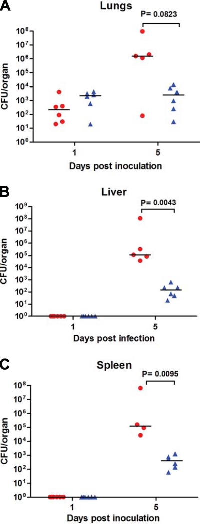
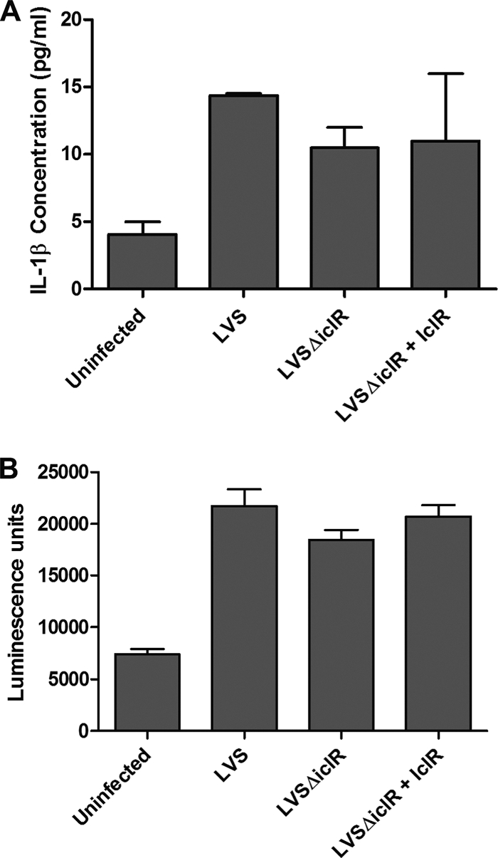
Francisella tularensis is a highly virulent Gram-negative bacterium and is the etiological agent of the disease tularemia. IclR, a presumed transcriptional regulator, is required for full virulence of the animal pathogen, F. tularensis subspecies novicida U112 (53). In this study, we investigated the contribution of IclR to the intracellular growth, virulence, and gene regulation of human pathogenic F. tularensis subspecies. Deletion of iclR from the live vaccine strain (LVS) and SchuS4 strain of F. tularensis subsp. holarctica and F. tularensis subsp. tularensis, respectively, did not affect their abilities to replicate within macrophages or epithelial cells. In contrast to F. tularensis subsp. novicida iclR mutants, LVS and SchuS4 ΔiclR strains were as virulent as their wild-type parental strains in intranasal inoculation mouse models of tularemia. Furthermore, wild-type LVS and LVSΔiclR were equally cytotoxic and induced equivalent levels of interleukin-1β expression by infected bone marrow-derived macrophages. Microarray analysis revealed that the relative expression of a limited number of genes differed significantly between LVS wild-type and ΔiclR strains. Interestingly, many of the identified genes were disrupted in LVS and SchuS4 but not in their corresponding F. tularensis subsp. novicida U112 homologs. Thus, despite the impact of iclR deletion on gene expression, and in contrast to the effects of iclR deletion on F. tularensis subsp. novicida virulence, IclR does not contribute significantly to the virulence or pathogenesis of F. tularensis LVS or SchuS4.


Figures

FIG. 1.
FIG. 1.
Comparison of iclR in three Francisella strains. (A) Synteny diagram of the genomic organization of the iclR locus in F. tularensis subsp. novicida U112 (FTN_0720), F. tularensis subsp. holarctica LVS (FTL_1364), and F. tularensis subsp. tularensis SchuS4 (FTT_0748). (B) Amino acid sequence alignment of F. tularensis subsp. novicida U112, F. tularensis subsp. holarctica LVS, and F. tularensis subsp. tularensis SchuS4 IclR. Alignment was created using VectorNTI software and iclR sequences uploaded from NCBI annotated genomes of each strain and translated using VectorNTI. Red letters highlight residues conserved between all three strains. Blue letters highlight the residues conserved between two strains.
FIG. 2.
FIG. 2.
Intracellular replication of LVS ΔiclR and SchuS4 ΔiclR in murine macrophages or lung epithelial cells. Gentamicin protection assays were performed by infecting J774A.1 murine macrophages (A), TC-1 murine lung epithelial cells (B), and bone marrow-derived macrophages (BMMs) (C) with wild-type LVS or LVS ΔiclR at an MOI of 100. (D) A gentamicin protection assay was performed using J774A.1 cells infected with wild-type SchuS4 or SchuS4 ΔiclR. Bars represent the standard deviations of three replicate wells, and each graph is representative of two separate experiments.
FIG. 3.
FIG. 3.
Recovery of LVS ΔiclR mutant in mice following i.n. or i.d. inoculation. (A and B) C57BL/6 mice were inoculated with either wild-type LVS (circles) or LVS ΔiclR (triangles) i.n. at a lethal dose of ∼1 × 10⁵ CFU (A) or a low dose of ∼1 × 10³ CFU (B). (C) C57BL/6 mice were inoculated with either wild-type LVS (circles) or LVS ΔiclR (triangles) i.d. at a dose of ∼3 × 10⁵ CFU. Each symbol represents data from a single mouse. There were no significant differences in recovery of mutant versus wild-type organisms from any organ at any time point as determined by the Mann-Whitney nonparametric test in the low-dose (B) and i.d. (C) experiments.
FIG. 4.
FIG. 4.
Recovery of the SchuS4 ΔiclR mutant in mice following i.n. inoculation. C57BL/6 mice were inoculated with either wild-type SchuS4 (circles) or SchuS4 ΔiclR (triangles) i.n. at a dose of ∼100 CFU. No differences in recovery of mutant versus wild-type organisms from any organ at any time point were significant based on the Mann-Whitney nonparametric test.
FIG. 5.
FIG. 5.
Recovery of the U112 iclR transposon mutant in mice following i.n. inoculation. C57BL/6 mice were inoculated with either wild-type U112 (circles) or U112 iclR mutant (triangles) i.n. at a dose of ∼10 CFU. Differences in recovery of mutant versus wild-type organisms at day 5 for the liver and spleen were significant based on the Mann-Whitney nonparametric test.
FIG. 6.
FIG. 6.
IL-1β release and cytotoxicity in murine bone marrow-derived macrophages infected with LVS ΔiclR. Infections were carried out at an MOI of 500 for wild-type LVS, LVS ΔiclR, and LVSΔiclR plus IclR (complementation). (A) IL-1β was quantified via ELISA, and (B) cytotoxicity was quantified via the ToxiLight bioassay (Lonza), both at 24 h postinfection. Graphs are representative of at least three separate experiments, with duplicate or triplicate wells for each strain per experiment. No differences were significant by any strain comparison based on Student's t test.
FIG. 7.
FIG. 7.
Transcript levels of genes found significantly changed in microarray analysis comparing LVS and LVS ΔiclR. RNA was isolated from wild-type F. tularensis subsp. holarctica LVS and LVSΔiclR (A) or wild-type F. tularensis subsp. novicida U112 and a U112 iclR transposon mutant (B) and used in qRT-PCR analysis for several genes that were significantly changed in the microarray. Data are presented as the relative log change for the wild type versus the respective iclR mutant after normalization to gyrA. The graphs are representative of two or three experiments.
FIG. 8.
FIG. 8.
Antibiotic sensitivity of LVS ΔiclR. Wild-type LVS and LVS ΔiclR were grown to mid-log phase, bacteria were spread on chocolate agar, and an antibiotic-containing paper disc was added to the center. Bacteria were grown for 36 h, and the diameter of the zone of inhibition was measured. The experiment was performed in triplicate, and the averages and standard deviations were calculated.

Similar articles

Cited by

References

    1. Anthony, L. D., R. D. Burke, and F. E. Nano. 1991. Growth of Francisella spp. in rodent macrophages. Infect. Immun. 59:3291-3296. - PMC - PubMed
    1. Birdsell, D. N., T. Stewart, A. J. Vogler, E. Lawaczeck, A. Diggs, T. L. Sylvester, J. L. Buchhagen, R. K. Auerbach, P. Keim, and D. M. Wagner. 2009. Francisella tularensis subsp. novicida isolated from a human in Arizona. BMC Res. Notes 2:223. - PMC - PubMed
    1. Bosio, C. M., and S. W. Dow. 2005. Francisella tularensis induces aberrant activation of pulmonary dendritic cells. J. Immunol. 175:6792-6801. - PubMed
    1. Centers for Disease Control and Prevention. 2005. Tularemia transmitted by insect bites—Wyoming, 2001-2003. MMWR Morb. Mortal. Wkly. Rep. 54:170-173. - PubMed
    1. Centers for Disease Control and Prevention. 2009. Tularemia—Missouri, 2000-2007. MMWR Morb. Mortal. Wkly. Rep. 58:744-748. - PubMed

Publication types

MeSH terms

Associated data