Review
. 2010 Dec 1;588(Pt 23):4621-39.
doi: 10.1113/jphysiol.2010.196030. Epub 2010 Oct 4.

Neuroeffector apparatus in gastrointestinal smooth muscle organs

Affiliations
Review

Neuroeffector apparatus in gastrointestinal smooth muscle organs

Kenton M Sanders et al. J Physiol. .

Abstract

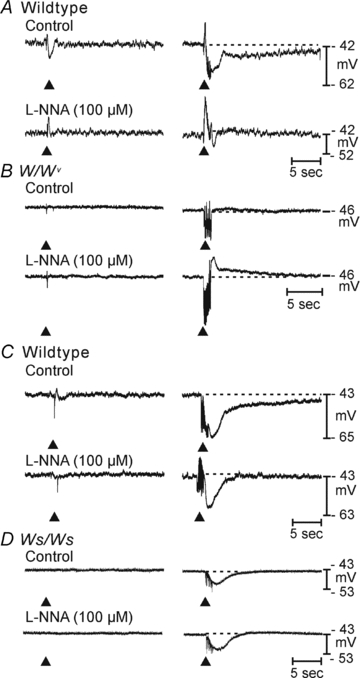
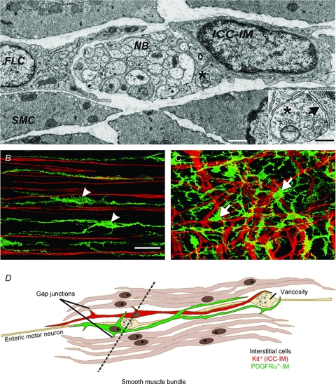
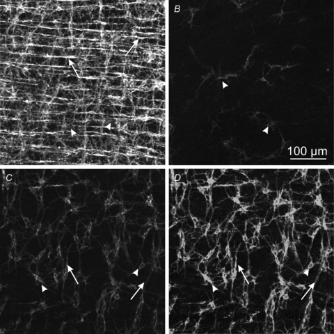
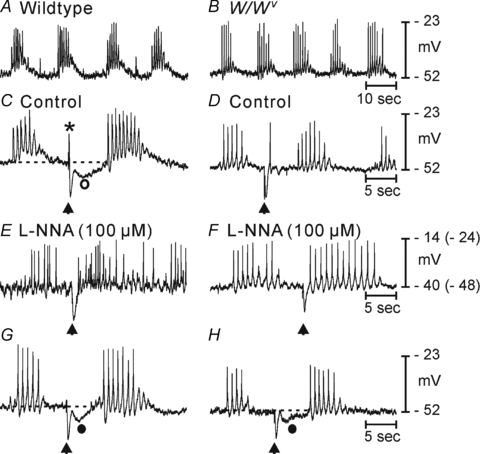
Control of gastrointestinal (GI) movements by enteric motoneurons is critical for orderly processing of food, absorption of nutrients and elimination of wastes. Work over the past several years has suggested that motor neurotransmission is more complicated than simple release of transmitter from nerve terminals and binding of receptors on smooth muscle cells. In fact the 'neuro-effector' junction in the tunica muscularis might consist of synaptic-like connectivity with specialized cells, and contributions from multiple cell types in integrated post-junctional responses. Interstitial cells of Cajal (ICC) were proposed as potential mediators in motor neurotransmission based on reduced post-junctional responses observed in W mutants that have reduced populations of ICC. More recent studies on W mutants have contradicted the original findings, and suggested that ICC may not be significant players in motor neurotransmission. This review examines the evidence for and against the role of ICC in motor neurotransmission and outlines areas for additional investigation that would help further resolve this controversy.


Figures

None
Professor Sanders received his PhD from the Department of Physiology in the UCLA School of Medicine in 1976. He further trained as a post-doctoral fellow at UCLA and the Mayo Foundation from 1976 to 1979. He became an Associate Professor at the University of Nevada in 1982 and head of Physiology and Cell Biology department in 1988. He has studied excitability mechanisms in smooth muscles and neural regulation of smooth muscles throughout his career. In 2004 he was a Carnegie Centenary Professor in Scotland and recently served on the National Commission for Digestive Diseases at the National Institutes of Health in Washington, DC. Professor Ward received his DPhil from the Department of Biology, University of Ulster in 1987. He spent a short time as a post-doctoral fellow in the Wellcome Laboratory, University College London before taking up a fellowship in Physiology and Cell Biology at the University of Nevada School of Medicine, where he is currently a Professor of Physiology. He has studied the excitability and structure of visceral organs, particularly the gastrointestinal tract and oviducts. In 2000 he was awarded the Sixth Annual American Gastroenterological Association in Gastroenterology for ‘Basic and Clinical Research’. In 2006 he was awarded a University of Nevada Foundation Professor, and in 2007 the Eighth International Award for Studies on Neurogastroenterology and Motility.
Figure 1
Figure 1. Comparison of responses in W/WV (A and B) and Ws/Ws (C and D) fundus muscles
A shows control responses of a wildtype circular fundus muscle strip to electrical field stimulation (EFS) (0.3 ms duration pulses): 1 pulse (at arrowheads, left traces all panels) and 20 pulses in 1 s (beginning at arrowheads, right traces in all panels). In the wildtype mouse, EFS elicited a small excitatory junction potential (EJP) followed by an inhibitory junction potential (IJP). 20 pulses increased these responses and a long-duration 2nd component is apparent. l-NNA (2nd line of traces in A enhanced the EJP and greatly reduced the IJP, including the long duration 2nd component. B, responses to single stimuli were minimal in W/WV muscles, and both the EJP and IJP to 20 pulses were greatly reduced. l-NNA had little effect on the single stimuli response but slightly increased the IJP and unmasked a more pronounced ‘rebound’ response to 20 stimuli in the W/WV muscle. C, nitrergic responses were not very pronounced in the rat. In wildtype animals, a small EJP and IJP were evoked by a single stimulus and a large amplitude hyperpolarization followed by a sustained 2nd component of hyperpolarization were evoked by 20 pulses. l-NNA had little effect on responses to single stimuli and reduced the duration of the response to 20 stimuli (see dotted lines in traces at right in C). D, little or no response was elicited by 1 stimulus in the Ws/Ws rat fundus. Small inhibitory junction potentials were elicited by 20 stimuli. Note the absence of the sustained 2nd component in the Ws/Ws muscle. l-NNA had very little effect, suggesting that nitrergic responses were reduced in these muscles.
Figure 2
Figure 2. Relationship of motor nerve varicosities to interstitial cells and smooth muscle cells
ICC of the circular muscle layer (ICC-IM) and fibroblast-like cells (PDGFRα+ cells) are commonly found closely associated with nerve bundles (NB), as in this section from rat stomach (A; scale bar is 0.5 μm; reproduced with kind permission from Springer Science+Business Media: Mitsui & Komuro, 2002, Cell & Tissue Res309, 219–227). Smooth muscle cells (SMC) surround these structures and form gap junctions with ICC-IM and PDGFRα+ cells (not shown in this image). PDGFRα+ cells have electron-lucent cytoplasm and dilated cisternae of rough endoplasmic reticulum. ICC-IM are frequently in very close contact with nerve varicosities containing many synaptic vesicles (*). Inset shows higher magnification of the varicosity (scale bar is 0.2 μm) denoted by the asterisk in A. The gap between membranes of the nerve varicosity and the ICC-IM (arrow) measures about 20 nm. Similar close associations between nerve varicosities and smooth muscle cells can also be found in the rat antrum (not shown in this image), but these appear to be more rare than junctions with ICC-IM in other species. B and C show double labelling of PDGFRα+ cells (green) and ICC (red) in murine stomach (images are reproduced with kind permission from Springer Science+Business Media: Iino et al. 2009a, Histochem Cell Biol131, 691–702). PDGFRα+-IM cells (green, arrowheads) of the gastric fundus are intermingled with ICC-IM (red). In the corpus multipolar PDGFRα+-MY cells (green; arrows) are closely associated with ICC-MY (red) in the plane of the myenteric plexus. These images indicate that ICC and PDGFRα+ cells have similar anatomical distributions in the tunica muscularis, but represent discrete populations of cells. Yellow pixels in these merged images are due to overlay of cells in stacks of optical sections not co-expression of antigens in single cells. Scale bar in B is 40 μm and applicable to C. D shows a cartoon of ICC-IM (red) and PDGFRα+-IM cells (green) within a muscle bundle. As demonstrated by ultrastructural and immunohistochemical studies, both of these cell types are found in close apposition to enteric motoneurons and make gap junctions with neighbouring smooth muscle cells. A section through the region denoted by the dotted line might generate an image similar to the electron micrograph in A. As discussed in the text, neuromuscular junctions in GI smooth muscles may reflect innervation of and post-junctional responses in all 3 classes of post-junctional cells. Transduction of neurotransmitter signals by ICC-IM and/or PDGFRα+-IM cells and activation of ionic conductances would be conducted electrotonically via gap junctions to surrounding smooth muscle cells and influence the excitability of the tunica muscularis and possibly the frequency of phasic activity (e.g. segmentation and/or peristalsis).
Figure 3
Figure 3. Incomplete lesions in ICC networks in colons of W/WV mice
A shows a digital reconstruction of a confocal Z-stack of ICC through the thickness of the muscularis in a wildtype mouse proximal colon. ICC form dense networks within the plane of the myenteric plexus region (ICC-MY; arrowheads) and within the circular and longitudinal muscle layers (ICC-IM; arrows). The density of ICC is greatly reduced and irregular in colons of W/WV mice, but loss of ICC is incomplete. B and C show ICC imaged at the same laser intensity, pixel time, detector gain and pinhole size as the image in A. The images in B and C are from a region where there were relatively few cells (B) and from another region where the ICC-MY network was considerably more intact (C). In some studies the extent of ICC lesions in W mutants may be overstated because the overall immunogenicity of ICC is reduced, most probably because of reduced Kit protein in these mutants. D demonstrates that increasing the detector gain from 643 to 750 to scan the same field as in C allows more complete resolution of ICC networks. Scale bar in B represents 100 μm and is the same in all panels.
Figure 4
Figure 4. Neural responses in a region of muscle with incomplete ICC lesion in W/WV mice
A and B, basal electrical activity is similar in wildtype (A) and W/WV (B) colonic muscles and consists of spike complexes with intermittent periods of quiescence. The frequency of spike complexes was greater in W/WV muscles (2.45 ± 0.26 cycles min−1 in control muscles (n= 11) vs. 4.9 ± 0.24 cycles min−1 for W/WV muscles (n= 12; P < 0.00001). We noted defects in post-junctional inhibitory responses to electrical field stimulation in many W/WV muscles (compare C and D). C is the wildtype control; D is the mutant muscle. C, in wildtype muscles EFS (stimulus applied at arrowhead; 1 pulse, 0.5 ms) evoked a single phase EJP (*) followed by an IJP that consisted of a fast and slow component (open circle). D, in W/WV muscles, EJPs were not evoked by EFS, and only the fast initial components of IJPs were resolved in 13 of 20 mice. E, the slow component is the nitrergic portion of the response and was blocked by l-NNA. The nitrergic component averaged 11 ± 0.7 mV (n= 18; P < 0.00001). l-NNA caused significant depolarization of wildtype muscles (see leftmost numbers on scale bars (F), but little depolarization was noted in W/WV muscles in response to l-NNA (numbers in brackets in F apply to the W/WV muscle after l-NNA). In the post-l-NNA W/WV muscle, a series of action potentials followed the IJP, possibly indicating phase advancement of the next action potential complex. G and H, in 7 of 20 W/WV animals, we observed small secondary components (filled circle) that were significantly smaller in amplitude than in wildtype colons and averaged 4 ± 0.6 mV (n= 7/20; P < 0.000001; 2 representative traces are shown in G and H).
Figure 5
Figure 5. Reduced tonic inhibition in W mutant animals
Another approach to evaluating nitrergic regulation in W mutants is to characterize the magnitude of tonic inhibitory drive that is normally imposed upon colonic muscles. l-NNA treatment of wildtype muscles caused dramatic depolarization (18.6 ± 2.3 mV; n= 6) and nearly continuous firing of action potentials (A), whereas l-NNA caused only a small degree of depolarization (3.0 ± 0.75 mV; n= 9; P < 0.00001) in W/WV colonic muscles and did not disrupt the pattern of action potential complexes (B). These data demonstrate many of the points elaborated upon in the text. Post-junctional neural responses are abnormal in W/WV muscles, but a portion of nitrergic regulation is retained. There are reduced numbers and heterogeneity in the distribution of ICC in the colons of W/WV muscles, which may explain the reduced and heterogenous post-junctional responses. The nitrergic component of the tonic inhibition normally imposed upon the murine colon is significantly depressed. We are currently testing how this partial loss of nitrergic responses affects colonic transit.
Figure 6
Figure 6. Comparison of electrical responses to exogenous transmitter and nerve evoked cholinergic excitation of circular muscles of the murine gastric antrum
A shows responses of wildtype, strain-matched control (top trace) and W/WV (bottom trace) antral muscles to exogenous carbachol (CCh, 100 nm; arrowheads). Both traces show spontaneous slow wave activity characteristic of antral muscles. CCh elicited depolarization and increased the frequency of electrical slow waves (chronotropic effect) in wildtype muscles. CCh caused depolarization in W/WV muscles, but no chronotropic effect was elicited in the absence of ICC-IM (Forrest et al. 2006). Both responses were blocked by atropine (1 μm; arrows). B, the responses to exogenous CCh are compared to responses to release of acetylcholine from nerves during EFS. EFS (5 Hz) was sustained through each trace in B. EFS produced positive chronotropic effects in wildtype muscles (2nd trace), but no response in W/WV muscles (not shown here but shown in Forrest et al. 2006, from which traces were redrawn). Chronotropic responses were blocked by 4-DAMP (3rd trace), an M3-specific antagonist, and no further effect was elicited by atropine (4th trace). Release of ACh from nerves did not depolarize cells as observed with the exogenous muscarinic agonist (A). EFS responses were due primarily to stimulation of M3 receptors, blocked by 4-DAMP (3rd trace) and not further affected by atropine (4th trace). C shows the direct response of CCh on a smooth muscle cell isolated from the murine antrum. As shown in many studies in the literature (e.g. Benham et al. 1985; Inoue & Isenberg, 1990), direct application of a muscarinic agonist to GI smooth muscles causes significant depolarization due to activation of non-selective cation channels encoded by Trpc4 and Trpc6 (Tsvilovskyy et al. 2009). Conclusions are summarized at the right of the traces. When ICC are present, nerve-evoked effects are mainly chronotropic. Muscarinic stimulation of smooth muscle cells causes depolarization, which is manifest when exogenous transmitter is applied to cells or tissues, but not elicited when transmitter is released from nerve terminals with constant EFS at 5 Hz. These data suggest that ACh released from neurons may be restricted to neuro-ICC junctions, most probably due to the action of acetylcholine esterases (see Ward et al. 2000).

Similar articles

Cited by

References

    1. Albertí E, Mikkelsen HB, Wang XY, Díaz M, Larsen JO, Huizinga JD, Jiménez M. Pacemaker activity and inhibitory neurotransmission in the colon of Ws/Ws mutant rats. Am J Physiol Gastrointest Liver Physiol. 2007;292:G1499–G1510. - PubMed
    1. Auintas LE, Noël F. Mechanisms of adaptive supersensitivity in vas deferens. Auton Neurosci. 2009;146:38–46. - PubMed
    1. Beckett EA, Horiguchi K, Khoyi M, Sanders KM, Ward SM. Loss of enteric motor neurotransmission in the gastric fundus of Sl/Sld mice. J Physiol. 2002;543:871–887. - PMC - PubMed
    1. Beckett EA, McGeough CA, Sanders KM, Ward SM. Pacing of interstitial cells of Cajal in the murine gastric antrum: neurally mediated and direct stimulation. J Physiol. 2003;553:545–559. - PMC - PubMed
    1. Beckett EA, Takeda Y, Yanase H, Sanders KM, Ward SM. Synaptic specializations exist between enteric motor nerves and interstitial cells of Cajal in the murine stomach. J Comp Neurol. 2005;493:193–206. - PubMed

Publication types

MeSH terms

LinkOut - more resources