. 2010 Dec 3;285(49):38710-9.
doi: 10.1074/jbc.M110.137414. Epub 2010 Oct 4.

Interaction of calcium-dependent activator protein for secretion 1 (CAPS1) with the class II ADP-ribosylation factor small GTPases is required for dense-core vesicle trafficking in the trans-Golgi network

Affiliations

Interaction of calcium-dependent activator protein for secretion 1 (CAPS1) with the class II ADP-ribosylation factor small GTPases is required for dense-core vesicle trafficking in the trans-Golgi network

Tetsushi Sadakata et al. J Biol Chem. .

Abstract

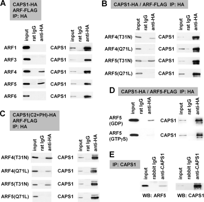
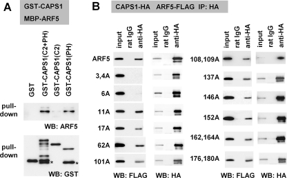
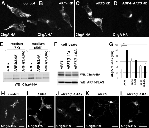
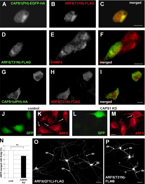
Ca(2+)-dependent activator protein for secretion (CAPS) regulates exocytosis of catecholamine- or neuropeptide-containing dense-core vesicles (DCVs) at secretion sites, such as nerve terminals. However, large amounts of CAPS protein are localized in the cell soma, and the role of somal CAPS protein remains unclear. The present study shows that somal CAPS1 plays an important role in DCV trafficking in the trans-Golgi network. The anti-CAPS1 antibody appeared to pull down membrane fractions, including many Golgi-associated proteins, such as ADP-ribosylation factor (ARF) small GTPases. Biochemical analyses of the protein-protein interaction showed that CAPS1 interacted specifically with the class II ARF4/ARF5, but not with other classes of ARFs, via the pleckstrin homology domain in a GDP-bound ARF form-specific manner. The pleckstrin homology domain of CAPS1 showed high affinity for the Golgi membrane, thereby recruiting ARF4/ARF5 to the Golgi complex. Knockdown of either CAPS1 or ARF4/ARF5 expression caused accumulation of chromogranin, a DCV marker protein, in the Golgi, thereby reducing its DCV secretion. In addition, the overexpression of CAPS1 binding-deficient ARF5 mutants induced aberrant chromogranin accumulation in the Golgi and consequently reduced its DCV secretion. These findings implicate a functional role for CAPS1 protein in the soma, a major subcellular localization site of CAPS1 in many cell types, in regulating DCV trafficking in the trans-Golgi network; this activity occurs via protein-protein interaction with ARF4/ARF5 in a GDP-dependent manner.


Figures

FIGURE 1.
FIGURE 1.
CAPS1 is associated with the Golgi membrane via the PH domain. A–C, accumulated expression of CAPS1(PH)-YPet-HA (the PH domain of CAPS1 C-terminally fused to the fluorescent protein YPet and the HA epitope tag) around Stx6-immunopositive Golgi structures of PC12 cells. A, CAPS1(PH)-YPet-HA (green); B, Stx6 (red); C, merged image. Scale bars, 20 μm. D–I, subcellular distribution of six deletion derivatives of CAPS1-YPet-HA expressed in PC12 cells; immunostaining for HA. D, wild-type CAPS1-YPet-HA; E, PH domain alone (PH)-YPet-HA; F, PH domain and Munc13-1-homologous domain (ΔN-Ter)-YPet-HA; G, the region from the first amino acid to the PH domain (ΔC-Ter)-YPet-HA; H, C2 and PH domain (ΔN/C-Ter)-YPet-HA; I, C2 domain-skipped CAPS1 (ΔC2)-YPet-HA. Scale bars, 20 μm.
FIGURE 2.
FIGURE 2.
Endogenous CAPS1 is associated with the Golgi membrane. A and B, electron micrograms of submicrosomal fractions affinity-purified using immune magnetic beads. Cerebellar microsomal fractions of P21 mice were incubated with magnetic beads coated with anti-CAPS1 antibody (A) or anti-VAMP2 antibody (B), and immunoaffinity-purified submicrosomal fractions were subjected to electron microscopic observation. Scale bars, 200 nm. C, Western blot analysis of submicrosomal fractions affinity-purified using normal rabbit IgG, anti-CAPS1 antibody, or anti-VAMP2 antibody immunomagnetic beads. Each immunopurified fraction was immunoblotted with anti-CAPS1, anti-VAMP2, anti-VAMP4, anti-SNAP25, anti-Bcl-2, anti-BiP/GRP78, anti-pan-ARF (1D9), anti-GM130, anti-GS28, anti-p115, anti-Stx5, anti-Stx6, anti-Stx16, and anti-Vti1a antibodies. Anti-pan-ARF (1D9) antibody recognizes all of the ARF family proteins (supplemental Fig. S3).
FIGURE 3.
FIGURE 3.
CAPS1 interacts with GDP-locked class II ARF4/5. A–D, protein-protein interaction between CAPS1-HA constructs and ARF-FLAG constructs coexpressed in COS-7 cells was analyzed by coimmunoprecipitation (IP) with anti-HA antibody followed by immunoblotting (WB) with anti-FLAG and anti-HA antibodies. A, coimmunoprecipitation of CAPS1-HA with FLAG-tagged ARF1, ARF3, ARF4, ARF5, and ARF6; B, coimmunoprecipitation of CAPS1-HA with the GDP-locked form (T31N) and the GTP-locked form (Q71L) of ARF4 and ARF5; C, coimmunoprecipitation of the HA-tagged C2 and PH domain of CAPS1 (CAPS1(C2+PH)-HA) with the GDP-locked form (T31N) and GTP-locked form (Q71L) of ARF4-FLAG and ARF5-FLAG; D, coimmunoprecipitation of CAPS1-HA with ARF5-FLAG in the presence of GDP or GTPγS in lysis and assay buffers. E, protein-protein interaction between CAPS1 and ARF5 in mouse cerebellum in vivo. Endogenous ARF5 was coimmunoprecipitated with endogenous CAPS1 by anti-CAPS1 antibody from cerebellar lysates of P21 mice. The blots were immunostained for ARF5 (left) and CAPS1 (right).
FIGURE 4.
FIGURE 4.
The PH domain of CAPS1 binds to the N-terminal region of ARF5. A, in vitro binding assay using bacterially expressed recombinant proteins reveals the involvement of the PH domain of CAPS1 in binding to ARF5. MBP-tagged ARF5 protein was pulled down by glutathione-Sepharose beads on which GST, GST-tagged C2 and PH domain (C2 + PH), C2 domain (C2), and PH domain (PH) proteins were immobilized, followed by Western blot analysis (IB) with anti-ARF5 (top) and anti-GST (bottom) antibodies. B, ARF5 binds CAPS1 via the N-terminal region. The region of ARF5 that binds to CAPS1 was screened by generating a series of Ala substitutions in ARF5(T31N)-FLAG at 16 amino acid positions, 3, 4, 6, 11, 17, 62, 101, 108, 109, 137, 146, 152, 162, 164, 176, and 180, all of which are unique to class II ARF4/5 among the ARF family of proteins. CAPS1-HA and Ala-substituted ARF5(T31N)-FLAG constructs were coexpressed in COS-7 cells, and cell lysates were subjected to coimmunoprecipitation (IP) with anti-HA antibody followed by Western blot analysis with anti-FLAG (top) and anti-HA (bottom) antibodies.
FIGURE 5.
FIGURE 5.
CAPS1 knockdown induces chromogranin accumulation in the Golgi complex. A, KD of CAPS1 expression in PC12 cells by siRNA. siRNA specific for CAPS1 was electroporated into PC12 cells, and the culture medium was concentrated and electrophoresed. Top, three bands were greatly reduced in the culture medium of the CAPS1 KD cells and were identified as chromogranin A (ChgA), chromogranin B (ChgB), and secretogranin II (SgII) by LC-MS/MS. Bottom, reduction of CAPS1 expression in PC12 cells by siRNA KD was assayed by immunoblotting of cell lysates with anti-CAPS1 antibody. B, high KCl-induced chromogranin A and secretogranin II release from CAPS1 KD PC12 cells. PC12 cells were transfected with either control siRNA or CAPS1 siRNA together with a ChgA-HA or SgII-HA expression plasmid. The culture media, treated with 5 mm KCl (5K) or 50 mm KCl (50K) (see “Experimental Procedures”), were collected and analyzed by immunoblotting with anti-HA antibody. C, statistical analyses of the effect of CAPS1 KD on ChgA-HA release from PC12 cells. PC12 cells were transfected with ChgA-HA together with the control (white bar) or CAPS1 siRNA (black bar). The amounts of ChgA-HA released into the culture media with 5 or 50 mm KCl stimulation were analyzed by Western blotting, followed by densitometric analysis (n = 6). There was no significant difference in the amount of ChgA-HA in cell lysates between the control and CAPS1 KD groups (n = 6). The signal intensities of the extracellular ChgA-HA bands of the culture media were normalized against those of the intracellular ChgA-HA bands of the cell lysates. AU, arbitrary unit. Error bars, S.E. **, p < 0.01 by Student's t test. D, subcellular localization of endogenous chromogranin A in NGF-differentiated PC12 cells without (left) and with (right) CAPS1 KD; immunostaining for chromogranin A. Scale bars, 20 μm. E, subcellular localization of ChgA-HA expressed in NGF-differentiated PC12 cells without (left) and with (right) CAPS1 KD; immunostaining for HA. Scale bars, 20 μm. F, subcellular localization of C-terminal HA-tagged TrkB expressed in NGF-differentiated PC12 cells without (left) and with (right) CAPS1 KD; immunostaining for HA. Scale bars, 20 μm.
FIGURE 6.
FIGURE 6.
Subcellular distribution and release of chromogranin is disrupted by either ARF4/5 siRNA knockdown or expression of CAPS1-binding-deficient ARF5. A–D, HA-tagged chromogranin (ChgA-HA) expressed in PC12 cells showed a Golgi-accumulated pattern following KD of ARF4/5 expression. Shown are immunocytochemical staining patterns of control (A), ARF4 KD (B), ARF5 KD (C), and ARF4/5 double KD (D) cells with anti-HA antibody. ARF4 and ARF5 siRNAs efficiently reduced the expression levels of ARF4 and ARF5 proteins, respectively (supplemental Fig. S3). Scale bar, 10 μm. E–G, ARF5(3,4A) and ARF5(3,4,6A) had a disrupted N-terminal CAPS1 binding site and showed a decrease in regulated release of ChgA-HA from PC12 cells. ChgA-HA and one of the ARF5 constructs (wild-type ARF5, ARF5(3,4A), or ARF5(3,4,5A)) were transfected into PC12 cells. Culture medium after treatment with 5 mm KCl (5K) or 50 mm KCl (50K) was examined by Western blot analysis with anti-HA antibody (E), and the cell lysates were analyzed by Western blotting (WB) with anti-HA antibody or anti-FLAG antibody (F). G, statistical analyses of the effect of ARF5 and CAPS1 binding-deficient ARF5 on ChgA-HA release from PC12 cells. PC12 cells were transfected with ChgA-HA together with wild-type ARF5, CAPS1 binding-deficient ARF5(3,4A), or ARF5(3,4,6A). The amount of ChgA-HA released into the culture media with 5 mm KCl (white bar) or 50 mm KCl (black bar) stimulation was analyzed by Western blotting followed by densitometric analysis (n = 4). The signal intensities of the extracellular ChgA-HA bands from the culture media were normalized against those of the intracellular ChgA-HA bands from the cell lysates. AU, arbitrary unit. Error bars, S.E. **, p < 0.01 by Student's t test. H–J, CAPS1 binding-deficient ARF5(3,4,6A) coexpressed in PC12 cells induces accumulation of ChgA-HA in the Golgi. Immunocytochemical staining with anti-HA antibody indicates that ChgA-HA is localized in the tips of processes and around the nuclei in control cells (H) and cells coexpressing wild-type ARF5 (I) but is accumulated in the Golgi in cells coexpressing ARF5(3,4,6A) (J). Scale bar, 25 μm. K and L, CAPS1 binding-deficient ARF5(3,4,6A) coexpressed in primary cultured hippocampal neurons (DIV8) induces accumulation of ChgA-HA in the Golgi. Immunocytochemistry with anti-HA antibody shows that expressed ChgA-HA is localized in neurites and in soma of cells coexpressing wild-type ARF5 (K), whereas it is accumulated in the Golgi in cells coexpressing ARF5(3,4,6A) (L). Scale bar, 50 μm.
FIGURE 7.
FIGURE 7.
The binding between the PH domain of CAPS1 and ARF5 induces the accumulation of GDP-locked ARF5 in the Golgi membrane. A–F, subcellular localization of the C-terminal EGFP-HA-tagged CAPS1(PH), the C-terminal FLAG-tagged GDP-locked ARF5(T31N), and endogenous VAMP4 in PC12 cells. A–C, immunostaining of CAPS1(PH)-EGFP-HA and ARF(T31N)-FLAG with anti-HA (A) and anti-FLAG (B) antibodies, respectively, and the merged image (C). D–F, immunostaining of ARF5(T31N)-FLAG and endogenous VAMP4 with anti-FLAG (D) and anti-VAMP4 (E) antibodies, respectively, and the merged image (F). Scale bars (C and F), 10 μm. G–I, subcellular localization of the C-terminal HA-tagged CAPS1(dPH) and the C-terminal FLAG-tagged ARF5(T31N) in PC12 cells. Immunostaining of CAPS1(dPH)-HA and ARF5(T31N)-FLAG with anti-HA (G) and anti-FLAG (H) antibodies, respectively, and the merged image (I). Scale bars, 10 μm. J–M, subcellular localization of endogenous ARF5 in PC12 cells transfected with EGFP together with either control (J and K) or CAPS1 siRNA (L and M). The arrows indicate EGFP-expressing siRNA-transfected cells. Shown is immunostaining of endogenous ARF5 with anti-ARF5 antibody (K and L). Scale bars (K and M), 10 μm. N, ARF5 immunoreactivity levels merged with immunoreactivity for the Golgi complex. PC12 cells were transfected with the Golgi marker B4galt1-tdTomato together with either control (white bar) or CAPS1 siRNA (black bar). The ratio of ARF5 immunoreactivity in the Golgi complex (merged with B4galt1-tdTomato fluorescence) and that in whole cell soma was qualified. **, p < 0.01 by Student's t test. Error bars, S.E. O and P, subcellular distribution of GTP-locked ARF5(Q71L)-FLAG (O) and GDP-locked ARF5(T31N)-FLAG (P) expressed in primary cultured mouse hippocampal neurons. Cultures were transfected at 6 days in vitro, fixed at 8 days in vitro, and immunostained with anti-FLAG antibody. ax, axon; de, dendrite. Scale bar, 50 μm.

Similar articles

Cited by

References

    1. Renden R., Berwin B., Davis W., Ann K., Chin C. T., Kreber R., Ganetzky B., Martin T. F., Broadie K. (2001) Neuron 31, 421–437 - PubMed
    1. Berwin B., Floor E., Martin T. F. (1998) Neuron 21, 137–145 - PubMed
    1. Tandon A., Bannykh S., Kowalchyk J. A., Banerjee A., Martin T. F., Balch W. E. (1998) Neuron 21, 147–154 - PubMed
    1. Speidel D., Varoqueaux F., Enk C., Nojiri M., Grishanin R. N., Martin T. F., Hofmann K., Brose N., Reim K. (2003) J. Biol. Chem. 278, 52802–52809 - PubMed
    1. Sadakata T., Mizoguchi A., Sato Y., Katoh-Semba R., Fukuda M., Mikoshiba K., Furuichi T. (2004) J. Neurosci. 24, 43–52 - PMC - PubMed

Publication types

MeSH terms