. 2010 Dec 10;285(50):39108-16.
doi: 10.1074/jbc.M110.151753. Epub 2010 Oct 4.

MST1 promotes apoptosis through phosphorylation of histone H2AX

Affiliations

MST1 promotes apoptosis through phosphorylation of histone H2AX

Weihong Wen et al. J Biol Chem. .

Abstract

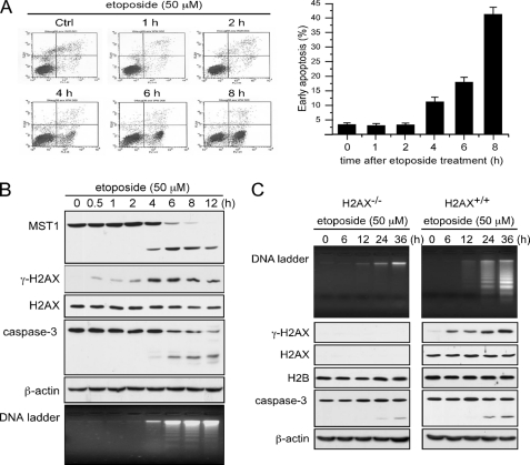
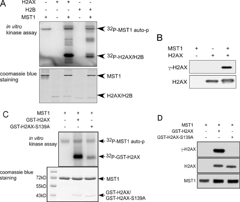
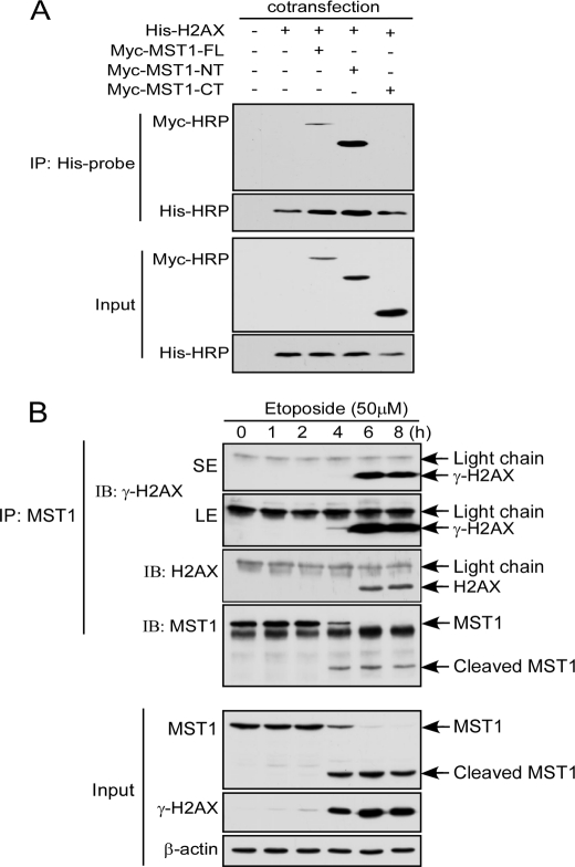
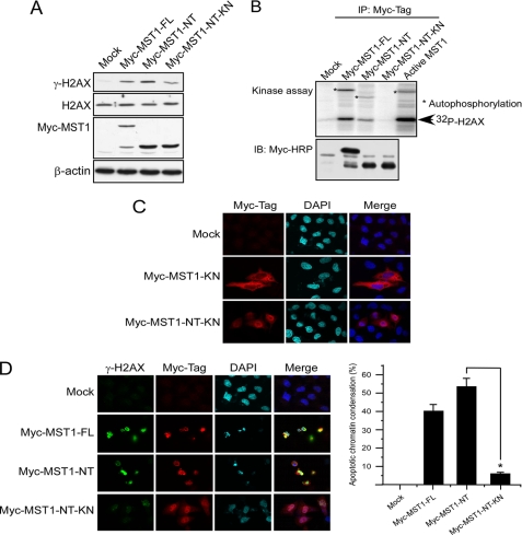
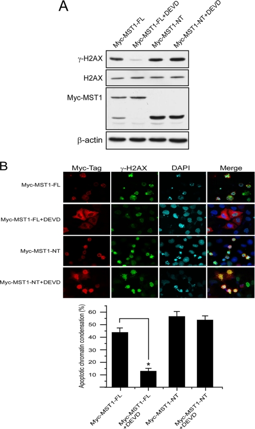
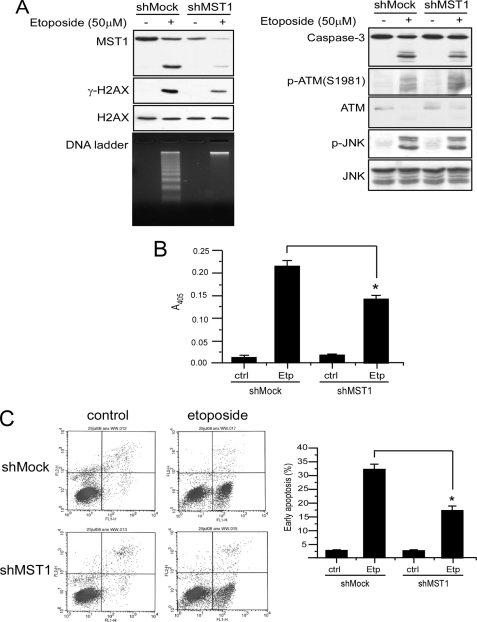
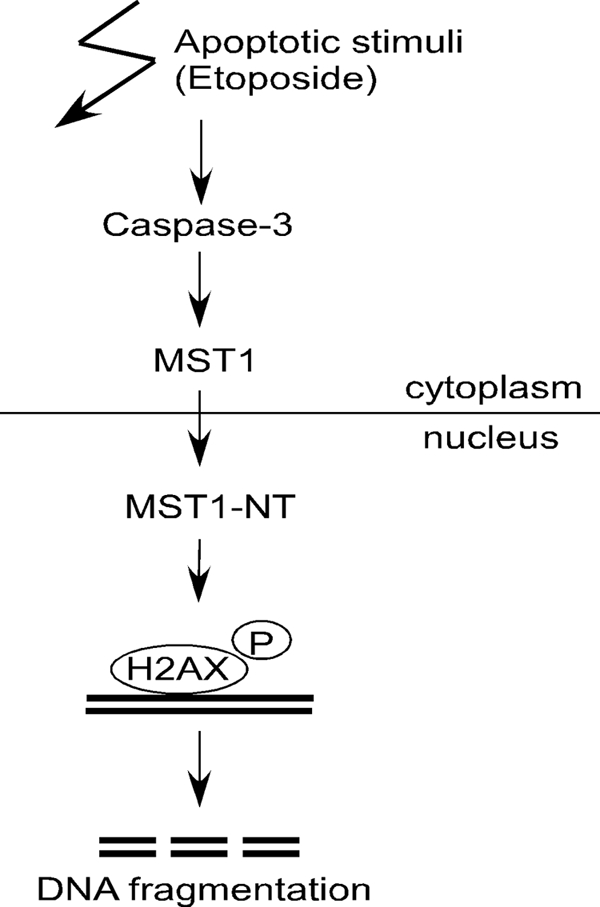
MST1 (mammalian STE20-like kinase 1) is a serine/threonine kinase that is cleaved and activated by caspases during apoptosis. Overexpression of MST1 induces apoptotic morphological changes such as chromatin condensation, but the mechanism is not clear. Here we show that MST1 induces apoptotic chromatin condensation through its phosphorylation of histone H2AX at Ser-139. During etoposide-induced apoptosis in Jurkat cells, the cleavage of MST1 directly corresponded with strong H2AX phosphorylation. In vitro kinase assay results showed that MST1 strongly phosphorylates histone H2AX. Western blot and kinase assay results with a mutant S139A H2AX confirmed that MST1 phosphorylates H2AX at Ser-139. Direct binding of MST1 and H2AX can be detected when co-expressed in HEK293 cells and was also confirmed by an endogenous immunoprecipitation study. When overexpressed in HeLa cells, both the MST1 full-length protein and the MST1 kinase domain (MST1-NT), but not the kinase-negative mutant (MST1-NT-KN), could induce obvious endogenous histone H2AX phosphorylation. The caspase-3 inhibitor benzyloxycarbonyl-DEVD-fluoromethyl ketone (Z-DEVD-fmk) attenuates phosphorylation of H2AX by MST1 but cannot inhibit MST1-NT-induced histone H2AX phosphorylation, indicating that cleaved MST1 is responsible for H2AX phosphorylation during apoptosis. Histone H2AX phosphorylation and DNA fragmentation were suppressed in MST1 knockdown Jurkat cells after etoposide treatment. Taken together, our data indicated that H2AX is a substrate of MST1, which functions to induce apoptotic chromatin condensation and DNA fragmentation.


Figures

FIGURE 1.
FIGURE 1.
MST1 cleavage corresponds with strong histone H2AX phosphorylation and DNA fragmentation during apoptosis. A, Jurkat cells were treated with etoposide (50 μm). Cells were harvested at the indicated times and subjected to flow cytometry after annexin V/propidium iodide staining to assess apoptosis. The means of three independent determinations ± S.D. are shown. Ctrl, control. B and C, Jurkat or H2AX+/+ and H2AX−/− murine embryonic fibroblasts were treated with etoposide (50 μm) and harvested at the indicated times. Lysates were prepared for the detection of MST1 and caspase-3. Histone proteins were extracted for the detection of γ-H2AX and total histone H2AX. DNA was extracted for the DNA ladder assay as described under “Experimental Procedures.”
FIGURE 2.
FIGURE 2.
MST1 phosphorylates H2AX at Ser-139 in vitro. A, histones H2AX and H2B were used as substrates for active MST1. Reactive products were resolved by SDS-PAGE followed by autoradiography or Coomassie blue staining. auto-p, autophosphorylation. B, the H2AX protein was used as a substrate for an in vitro kinase assay. Reactive products were subjected to SDS-PAGE and Western blot to detect histone H2AX and γ-H2AX. C, equal molar amounts of GST-H2AX or GST-H2AX-S139A were used as substrates for MST1 in an in vitro kinase assay. Reactive products were resolved by SDS-PAGE followed by autoradiography or Coomassie Blue staining. D, the same in vitro kinase assay as shown in C was conducted, and reactive products were subjected to SDS-PAGE and Western blot to detect histone H2AX, γ-H2AX, and MST1.
FIGURE 3.
FIGURE 3.
MST1 interacts with H2AX ex vivo. A, HEK293 cells were co-transfected with pCE-MST1-FL, pCE-MST1-NT, or pCE-MST1-CT and pCDNA4-H2AX. At 36 h after transfection, cells were harvested, and the overexpressed H2AX was immunoprecipitated (IP) with anti-His. The expression level of MST1-FL or MST1-NT or MST1-CT was detected with anti-Myc-Tag, and the expression level of H2AX was detected by anti-His. B, Jurkat cells were treated with etoposide and harvested at different times. MST1 was immunoprecipitated with anti-MST1, and H2AX and γ-H2AX were detected with the appropriate antibody.
FIGURE 4.
FIGURE 4.
MST1 overexpression induces apoptotic H2AX phosphorylation in HeLa cells. A, HeLa cells were transfected with pCE-MST1-FL, pCE-MST1-NT, or pCE-MST1-NT-KN. At 36 h after transfection, cells were harvested, and lysates and histone proteins were prepared. The expression level of MST1-FL or MST1-NT was detected with anti-Myc-Tag, and the level of γ-H2AX was detected in the acid-extracted histones. B, HeLa cells were transfected with pCE-MST1-WT, pCE-MST1-NT, or pCE-MST1-NT-KN. At 36 h after transfection, cells were harvested, the overexpressed MST1 was immunoprecipitated (IP) with anti-Myc-Tag, and an in vitro kinase assay was conducted as described under “Experimental Procedures.” IB, immunoblot. C, HeLa cells growing on a slide chamber were transfected with pCE-MST1-KN or pCE-MST1-NT-KN. At 36 h after transfection, cells were fixed with paraformaldehyde, stained for Myc-Tag (MST1, red) and DAPI (blue), and observed by confocal immunofluorescence microscopy. D, HeLa cells growing on a slide chamber were transfected with pCE-MST1-FL, pCE-MST1-NT, or pCE-MST1-NT-KN. At 36 h after transfection, cells were fixed with paraformaldehyde, stained for Myc-Tag (MST1, red), γ-H2AX (green), and Hoechst 33342 (blue), and observed by confocal immunofluorescence microscopy. The bar graphs represent the percentage of apoptotic chromatin condensation in transfected cells from three independent experiments (±S.D.). At least 200 cells were counted each time. The asterisk indicates p < 0.05.
FIGURE 5.
FIGURE 5.
MST1-induced histone H2AX phosphorylation is dependent on its cleavage in HeLa cells. A, HeLa cells were transfected with the pCE-MST1-FL or pCE-MST1-NT plasmid. The caspase-3 inhibitor Z-DEVD-fmk (20 μm) was added to the medium immediately after transfection. At 36 h after transfection, cells were harvested, and lysates and histone proteins were prepared. The expression level of MST1-FL or MST1-NT was detected with anti-Myc-Tag, and the level of γ-H2AX expression was detected in the acid-extracted histones. B, HeLa cells growing on a slide chamber were transfected with the pCE-MST1-FL or pCE-MST1-NT plasmid (NT). The caspase-3 inhibitor Z-DEVD-fmk (20 μm) was added to the medium immediately after transfection. At 36 h after transfection, cells were fixed with paraformaldehyde, stained for Myc-Tag (MST1, red), γ-H2AX (green), and Hoechst 33342 (blue), and observed by confocal immunofluorescence microscopy. The bar graphs represent the percentage of apoptotic chromatin condensation in transfected cells from three independent experiments (±S.D.). At least 200 cells were counted each time. The asterisk indicates p < 0.05.
FIGURE 6.
FIGURE 6.
MST1 is required for histone H2AX phosphorylation and DNA fragmentation during apoptosis. A, mock and MST1 knockdown Jurkat transfectant cells (shMock and shMST1) were treated with 50 μm etoposide for 6 h. Each cell type was divided into three groups: one was used to extract histones to detect γ-H2AX and H2AX; one was prepared as a cell lysate to detect MST1, caspase-3, phosphorylated JNKs and total JNKs, and phosphorylated ATM and total ATM; and the other was used for the DNA ladder assay. B, mock and MST1 knockdown transfectant Jurkat cells were treated as in A. Cell lysates were prepared, and 50 μg were used for the caspase-3 activity assay. Ctrl, control; Etp, etoposide. C, mock and MST1 knockdown Jurkat cells (shMock and shMST1) were treated with 50 μm etoposide for 6 h. Cells were harvested and subjected to flow cytometry analysis after annexin V/propidium iodide staining to assess apoptosis. The percentage of early apoptosis is shown in the bar graphs. The means of three independent determinations ±S.D. are shown, and the asterisk indicates p < 0.05.
FIGURE 7.
FIGURE 7.
Model of the proposed function of MST1 in histone H2AX phosphorylation and DNA fragmentation during apoptosis. Once caspase-3 is activated during etoposide-induced apoptosis, MST1 will be cleaved and activated. The cleaved N-terminal kinase domain translocates into the nucleus and mediates the phosphorylation (P) of histone H2AX, and the phosphorylated H2AX then recruits caspase-activated DNase to mediate DNA fragmentation.

Similar articles

Cited by

References

    1. Creasy C. L., Chernoff J. (1995) Gene 167, 303–306 - PubMed
    1. Taylor L. K., Wang H. C., Erikson R. L. (1996) Proc. Natl. Acad. Sci. U.S.A. 93, 10099–10104 - PMC - PubMed
    1. Graves J. D., Gotoh Y., Draves K. E., Ambrose D., Han D. K., Wright M., Chernoff J., Clark E. A., Krebs E. G. (1998) EMBO J. 17, 2224–2234 - PMC - PubMed
    1. Creasy C. L., Ambrose D. M., Chernoff J. (1996) J. Biol. Chem. 271, 21049–21053 - PubMed
    1. Graves J. D., Draves K. E., Gotoh Y., Krebs E. G., Clark E. A. (2001) J. Biol. Chem. 276, 14909–14915 - PubMed

Publication types

LinkOut - more resources