. 2010 Oct 25;207(11):2323-30.
doi: 10.1084/jem.20101235. Epub 2010 Oct 4.

A critical role for regulatory T cell-mediated control of inflammation in the absence of commensal microbiota

Affiliations

A critical role for regulatory T cell-mediated control of inflammation in the absence of commensal microbiota

Takatoshi Chinen et al. J Exp Med. .

Abstract

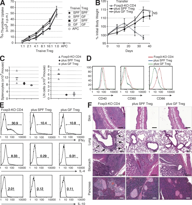
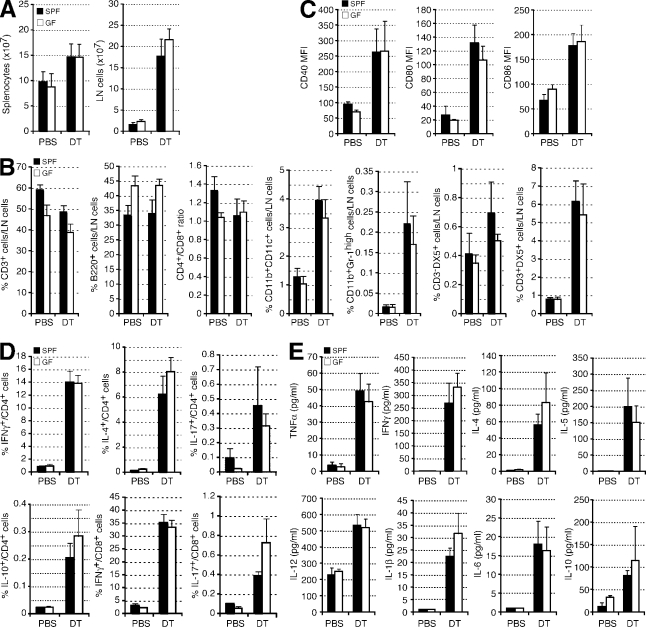
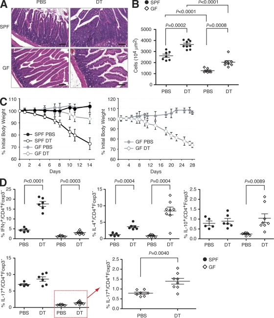
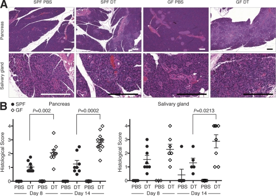
Suppression mediated by regulatory T cells (T reg cells) represents a unique, cell-extrinsic mechanism of in-trans negative regulation that restrains multiple types of immune cells. The loss of T reg cells leads to fatal, highly aggressive, and widespread immune-mediated lesions. This severe autoimmunity may be driven by commensal microbiota, the largest source of non-self ligands activating the innate and adaptive immune systems. Alternatively, T reg cells may primarily restrain T cells with a diverse self-major histocompatibility complex (MHC)-restricted T cell receptor repertoire independently of commensal microbiota. In this study, we demonstrate that in germ-free (GF) mice, ablation of the otherwise fully functional T reg cells resulted in a systemic autoimmune lympho- and myeloproliferative syndrome and tissue inflammation comparable with those in T reg cell-ablated conventional mice. Importantly, there were two exceptions: in GF mice deprived of T reg cells, the inflammation in the small intestine was delayed, whereas exocrine pancreatitis was markedly accelerated compared with T reg cell-ablated conventional mice. These findings suggest that the main function of T reg cells is restraint of self-MHC-restricted T cell responsiveness, which, regardless of the presence of commensal microbiota, poses a threat of autoimmunity.


Figures

Figure 1.
Figure 1.
The suppressor activity of T reg cells from SPF and GF mice is comparable. (A) In vitro suppressor activity of sorted SPF and GF CD4+Foxp3+ T reg cells was assessed by [3H]thymidine incorporation in co-cultures with SPF and GF CD4+Foxp3CD62LhighCD44low naive T cells (Tnaive) and irradiated T cell–depleted SPF splenocytes (APC) in the presence of anti-CD3. The data are representative of two independent experiments. (B) Analysis of the body weight of Tcrb−/−Tcrd−/− recipients of effector CD4+ T cells from Foxp3 mice alone (Foxp3-KO CD4) or together with T reg cells from SPF (plus SPF T reg) or GF (plus GF T reg) mice (n = 6 per group). (A and B) Error bars represent SD. (C) Spleen and LN cellularity of Tcrb−/−Tcrd−/− mice 35 d after the transfer of the indicated cell subsets. The horizontal bars indicate the mean values for each group. (D and E) Flow cytometric analyses of splenocytes of Tcrb−/−Tcrd−/− mice 35 d after the transfer of effector CD4+ T cells from Foxp3 mice with or without SPF or GF T reg cells. Two mice per group were analyzed in two independent experiments, and representative data are shown. The surface expression of the indicated co-stimulatory molecules on B cells (D) and the frequencies of the cytokine-producing cells among CD4+Foxp3 cells determined by flow cytometry after T cell stimulation with anti-CD3 and anti-CD28 (E) are shown. (F) Representative histopathology of the Tcrb−/−Tcrd−/− mice 35 d after the transfer of the indicated cell subsets. Inflamed area of the lung is marked by arrows. Bars, 100 µm.
Figure 2.
Figure 2.
T reg cell–depleted GF mice developed severe inflammatory disease. SPF and GF Foxp3DTR mice were treated with either PBS or DT and analyzed at day 8 after the start of the treatment. (A) The numbers of cells in the spleen and LNs in the indicated mice (n = 7–16 per group). (B and C) Flow cytometric analyses of the cellular composition of LNs in the indicated mice (n = 7–16 per group). (B) Percentage of cells expressing the indicated surface markers. (C) Mean fluorescence intensity (MFI) of the indicated co-stimulatory molecules on CD11c+ MHC class IIhigh DCs. (D) Splenocytes isolated from the indicated mice were stimulated in vitro with anti-CD3 and anti-CD28, and production of the indicated cytokine was measured by flow cytometry. Frequencies of the cytokine-producing cells among CD4+ or CD8+ T cells were determined (n = 3–9 per group). (E) The serum cytokine levels (n = 5–8 per group) were measured by Luminex multiplex bead cytokine assay. (A–E) Error bars represent SD.
Figure 3.
Figure 3.
Inflammation in the small intestine of T reg cell–depleted SPF mice was more severe than in GF mice. (A) Representative histopathology of the small intestine of SPF and GF Foxp3DTR mice 8 d after the start of the treatment with PBS or DT. Bars, 100 µm. (B) The cell numbers in the lamina propria of the small intestine in the indicated mice were determined by morphometry. (C) Analysis of the body weight of SPF and GF Foxp3DTR mice treated with DT or PBS (control). The left panel shows changes in body weights between days 0 and 14 (n = 13 for each group). The right panel shows changes in body weights between days 0 and 28 (n = 4 for each group). Error bars represent SD. (D) Leukocytes isolated from the small intestinal lamina propria of the indicated mice were stimulated in vitro with anti-CD3 and anti-CD28 in the presence of monensin, and intracellular staining for the indicated cytokines was performed. The frequencies of cytokine-producing cells among CD4+Foxp3 cells were determined by flow cytometric analysis. Absolute numbers of the cells are shown in Fig. S2. Data are representative of two independent experiments. (B and D) The horizontal lines indicate the mean ± SEM.
Figure 4.
Figure 4.
Commensal microbiota contributed to protection from autoimmunity in the pancreas and the salivary gland. (A) Representative histopathology of the pancreas and the salivary gland of SPF and GF Foxp3DTR mice 14 d after the start of the treatment with DT or PBS (control). Bars, 200 µm. (B) Histological scores of the pancreas and salivary gland of the indicated mice. The horizontal lines indicate the mean ± SEM.

References

    1. Bennett C.L., Christie J., Ramsdell F., Brunkow M.E., Ferguson P.J., Whitesell L., Kelly T.E., Saulsbury F.T., Chance P.F., Ochs H.D. 2001. The immune dysregulation, polyendocrinopathy, enteropathy, X-linked syndrome (IPEX) is caused by mutations of FOXP3. Nat. Genet. 27:20–21 10.1038/83713 - DOI - PubMed
    1. Brunkow M.E., Jeffery E.W., Hjerrild K.A., Paeper B., Clark L.B., Yasayko S.A., Wilkinson J.E., Galas D., Ziegler S.F., Ramsdell F. 2001. Disruption of a new forkhead/winged-helix protein, scurfin, results in the fatal lymphoproliferative disorder of the scurfy mouse. Nat. Genet. 27:68–73 10.1038/83784 - DOI - PubMed
    1. Chatila T.A., Blaeser F., Ho N., Lederman H.M., Voulgaropoulos C., Helms C., Bowcock A.M. 2000. JM2, encoding a fork head-related protein, is mutated in X-linked autoimmunity-allergic disregulation syndrome. J. Clin. Invest. 106:R75–R81 10.1172/JCI11679 - DOI - PMC - PubMed
    1. Feuerer M., Shen Y., Littman D.R., Benoist C., Mathis D. 2009. How punctual ablation of regulatory T cells unleashes an autoimmune lesion within the pancreatic islets. Immunity. 31:654–664 10.1016/j.immuni.2009.08.023 - DOI - PMC - PubMed
    1. Fontenot J.D., Gavin M.A., Rudensky A.Y. 2003. Foxp3 programs the development and function of CD4+CD25+ regulatory T cells. Nat. Immunol. 4:330–336 10.1038/ni904 - DOI - PubMed

Publication types

MeSH terms