. 2010 Oct 25;207(11):2395-405.
doi: 10.1084/jem.20100995. Epub 2010 Oct 4.

A requirement for FcγR in antibody-mediated bacterial toxin neutralization

Affiliations

A requirement for FcγR in antibody-mediated bacterial toxin neutralization

Nareen Abboud et al. J Exp Med. .

Abstract

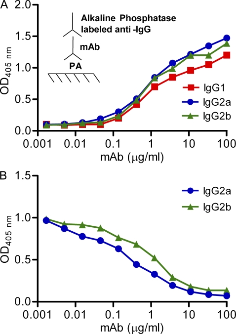
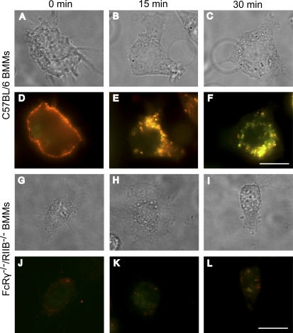
One important function of humoral immunity is toxin neutralization. The current view posits that neutralization results from antibody-mediated interference with the binding of toxins to their targets, a phenomenon viewed as dependent only on antibody specificity. To investigate the role of antibody constant region function in toxin neutralization, we generated IgG2a and IgG2b variants of the Bacillus anthracis protective antigen-binding IgG1 monoclonal antibody (mAb) 19D9. These antibodies express identical variable regions and display the same specificity. The efficacy of antibody-mediated neutralization was IgG2a > IgG2b > IgG1, and neutralization activity required competent Fcγ receptor (FcγR). The IgG2a mAb prevented lethal toxin cell killing and mitogen-activated protein kinase/extracellular signal-regulated kinase kinase cleavage more efficiently than the IgG1 mAb. Passive immunization with IgG1 and IgG2a mAb protected wild-type mice, but not FcγR-deficient mice, against B. anthracis infection. These results establish that constant region isotype influences toxin neutralization efficacy of certain antibodies through a mechanism that requires engagement of FcγR. These findings highlight a new parameter for evaluating vaccine responses and the possibility of harnessing optimal FcγR interactions in the design of passive immunization strategies.


Figures

Figure 1.
Figure 1.
Specific and competitive binding of IgG1, IgG2a, and IgG2b mAbs to PA. (A) IgG1, IgG2a, and IgG2b mAbs were added at different concentrations to PA bound to polystyrene plates. Binding was measured by ELISA. (B) A constant amount of IgG1 (2 µg/ml) and increasing concentrations of IgG2a and IgG2b were allowed to compete for binding to PA bound to polystyrene plates. Data shown are representative of three independent experiments.
Figure 2.
Figure 2.
LeTx-neutralizing activity of IgG subclasses on RAW264.7 macrophages. Cells were treated with or without LeTx (100 ng/ml PA and 100 ng/ml LF) in the presence or absence of varying amounts of mAb (A), or treated with or without 10 µg/ml mAb in the presence or absence of varying amounts of LF (PA at fixed saturating concentrations, 100 ng/ml; B) or PA (LF at fixed saturating concentrations, 100 ng/ml; C). Viability was established by MTT assay 4 h after LT treatment. Cells treated with IgG2a and IgG2b mAbs showed statistically significant elevation in protection with cells treated with IgG1 mAb. Significance was determined by Student’s t test. Data shown are representative of three independent experiments. Means and standard deviations of triplicates are shown from one representative experiment out of three (A).
Figure 3.
Figure 3.
LeTx-neutralizing activity of IgG1, IgG2a, and IgG2b mAbs is dependent on FcR interactions. (A and B) BMMs from C57BL/6 (A) and FcRγ−/−/RIIB−/− (B) mice were plated in 96-well plates at a density of 8 × 10⁴ cells per well in maintenance medium supplemented with FCS, 2 h before treatment with LeTx (100 ng/ml PA and 100 ng/ml LF). The indicated concentrations of mAb were added at the same time with LeTx. After 72 h, cell death was assayed with MTT (3,(4,5-dimethylthiazol-2-yl) 2,5-diphenyltetrazolium bromide; controls were cells treated with LeTx only or with media alone). Representative data are shown from three independent experiments. Significance was determined by a Student’s t test. (C) BMMs from C57BL/6 mice were plated as described in A and B, and the indicated dilutions of each mAb were used to neutralize LeTx in the presence of anti-FcRII/III (mAb 2.4G2). (D) RAW264.7 macrophages were treated with toxin, as described in A and B, and 10 µg/ml IgG2a mAb in the presence of blocking mAb to FcγRIII (2.4G2), FcγRIV (9E9), and/or a competitor inhibitor to block FcγRI (IgG2a Std.). (E) RAW264.7 macrophages were gated and analyzed by flow cytometric analysis for FcγRI, FcγRII, FcγRIII, and FcγRIV expression with specific antibodies. Histograms shaded in gray represent the specific FcγR, and histograms shaded in black indicates unstained cells used as a control. (F and G) C57BL/6 and FcRγ−/−/RIIB−/− were treated with 100 ng/ml LeTx and 10 µg/ml mAb. MEK-3 cleavage was determined by Western blotting with anti–MEK-3. Data shown are representative of three independent experiments. Means and standard deviations of triplicates are shown from one representative experiment out of three (A and D).
Figure 4.
Figure 4.
IgG1 and IgG2a mAbs protect wild-type but not FcRγ−/−/RIIB−/− or FcRγ−/− mice from B. anthracis infection. (A) Kaplan-Meier analysis of survival rate of FcRγ−/−/RIIB−/− or wild-type mice treated with IgG1 and IgG2a mAbs or isotype control antibodies (n = 10). (B) Kaplan-Meier analysis of survival rate of FcRγ−/− or wild-type mice treated with IgG1 and IgG2a mAbs or isotype control antibodies (n = 10). The log-rank test was used to determine significance between wild-type and FcRγ−/−/RIIB−/− and FcRγ−/− mouse groups treated with IgG1 and IgG2a mAbs. Data shown are representative of two independent experiments.
Figure 5.
Figure 5.
Colocalization between IgG2a mAb and PA. Wild-type and FcRγ−/−/RIIB−/− BMMs were cooled to 4°C, incubated with Alexa Fluor 488–conjugated IgG2a (red) and Alexa Fluor 568–conjugated PA (green), washed, and warmed to 37°C for the indicated time points. The cells were then fixed and analyzed by fluorescence microscopy. Images in A–C were acquired in a bright field and correspond to D–F, respectively, whereas G–I correspond to J–L, respectively. Data shown are representative of three independent experiments. Bars, 10 µm.
Figure 6.
Figure 6.
Inhibition of phagocytosis by cytochalasin d and up-regulation of FcγRs by IgG1 control antibody on RAW264.7 macrophages. (A) RAW264.7 macrophages that were treated with LeTx and mAb or LeTx, mAb, and 2 µM cytochalasin d (Cyto D). Cells were preincubated for 45 min with cytochalasin d. Cell viability was determined as indicated in the Cell viability assays section of Materials and methods. (B) RAW264.7 macrophages were either not preincubated or preincubated overnight with an IgG1 control antibody (used to preactivate FcRs) and next-day treated with toxin and mAb. Data shown are representative of two independent experiments. Means and standard deviations of triplicates are shown from one representative experiment out of two (A).

References

    1. Abboud N., Casadevall A. 2008. Immunogenicity of Bacillus anthracis protective antigen domains and efficacy of elicited antibody responses depend on host genetic background. Clin. Vaccine Immunol. 15:1115–1123 10.1128/CVI.00015-08 - DOI - PMC - PubMed
    1. Abboud N., De Jesus M., Nakouzi A., Cordero R.J., Pujato M., Fiser A., Rivera J., Casadevall A. 2009. Identification of linear epitopes in Bacillus anthracis protective antigen bound by neutralizing antibodies. J. Biol. Chem. 284:25077–25086 10.1074/jbc.M109.022061 - DOI - PMC - PubMed
    1. Abrami L., Liu S., Cosson P., Leppla S.H., van der Goot F.G. 2003. Anthrax toxin triggers endocytosis of its receptor via a lipid raft-mediated clathrin-dependent process. J. Cell Biol. 160:321–328 10.1083/jcb.200211018 - DOI - PMC - PubMed
    1. Abrami L., Lindsay M., Parton R.G., Leppla S.H., van der Goot F.G. 2004. Membrane insertion of anthrax protective antigen and cytoplasmic delivery of lethal factor occur at different stages of the endocytic pathway. J. Cell Biol. 166:645–651 10.1083/jcb.200312072 - DOI - PMC - PubMed
    1. Aderem A., Underhill D.M. 1999. Mechanisms of phagocytosis in macrophages. Annu. Rev. Immunol. 17:593–623 10.1146/annurev.immunol.17.1.593 - DOI - PubMed

Publication types

MeSH terms