. 2010 Oct 25;207(11):2479-91.
doi: 10.1084/jem.20101376. Epub 2010 Oct 4.

A novel subset of CD4(+) T(H)2 memory/effector cells that produce inflammatory IL-17 cytokine and promote the exacerbation of chronic allergic asthma

Affiliations

A novel subset of CD4(+) T(H)2 memory/effector cells that produce inflammatory IL-17 cytokine and promote the exacerbation of chronic allergic asthma

Yui-Hsi Wang et al. J Exp Med. .

Abstract

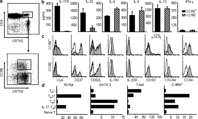
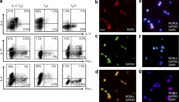
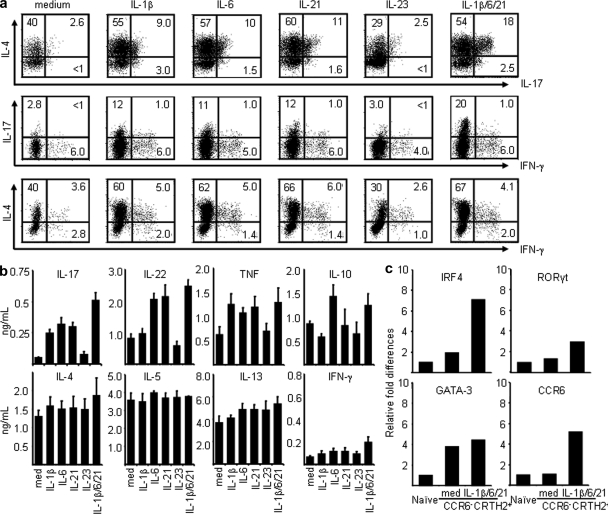
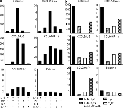
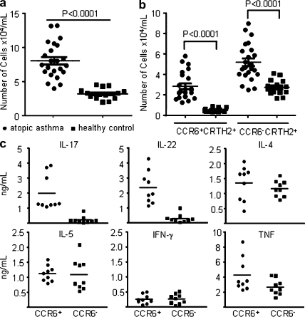
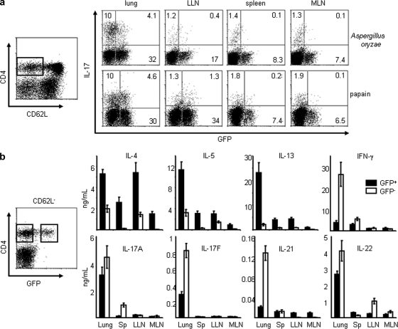
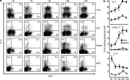
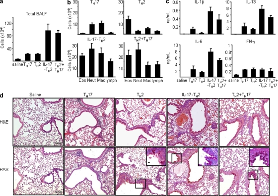
The inflammatory cytokine interleukin (IL)-17 is involved in the pathogenesis of allergic diseases. However, the identity and functions of IL-17-producing T cells during the pathogenesis of allergic diseases remain unclear. Here, we report a novel subset of T(H)2 memory/effector cells that coexpress the transcription factors GATA3 and RORγt and coproduce T(H)17 and T(H)2 cytokines. Classical T(H)2 memory/effector cells had the potential to produce IL-17 after stimulation with proinflammatory cytokines IL-1β, IL-6, and IL-21. The number of IL-17-T(H)2 cells was significantly increased in blood of patients with atopic asthma. In a mouse model of allergic lung diseases, IL-17-producing CD4(+) T(H)2 cells were induced in the inflamed lung and persisted as the dominant IL-17-producing T cell population during the chronic stage of asthma. Treating cultured bronchial epithelial cells with IL-17 plus T(H)2 cytokines induced strong up-regulation of chemokine eotaxin-3, Il8, Mip1b, and Groa gene expression. Compared with classical T(H)17 and T(H)2 cells, antigen-specific IL-17-producing T(H)2 cells induced a profound influx of heterogeneous inflammatory leukocytes and exacerbated asthma. Our findings highlight the plasticity of T(H)2 memory cells and suggest that IL-17-producing T(H)2 cells may represent the key pathogenic T(H)2 cells promoting the exacerbation of allergic asthma.


Figures

Figure 1.
Figure 1.
Phenotypic and functional characteristics of CRTH2+CD4+ TH2 cell subsets. (a) Enriched human CRTH2hiCD4+ T cells could be divided into two subsets based on their surface expression of CCR6. Purified resting CCR6+ and CCR6 of CRTH2+CD4+ TH2 cells were stimulated with anti-CD3/CD28 mAbs for the measurements of cytokines in the culture supernatants by ELISA (b) or analyzed for the expression of other surface markers (c). Filled histograms represent the staining of indicated cell subset with markers shown below the histogram; open histograms represent the isotype. (d) cDNA templates made from the indicated T cell subsets were prepared as described in the Materials and methods. Expression levels of indicated genes that are involved in T cell differentiation were measured by real-time PCR as described in methods. Fold differences in gene expression level between cell types marked in the left panel are indicated in horizontal axis. The results in a–d are from separate experiments. Data represent the mean (±SD) of five experiments (b). Data are from one of three independent experiments (c and d). IL-17-TH2, IL-17–producing TH2 cells.
Figure 2.
Figure 2.
The novel human CCR6+CRTH2+CD4+ TH2 cell subset displays features of both TH2 and TH17 cell lineages. Sorted CCR6+ (b-e) and CCR6 (f) subsets of CRTH2+CD4+ TH2 and CCR6+CRTH2CD4+ TH17 (g) cells were cultured in the homeostatic cytokines IL-7/IL-15 in the presence of anti–IL-4 and anti–IFN-γ mAbs for 3 d. Expanded cells were collected and restimulated with PMA plus ionomycin for analysis of intracellular cytokine production (a) or activated with anti-CD3 before two-color immunofluorescent staining with anti–human RORγt (red) or GATA-3 (green) mAb (b-g). Cell nuclei were identified with DAPI (blue). Numbers within the quadrants indicate the percentage of cultured cells that stained positive for each respective cytokine. Bars, 5 µm. Data are from one of three independent experiments (a–g). IL-17-TH2, IL-17–producing TH2 cells.
Figure 3.
Figure 3.
Proinflammatory stimuli induce classical CD4+ TH2 cells to produce IL-17 cytokine. Sorted CCR6CRTH2+CD4+ TH2 cells were maintained with homeostatic cytokines IL-7/IL-15 plus anti–IL-4 and anti–IFN-γ mAbs in the presence or absence of the indicated proinflammatory cytokines for 6 d. Expanded cells were restimulated with PMA plus ionomycin for the analysis of intracellular cytokine production (a) or anti-CD3/CD28 for measurement of cytokines in the culture supernatants by ELISA (b) or were used as cDNA templates for the indicated gene expression analysis by real-time PCR (c). Fold differences in gene expression level between cell/treatment groups marked in the horizontal axis are indicated in the left panel. Data represent the mean (±SD) of five experiments (b). Data are from one of three independent experiments (a and c).
Figure 4.
Figure 4.
Combinations of IL-17 and TH2 cytokines induce profound up-regulation of chemokine gene expression. cDNA templates made from cultured normal human bronchial epithelial cells treated with or without the indicated cytokines (a) or supernatants collected from indicated T helper subsets activated by anti-CD3/CD28 mAbs for 24 h in the presence or absence of anti–IL-17 neutralizing antibodies (b). Expression levels of indicated chemokine genes were measured by real-time PCR as described in the Materials and methods. Fold differences in gene expression level between treatment groups marked in the horizontal axis are indicated in the left panel. Data are from one of three independent experiments (a and b). IL-17-TH2, IL-17–producing TH2 cells.
Figure 5.
Figure 5.
An increased number of circulating IL-17–producing TH2 cells is associated with patients with allergic asthma. Total CD4+ T cells from peripheral blood of healthy donors (n = 16) or subjects with allergic asthma (n = 23) were enriched and counted by hemacytometer. Cell numbers of total CRTH2+CD4+ TH2 cells (a) and of CCR6+ and CCR6 subsets of CRTH2+CD4+ TH2 cells (b) were obtained based on their the percentage within enriched CD4+ T cells by multicolor immunofluorescence analyses. (c) Sorted CCR6+ or CCR6 subsets of CRTH2+CD4+ TH2 cells from subjects with atopic asthma (n = 9) were restimulated with anti-CD3/CD28 mAbs for 24 h before measurement of the secreted cytokines in the supernatants by ELISA.
Figure 6.
Figure 6.
The induction of IL-17–producing TH2 cells occurred in the inflamed lung after allergen exposure. 4GET mice (n = 4) were challenged with indicated allergens plus OVA intranasally every other day for a total of six times before sacrifice. Sorted total (a) or GFP+ and GFP subsets (b) of CD4+CD62LCD44hi memory/effector T cells from lung or indicated lymphoid tissues were restimulated with PMA plus ionomycin for the analysis of intracellular cytokine production (a) or with anti-CD3/CD28 mAbs for 24 h before measurement of cytokines in the supernatants by ELISA (b). Data are from one of three independent experiments (a and b). Data represented as the mean (±SD); four mice per group. LLN, lung draining LNs; MLN, intestine mesenteric LNs.
Figure 7.
Figure 7.
Inflammatory IL-17–producing TH2 cells persist in the inflamed lung during the chronic phase of allergic asthma. 4GET mice (n = 4) were challenged with Aspergillus Orazae plus OVA intranasally every other day for a total of six times (a and b). After the last challenge, mice rested for the indicated time frame (a and b; horizontal axis) before sacrifice. Purified CD4+CD62LCD44hi memory/effector T cells from lung or other lymphoid tissues were restimulated with PMA plus ionomycin for the analysis of intracellular cytokine production (a). The percentage of indicated cytokine producing cells within the total CD4+ memory/effector T cell pool were numerated as shown on the left axis (b). Data are representative of two independent experiments. Data represented as the mean (±SD); four mice per group.
Figure 8.
Figure 8.
Antigen-specific inflammatory IL-17–producing TH2 cells promote the exacerbation of allergic asthma. Five groups of BALB/c mice were intranasally challenged once a day for 2 d with OVA 24 h after being adoptive transferred with 0.9% saline as a control or OVA-specific IL-17–producing TH2, classical TH2, classical TH17, or classical TH2 and TH17 cells generated in vitro, as described in Materials and methods. BALF of individual mice of each group were collected for the measurement of total cell counts (a) and differential cell counts (b), indicating that the total numbers of individual inflammatory cells in each group or (c) concentrations of indicated cytokines by ELISA. (d) Histological analysis of representative lung bronchovascular bundles stained with hematoxylin and eosin (H&E; top) or stained with periodic acid Schiff (PAS; bottom). The insets at the corner depict higher magnification images of the airway epithelium stained with PAS, showing that much more abundant mucus-producing cells (pink cytoplasm) are lining the airway epithelium of mice receiving indicated T helper cell subsets. Data are representative of three independent experiments. Data represented as the mean (±SD); four mice per group. Bars: (capped) 100 µm; (uncapped) 10 µm.

References

    1. Aarvak T., Chabaud M., Miossec P., Natvig J.B. 1999. IL-17 is produced by some proinflammatory Th1/Th0 cells but not by Th2 cells. J. Immunol. 162:1246–1251 - PubMed
    1. Acosta-Rodriguez E.V., Rivino L., Geginat J., Jarrossay D., Gattorno M., Lanzavecchia A., Sallusto F., Napolitani G. 2007. Surface phenotype and antigenic specificity of human interleukin 17-producing T helper memory cells. Nat. Immunol. 8:639–646 10.1038/ni1467 - DOI - PubMed
    1. Anderson G.P. 2008. Endotyping asthma: new insights into key pathogenic mechanisms in a complex, heterogeneous disease. Lancet. 372:1107–1119 10.1016/S0140-6736(08)61452-X - DOI - PubMed
    1. Annunziato F., Cosmi L., Santarlasci V., Maggi L., Liotta F., Mazzinghi B., Parente E., Filì L., Ferri S., Frosali F., et al. 2007. Phenotypic and functional features of human Th17 cells. J. Exp. Med. 204:1849–1861 10.1084/jem.20070663 - DOI - PMC - PubMed
    1. Bates C.A., Silkoff P.E. 2003. Exhaled nitric oxide in asthma: from bench to bedside. J. Allergy Clin. Immunol. 111:256–262 - PubMed

Publication types

MeSH terms