. 2010 Oct 19;107(42):18079-84.
doi: 10.1073/pnas.1000467107. Epub 2010 Oct 4.

Characterization of mononuclear phagocytic cells in medaka fish transgenic for a cxcr3a:gfp reporter

Affiliations

Characterization of mononuclear phagocytic cells in medaka fish transgenic for a cxcr3a:gfp reporter

Narges Aghaallaei et al. Proc Natl Acad Sci U S A. .

Abstract

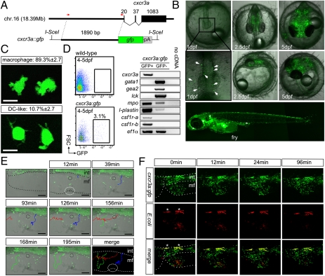
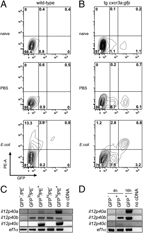
Chemokines and chemokine receptors are key evolutionary innovations of vertebrates. They are involved in morphogenetic processes and play an important role in the immune system. Based on an analysis of the chemokine receptor gene family in teleost genomes, and the expression patterns of chemokine receptor genes during embryogenesis and the wounding response in young larvae of Oryzias latipes, we identified the chemokine receptor cxcr3a as a marker of innate immune cells. Cells expressing cxcr3a were characterized in fish transgenic for a cxcr3a:gfp reporter. In embryos and larvae, cxcr3a-expressing cells are motile in healthy and damaged tissues, and phagocytic; the majority of these cells has the morphology of tissue macrophages, whereas a small fraction has a dendritic phenotype. In adults, cxcr3a-positive cells continue to specifically express myeloid-associate markers and genes related to antigen uptake and presentation. By light microscopy and ultrastructural analysis, the majority of cxcr3a-expressing cells has a dendritic phenotype, whereas the remainder resembles macrophage-like cells. After challenge of adult fish with bacteria or CpG oligonucleotides, phagocytosing cxcr3a-positive cells in the blood up-regulated il12p40 genes, compatible with their function as part of the mononuclear phagocytic system. Our results identify a marker of teleost mononuclear phagocytic cells and suggest a surprising degree of morphological and functional similarity between the innate immune systems of lower and higher vertebrates.


Conflict of interest statement

The authors declare no conflict of interest.

Figures

Fig. 1.
Fig. 1.
The chemokine receptor gene family in two teleost species. (A) Phylogenetic tree based on derived protein sequences of chemokine receptor genes of human (prefix Hs), mouse (prefix Mm), medaka (prefix Ol), and tetraodon (prefix Tn) genomes. The sequence for the human formyl-peptide receptor FPR1 was used as an outgroup. (B) Expression of chemokine receptor genes in medaka from 5 dpf until larval stage. White box indicates no expression, filled box indicates expression detected by WISH.
Fig. 2.
Fig. 2.
Characterization of cxcr3a-expressing cells in O. latipes embryos and larvae. (A) Schematic diagram of the transgenic construct indicating the position of the 1.9-kb promoter fragment. (B) Fluorescence microscopy of transgenic embryos at the indicated time points (in dpf). Positive cells identifiable at 1 dpf are indicated. In the panel depicting the fry, merged from four pictures, regions with autofluorescent pigment cells are indicated by an asterisk. Note that the pattern of fluorescence is similar to the distribution of cxcr3a-mRNA as determined by whole-mount in situ hybridization (Fig. S1). (C) Morphology of GFP-positive cells. A total of 664 cells from 13 embryos (51 ± 17 cells per embryo) were observed by live imaging of 5 dpf embryos and categorized as macrophages (two representative examples are shown, Upper) or dendritic-like cells (Lower). (D) Flow cytometric profiles of WT and transgenic embryos (Left) and representative results from RT-PCR reactions. GFP-positive cells express myeloid-associated markers, whose expression is greatly reduced or not detectable in the GFP-negative fraction. By contrast, genes specifically expressed in the erythroid (gata1, embryonic alpha globin 2 [gea2]) and T cell (lck) lineages are exclusively expressed in GFP-negative cells. Note that the flow cytometric analysis is not quantitative, because only mechanical disruption but no enzymatic digestion of tissues was used. Results are representative of two independent cell sortings from pooled embryos and subsequent duplicate cDNA analyses. (E) Still photographs taken at various time points after wounding in the membranous fin (mf) of transgenic larvae. The region of insult is marked by a white dotted circle. Three different migration behaviors are depicted by different colors. Blue and green cells migrate from the intestine (int) region into the insult (Movie S2). (Scale bar, 100 μm.) (F) Time course of bacterial clearance in transgenic larvae. Regions of autofluorescent cells are indicated by an asterisk. Note the appearance of yellow cells, indicative of phagocytosis by GFP-positive cells; also see Movies S3 and S4.
Fig. 3.
Fig. 3.
Gene expression profiles and morphology of cxcr3a-positive cells in adult O. latipes. (A) Light-scatter properties of adult GFP-expressing cells isolated from whole kidney marrow. Note that the majority of cells (green dots) falls into the myelomonocytic gate (red circle). Similar results were obtained for GFP-expressing cells isolated from blood and spleen. Results are representative of three independent experiments from pooled tissues. (B) Cells expressing cxcr3a are confined to the cxcr3a:gfp-positive fraction of blood cells from adult fish. Single myelomonocytic cells were separated according to fluorescence and examined for cxcr3a-expression by RT-PCR. (C) Expression profiles of GFP-positive and GFP-negative cells isolated from whole kidney marrow of adult cxcr3a:gfp medaka fish. Results are representative of two independent cell sorting experiments from pooled tissues and subsequent duplicate cDNA analyses. (D) Expression profiles of GFP-positive and GFP-negative cells isolated from blood of adult cxcr3a:gfp medaka. Results are representative of duplicate cDNA analyses. (E) Distribution of cell types (mean ± SD) among GFP-positive and GFP-negative cells isolated from blood of adult cxcr3a:gfp medaka as revealed by Wright-Giemsa staining. A total of 805 GFP-positive cells (from a total of five fish; i.e., five independent cell sorting experiments) and 227 GFP-negative cells of myelomonocytic morphology (from three fish; i.e., three independent cell sorting experiments) were evaluated from cytospin preparations. (Scale bar, 3 μm.) (F) EM images of GFP-expressing cells obtained from myelomonocytic cells of kidney and spleen (Left) and blood (Right) of adult fish processed according to the two protocols described in SI Materials and Methods. Note the presence of electron-dense cytoplasmic extensions and large numbers of cytoplasmic vacuoles. (Scale bars, 1 μm.)
Fig. 4.
Fig. 4.
Functional analysis of cxcr3a-positive cells. (A) WT adult fish received intracoelomic injections of red fluorescently labeled bacteria (or vehicle alone), and 2 h later, blood cells were analyzed by flow cytometry (gated for cells with light scatter characteristics of myelomonocytic cells). The proportions of cells are indicated (representative patterns for three animals are shown). Results are representative of at least three independent experiments. (B) cxcr3a:gfp transgenic adult fish received intracoelomic injections of red fluorescently labeled bacteria (or vehicle alone), and 2 h later, blood cells were analyzed by flow cytometry as in A. The proportions of myelomonocytic cells in the individual gates are indicated (representative patterns for at least three animals are shown). Note that the proportions of both GFPlow and GFPhigh cells increase in injected fish and that the majority of phagocytosing cells are GFP-positive. (C) Phagocytosis induces expression of il12p40 genes. Adult cxcr3a:gfp transgenic fish received intracoelomic injections of red fluorescently labeled bacteria, and 2 h later, blood cells were separated by flow cytometry according to their fluorescence characteristics. GFP-negative cells express erythroid (gata1) and lymphocyte (lck) lineage markers, whereas GFP-positive cells do not (cf. Fig. 3C). GFP-positive cells are separated into those expressing low (lo) and high (hi) levels and whether they have phagocytosed red fluorescently labeled bacteria (PE+ or PE−). Results for the three il12p40 paralogues (Fig. S7) are shown. Results are representative of two independent experiments (i.e., cell sorting followed by duplicate cDNA analyses). (D) CpG oligonucleotides induce il12p40 gene expression. Adult cxcr3a:gfp fish were given intracoelomic injections; GFP-negative and GFP-positive blood cells were isolated by flow cytometry 4 h after injection and processed for RT-PCR analysis. For GFP-positive cells, the results for the 16-h time point after injection are also shown. Results are representative of two independent experiments (i.e., cell sorting followed by duplicate cDNA analyses).

References

    1. Bajoghli B, et al. Evolution of genetic networks underlying the emergence of thymopoiesis in vertebrates. Cell. 2009;138:186–197. - PubMed
    1. de Jong JLO, Zon LI. Use of the zebrafish system to study primitive and definitive hematopoiesis. Annu Rev Genet. 2005;39:481–501. - PubMed
    1. Meeker ND, Trede NS. Immunology and zebrafish: Spawning new models of human disease. Dev Comp Immunol. 2008;32:745–757. - PubMed
    1. Zapata A, Diez B, Cejalvo T, Gutiérrez-de Frías C, Cortés A. Ontogeny of the immune system of fish. Fish Shellfish Immunol. 2006;20:126–136. - PubMed
    1. Li M, Ransohoff RM. Multiple roles of chemokine CXCL12 in the central nervous system: A migration from immunology to neurobiology. Prog Neurobiol. 2008;84:116–131. - PMC - PubMed

Publication types

Substances

LinkOut - more resources