Epithelial cell proliferation in the developing zebrafish intestine is regulated by the Wnt pathway and microbial signaling via Myd88
- PMID: 20921418
- PMCID: PMC3063593
- DOI: 10.1073/pnas.1000072107
Epithelial cell proliferation in the developing zebrafish intestine is regulated by the Wnt pathway and microbial signaling via Myd88
Abstract
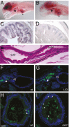
Rates of cell proliferation in the vertebrate intestinal epithelium are modulated by intrinsic signaling pathways and extrinsic cues. Here, we report that epithelial cell proliferation in the developing zebrafish intestine is stimulated both by the presence of the resident microbiota and by activation of Wnt signaling. We find that the response to microbial proliferation-promoting signals requires Myd88 but not TNF receptor, implicating host innate immune pathways but not inflammation in the establishment of homeostasis in the developing intestinal epithelium. We show that loss of axin1, a component of the β-catenin destruction complex, results in greater than WT levels of intestinal epithelial cell proliferation. Compared with conventionally reared axin1 mutants, germ-free axin1 mutants exhibit decreased intestinal epithelial cell proliferation, whereas monoassociation with the resident intestinal bacterium Aeromonas veronii results in elevated epithelial cell proliferation. Disruption of β-catenin signaling by deletion of the β-catenin coactivator tcf4 partially decreases the proliferation-promoting capacity of A. veronii. We show that numbers of intestinal epithelial cells with cytoplasmic β-catenin are reduced in the absence of the microbiota in both WT and axin1 mutants and elevated in animals' monoassociated A. veronii. Collectively, these data demonstrate that resident intestinal bacteria enhance the stability of β-catenin in intestinal epithelial cells and promote cell proliferation in the developing vertebrate intestine.
Conflict of interest statement
The authors declare no conflict of interest.
Figures


Similar articles
-
H. pylori virulence factor CagA increases intestinal cell proliferation by Wnt pathway activation in a transgenic zebrafish model.Dis Model Mech. 2013 May;6(3):802-10. doi: 10.1242/dmm.011163. Epub 2013 Mar 1. Dis Model Mech. 2013. PMID: 23471915 Free PMC article.
-
Phases of canonical Wnt signaling during the development of mouse intestinal epithelium.Gastroenterology. 2007 Aug;133(2):529-38. doi: 10.1053/j.gastro.2007.04.072. Epub 2007 May 3. Gastroenterology. 2007. PMID: 17681174
-
CCAAT/enhancer-binding protein-β functions as a negative regulator of Wnt/β-catenin signaling through activation of AXIN1 gene expression.Cell Death Dis. 2018 Oct 3;9(10):1023. doi: 10.1038/s41419-018-1072-1. Cell Death Dis. 2018. PMID: 30283086 Free PMC article.
-
[Dual-role regulations of canonical Wnt/beta-catenin signaling pathway].Beijing Da Xue Xue Bao Yi Xue Ban. 2010 Apr 18;42(2):238-42. Beijing Da Xue Xue Bao Yi Xue Ban. 2010. PMID: 20396373 Review. Chinese.
-
Mechanisms of epithelial growth and development in the zebrafish intestine.Biochem Soc Trans. 2023 Jun 28;51(3):1213-1224. doi: 10.1042/BST20221375. Biochem Soc Trans. 2023. PMID: 37293990 Review.
Cited by
-
Conserved anti-inflammatory effects and sensing of butyrate in zebrafish.Gut Microbes. 2020 Nov 9;12(1):1-11. doi: 10.1080/19490976.2020.1824563. Gut Microbes. 2020. PMID: 33064972 Free PMC article.
-
Investigating bacterial-animal symbioses with light sheet microscopy.Biol Bull. 2012 Aug;223(1):7-20. doi: 10.1086/BBLv223n1p7. Biol Bull. 2012. PMID: 22983029 Free PMC article.
-
Microgavage of zebrafish larvae.J Vis Exp. 2013 Feb 20;(72):e4434. doi: 10.3791/4434. J Vis Exp. 2013. PMID: 23463135 Free PMC article.
-
Regulation of immunity and disease resistance by commensal microbes and chromatin modifications during zebrafish development.Proc Natl Acad Sci U S A. 2012 Sep 25;109(39):E2605-14. doi: 10.1073/pnas.1209920109. Epub 2012 Sep 4. Proc Natl Acad Sci U S A. 2012. PMID: 22949679 Free PMC article.
-
Study of host-microbe interactions in zebrafish.Methods Cell Biol. 2011;105:87-116. doi: 10.1016/B978-0-12-381320-6.00004-7. Methods Cell Biol. 2011. PMID: 21951527 Free PMC article.
References
-
- van der Flier LG, Clevers H. Stem cells, self-renewal, and differentiation in the intestinal epithelium. Annu Rev Physiol. 2009;71:241–260. - PubMed
-
- Abrams GD, Bauer H, Sprinz H. Influence of the normal flora on mucosal morphology and cellular renewal in the ileum. A comparison of germ-free and conventional mice. Lab Invest. 1963;12:355–364. - PubMed
-
- Uribe A, Alam M, Johansson O, Midtvedt T, Theodorsson E. Microflora modulates endocrine cells in the gastrointestinal mucosa of the rat. Gastroenterology. 1994;107:1259–1269. - PubMed
Publication types
MeSH terms
Substances
Grants and funding
LinkOut - more resources
Full Text Sources
Other Literature Sources
Molecular Biology Databases

