. 2010 Oct 19;107(42):18143-8.
doi: 10.1073/pnas.1003919107. Epub 2010 Oct 4.

Inhibition of brain tumor growth by intravenous poly (β-L-malic acid) nanobioconjugate with pH-dependent drug release [corrected]

Affiliations

Inhibition of brain tumor growth by intravenous poly (β-L-malic acid) nanobioconjugate with pH-dependent drug release [corrected]

Hui Ding et al. Proc Natl Acad Sci U S A. .

Erratum in

  • Proc Natl Acad Sci U S A. 2010 Nov 9;107(45):19603

Abstract

Effective treatment of brain neurological disorders such as Alzheimer's disease, multiple sclerosis, or tumors should be possible with drug delivery through blood-brain barrier (BBB) or blood-brain tumor barrier (BTB) and targeting specific types of brain cells with drug release into the cell cytoplasm. A polymeric nanobioconjugate drug based on biodegradable, nontoxic, and nonimmunogenic polymalic acid as a universal delivery nanoplatform was used for design and synthesis of nanomedicine drug for i.v. treatment of brain tumors. The polymeric drug passes through the BTB and tumor cell membrane using tandem monoclonal antibodies targeting the BTB and tumor cells. The next step for polymeric drug action was inhibition of tumor angiogenesis by specifically blocking the synthesis of a tumor neovascular trimer protein, laminin-411, by attached antisense oligonucleotides (AONs). The AONs were released into the target cell cytoplasm via pH-activated trileucine, an endosomal escape moiety. Drug delivery to the brain tumor and the release mechanism were both studied for this nanobiopolymer. Introduction of a trileucine endosome escape unit resulted in significantly increased AON delivery to tumor cells, inhibition of laminin-411 synthesis in vitro and in vivo, specific accumulation in brain tumors, and suppression of intracranial glioma growth compared with pH-independent leucine ester. The availability of a systemically active polymeric drug delivery system that passes through the BTB, targets tumor cells, and inhibits glioma growth gives hope for a successful strategy of glioma treatment. This delivery system with drug release into the brain-specific cell type could be useful for treatment of various brain pathologies.


Conflict of interest statement

The authors declare no conflict of interest.

Figures

Fig. 1.
Fig. 1.
Nanoconjugates with polymalic acid platform and their ζ potentials. (A) Cartoon of PMLA-based nanoconjugate. (Left to Right) endosome escape unit (LLL or LOEt), AONs to laminin-411 α4 and β1 chains, disulfide linkages cleaved by cytoplasmic glutathione, capped unused sulfhydryl, mAb (Ms) targeting BTB endothelium (mouse TfR), mAb (Hu) targeting tumor cells (human TfR), tracking dye Alexa Fluor 680, pendant carboxylates for water solubility. (B) Table listing nanoconjugates and their molecular weight and composition. (C) ζ potential for P/LLL and P/LOEt at variable pH. ζ potential in the absence or presence of liposomes was measured under conditions when membrane disruption and leakage had been completed within 5–10 min. P/LOEt potential was significantly shifted in the presence of liposomes but only a marginal shift was seen for P/LLL. This indicated that most P/LOEt was on the liposomes, but most P/LLL was in the free solute state. Measurements were performed with 200 μg/mL P/LLL or P/LOEt and in the presence of 160 μM lipid (liposomes).
Fig. 2.
Fig. 2.
Membrane disruption activity of P/LLL and P/LOEt and their binding to liposomes. (A) Concentration dependence of P/LLL and P/LOEt membrane disruption activity at pH 5.0 measured by the liposome leakage assay. The degree of leakage refers to complete leakage in the presence of 0.25% (vol/vol) Triton-X 100. (B) Membrane disruption activity for nanoconjugates P/LLL/AON/IgG and P/LOEt/AON/IgG at pH 5. It is not abolished over the range of concentrations by the conjugation of AON and antibody. (C) Retention of membrane-disrupting activity at pH 7.4 by P/LOEt and its loss by P/LLL. (D) Membrane disruption for P/LLL and P/LOEt (each 50 μg/mL) as a function of pH. Only P/LLL membrane disruption activity is pH-dependent following an apparent pKa 5.5. (E) Binding of P/LOEt and P/LLL to liposomes at neutral pH. Confocal microscopy showing colocalization of P/LOEt and giant artificial liposomes. P/LLL and P/LOEt were conjugated with rhodamine (red). Giant liposomes were labeled with NBD [N(1)-(7-nitrobenzo[c][1,2,5]oxadiazol-4-yl)propane-1,3-diamine] (green). (Upper) Large amounts of P/LOEt stuck to the vesicle membrane; liposomes and P/LOEt colocalized at pH 7.4 (yellow). (Lower) Binding of rhodamine-labeled P/LLL to liposomes could not be detected. Concentrations of conjugates were 20 μg/mL. The data correlate well with ζ potential measurement (Fig. 1C).
Fig. 3.
Fig. 3.
Human U87MG and T98G glioma cell treatment in vitro with nanoconjugates containing pH-dependent or pH-independent endosome escape units. (A) Effects of LLL and LOEt endosomal escape units on U87MG cell viability. Arrows relate to microscopic views (20× magnification) after 24 h treatment with 0.5 mg of either nanoconjugate. The cells treated with P/LOEt unlike P/LLL had low viability at high concentrations (Left) and were in early apoptosis (Center). (Right) Representative FACS analysis of cell death after double staining of cells with propidium iodide and FITC Annexin V. Note markedly increased fraction of apoptotic cells after treatment of cells with P/LOEt as compared with P/LLL. (B) Inhibition of laminin-411 α4 and β1 chain synthesis in human U87MG and T98G glioma cells after treatment with PBS, AON, P/AON/Hu (lacking endosomal escape unit), P/LOEt/AON/Hu, and P/LLL/AON/Hu (1.4 μM with regard to AON). Samples were prepared from the culture supernatant at the end of the treatment and subjected to Western blot analysis. P/LLL/AON/Hu was the most effective in inhibiting the synthesis of both laminin-411 α4 and β1 chains. Secreted fibronectin was used to normalize gel loading.
Fig. 4.
Fig. 4.
Imaging analysis of U87MG human brain tumor implanted in mice 24 h after i.v. injection of nanoconjugate variants. Tumor cells (105) were implanted intracranially and animals were treated with nanoconjugates after 21 d. Brains were isolated and PBS perfused. The nanoconjugates were P/LLL/AON/Ms (Ms = anti-mouse TfR), P/LLL/AON/Hu (Hu = anti-human TfR), P/LLL/AON/Hu/Ms, P/LOEt/AON/Hu/Ms, and P/LLL/AON/IgG (unrelated IgG). Representative results are shown. (A, Upper) Alexa Fluor 680-labeled nanoconjugates with Ms, Hu, and Ms/Hu were injected i.v. Different experiments are shown in Upper and Lower. (Upper) the nanoconjugate with both Ms and Hu mAbs showed the highest tumor accumulation. P/LLL/AON/Hu accumulated markedly better than P/LLL/AON/Ms, although Hu in contrast to Ms would not support transcytosis through mouse endothelium. This effect was ascribed to efficient drug withdrawal into the tumor cells after low-level EPR-mediated delivery into the tumor interstitium. The withdrawal effect could account for high accumulation. (Lower) LLL presence ensured higher drug tumor accumulation than LOEt, whereas a control nanoconjugate with unrelated IgG showed low accumulation (Xenogen IVIS 200 imaging). (B) Drug accumulation in A, Lower, representing the signal with subtracted background was quantitated by (F − Fo)/Fo. Averaged intensities F and Fo refer to equally sized areas of the tumor and the reference nontumor area, respectively. Means ± SD of three independent measurements are shown. There is significantly higher accumulation of P/LLL/AON/Hu/Ms than of P/LOEt/AON/Hu/Ms in the tumor tissue (P < 0.03). (C) Confocal microscopy of brain cryostat sections after i.v. injection of double-labeled P/LLL/AON/Hu/Ms nanoconjugate in vivo. The PMLA platform was labeled with Alexa Fluor 680 (magenta) and the AONs with Lissamine (red). Vessels were revealed by immunostaining for vWF (green). (Upper) There is little signal of PMLA and AON in the normal brain tissue contralateral to the tumor. (Lower) Both PMLA and AON show distinct accumulation in the tumor tissue. They display significant colocalization in the tumor cell cytoplasm (purple, Right).
Fig. 5.
Fig. 5.
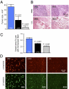
Efficacy of different pH-dependent or -independent endosomal escape units in inhibiting brain tumor growth, vascularity, and target protein expression. P/LLL/AON/Hu/Ms and P/LOEt/AON/Hu/Ms were injected i.v. at 5 mg/kg morpholino AONs to laminin-411 α4 and β1 chains. (A) Tumor size quantitation after treatment with P/LLL/AON/Hu/Ms or P/LOEt/AON/Hu/Ms. Both nanoconjugates significantly decreased tumor volume. However, volume decrease for P/LLL/AON/Hu/Ms (LLL) with pH-dependent unit was significantly greater than for P/LOEt/AON/Hu/Ms (LOEt) with pH-independent unit. (B) H&E-stained sections of tumors treated by either PBS or the lead drug P/LLL/AON/Hu/Ms (LLL). Two different animals represent each group. In PBS-injected mice (#35 and #37), invasively growing intact tumors are seen. In the LLL-treated animals (#22 and #23), massive necrosis is visible with some tumor remnants. (C) Morphometric analysis of microvessel area after various treatments. Both P/LOEt/AON/Hu/Ms (LOEt) and P/LLL/AON/Hu/Ms (LLL) significantly reduced vessel area (most pronounced in the latter group), compared with PBS. In the LLL group, the reduction was significantly greater than in the LOEt group (P < 0.05). Data are from 25 nonoverlapping fields of view per group (field area = 0.245 μm2) using 20× objective (five per tumor, five tumors per group). Percentage of area occupied by vessels (revealed by laminin β1 chain immunostaining) to total field area is shown. (D) Immunostaining of tumor sections for laminin α4 and β1 chains upon nanoconjugate treatment. In the PBS group, vessels stained brightly for both chains and many large vessels with irregular shapes were seen. Upon treatment with P/LOEt/AON/Hu/Ms (LOEt) and especially P/LLL/AON/Hu/Ms (LLL), tumor staining intensity for both chains was diminished and vessels became smaller, more similar to normal brain vessels. Representative pictures for each group are shown. For each antigen, exposure times were the same among groups.

Comment in

Similar articles

Cited by

References

    1. Brandsma D, van den Bent MJ. Molecular targeted therapies and chemotherapy in malignant gliomas. Curr Opin Oncol. 2007;19:598–605. - PubMed
    1. Folkman J. Angiogenesis: An organizing principle for drug discovery? Nat Rev Drug Discov. 2007;6:273–286. - PubMed
    1. Benny O, et al. An orally delivered small-molecule formulation with antiangiogenic and anticancer activity. Nat Biotechnol. 2008;26:799–807. - PMC - PubMed
    1. Chi AS, Norden AD, Wen PY. Antiangiogenic strategies for treatment of malignant gliomas. Neurotherapeutics. 2009;6:513–526. - PMC - PubMed
    1. Ljubimova JY, et al. Overexpression of alpha4 chain-containing laminins in human glial tumors identified by gene microarray analysis. Cancer Res. 2001;61:5601–5610. - PubMed

Publication types