. 2010 Nov;56(5):964-72.
doi: 10.1161/HYPERTENSIONAHA.110.152298. Epub 2010 Oct 4.

Candidate genes and mechanisms for 2-methoxyestradiol-mediated vasoprotection

Affiliations

Candidate genes and mechanisms for 2-methoxyestradiol-mediated vasoprotection

Federica Barchiesi et al. Hypertension. 2010 Nov.

Erratum in

  • Correction.
    [No authors listed] [No authors listed] Hypertension. 2016 May;67(5):e21. doi: 10.1161/HYP.0000000000000045. Hypertension. 2016. PMID: 27075470 No abstract available.

Abstract

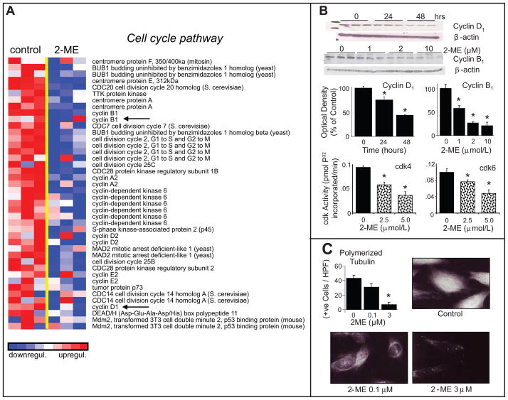
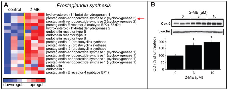
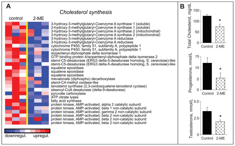
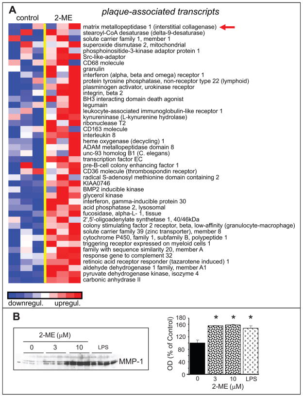
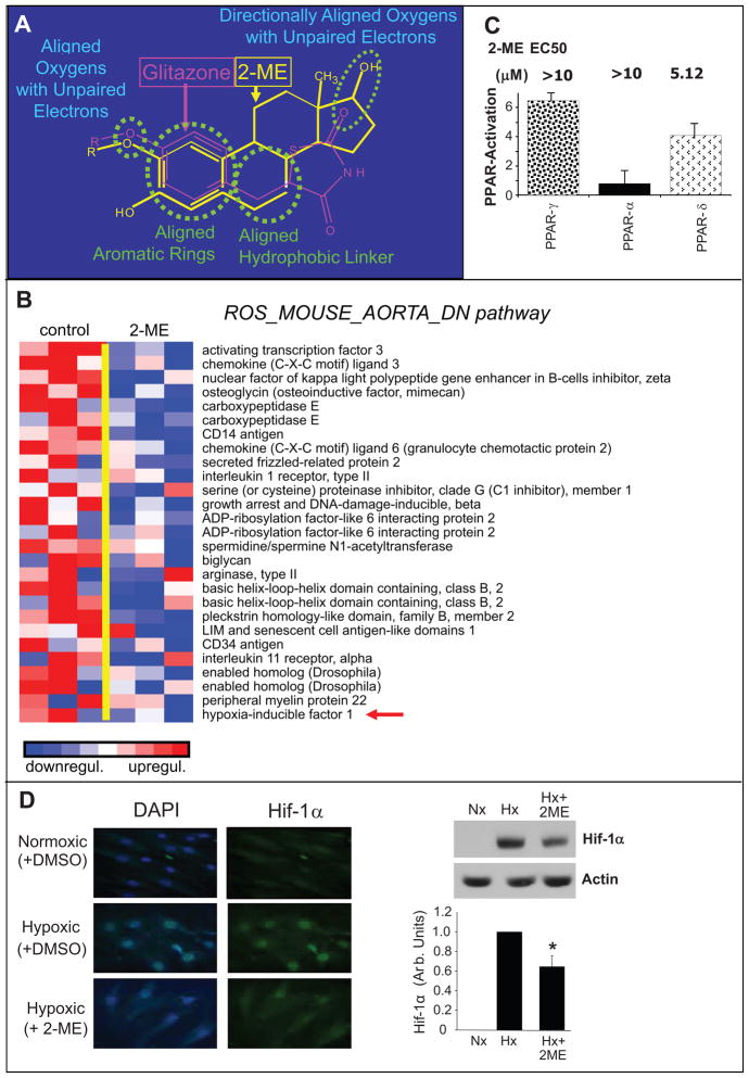
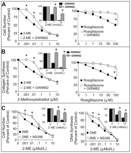
2-Methoxyestradiol (2-ME; estradiol metabolite) inhibits vascular smooth muscle cell (VSMC) growth and protects against atherosclerosis and vascular injury; however, the mechanisms by which 2-ME induces these actions remain obscure. To assess the impact of 2-ME on biochemical pathways regulating VSMC biology, we used high-density oligonucleotide microarrays to identify differentially expressed genes in cultured human female aortic VSMCs treated with 2-ME acutely (4 hours) or long term (30 hours). Both single gene analysis and Gene Set Enrichment Analysis revealed 2-ME-induced downregulation of genes involved in mitotic spindle assembly and function in VSMCs. Also, Gene Set Enrichment Analysis identified effects of 2-ME on genes regulating cell-cycle progression, cell migration/adhesion, vasorelaxation, inflammation, and cholesterol metabolism. Transcriptional changes were associated with changes in protein expression, including inhibition of cyclin D1, cyclin B1, cyclin-dependent kinase 6, cyclin-dependent kinase 4, tubulin polymerization, cholesterol and steroid synthesis, and upregulation of cyclooxygenase 2 and matrix metalloproteinase 1. Microarray data suggested that 2-ME may activate peroxisome proliferator-activated receptors (PPARs) in VSMCs, and 2-ME has structural similarities with rosiglitazone (PPARγ agonist). However, our finding of weak activation and lack of binding of 2-ME to PPARs suggests that 2-ME may modulate PPAR-associated genes via indirect mechanisms, potentially involving cyclooxygenase 2. Indeed, the antimitogenic effects of 2-ME at concentrations that do not inhibit tubulin polymerization were blocked by the PPAR antagonist GW9662 and the cyclooxygenase 2 inhibitor NS398. Finally, we demonstrated that 2-ME inhibited hypoxia-inducible factor 1α. Identification of candidate genes that are positively or negatively regulated by 2-ME provides important leads to investigate and better understand the mechanisms by which 2-ME induces its vasoprotective actions.


Figures

Figure 1
Figure 1
Inhibitory effects of 2-ME on serum-induced activation of pathways regulating cell cycle. Panel-A depicts the microarray analysis of transcriptional changes in HASMCs treated with 2-ME (3 μmol/L) for 30-hours. Expression levels are depicted as color scale from red (up-regulation) to blue (down-regulation). Gene Set Enrichment Analysis (GSEA) revealed down-regulation of key genes known to induce cytokinesis. Panel-B shows Western-blots and bar graph (changes in optical density; mean±SEM) depicting the significant inhibitory effects of 2-ME on cyclin-D1and cyclin-B1 expression and on cdk4 and cdk6 activity in HASMCs treated with 2-ME. Panel-C illustrates representative photomicrographs and bar graph depicting the inhibitory effects of 2-ME on tubulin polymerization in proliferating HASMCs. The tubulin polymerization experiments were conducted in triplicate. Western blotting and cdk activity data are presented as mean±SEM (n=3). *p<0.05 versus vehicle-treated control. Arrows indicate the genes for which protein expression was confirmed.
Figure 2
Figure 2
Stimulatory effects of 2-ME on prostaglandin synthesis in HASMCs. Panel-A depicts the microarray analysis of transcriptional changes in HASMCs treated with 2-ME (3μmol/L) for 30 hours. Expression levels are depicted as color scale from red (up-regulation) to blue (down-regulation). Gene Set Enrichment Analysis (GSEA) revealed induction of key proteins known to facilitate prostaglandin synthesis. Panel-B shows the significant stimulatory effects of 2-ME on COX-2 protein expression in HASMCs treated with 2-ME. Data are presented as mean±SEM (n=3). *p<0.05 versus vehicle-treated control. Arrows indicate the genes for which protein expression was confirmed.
Figure 3
Figure 3
Inhibitory effects of 2-ME on pathways regulating cholesterol synthesis. Panel-A depicts the microarray analysis of transcriptional changes in HASMCs treated with 2-ME (3μmol/L) for 30-hours. Expression levels are depicted as color scale from red (up-regulation) to blue (down-regulation). Gene Set Enrichment Analysis (GSEA) revealed down-regulation of key genes known to facilitate cholesterol synthesis. Panel-B shows the significant inhibitory effects of 2-ME on circulating levels of cholesterol, progesterone and testosterone in male rats treated for 14-days with 2-ME. Data for cholesterol levels represent mean±SEM (n=7 placebo; and n=7 2-ME treated). *p<0.05 versus vehicle-treated control.
Figure 4
Figure 4
Stimulatory effects of 2-ME on plaque-associated regulatory molecules in HASMCs. Panel-A depicts the transcriptional changes in HASMCs treated with 2-ME (3μmol/L) for 30-hours. Expression levels are depicted as color scale from red (up-regulation) to blue (down-regulation). Gene Set Enrichment Analysis (GSEA) revealed up-regulation of key plaque-associated transcripts. Panel-B shows the significant stimulatory effects of 2-ME on MMP-1 expression in HASMCs treated with 2-ME. HASMCs treated with LPS served as a positive control. Data for microarray analysis and Western-blotting represent mean±SEM (n=3). *p<0.05 versus vehicle-treated control. Arrows indicate the genes for which protein expression was confirmed.
Figure 5
Figure 5
Inhibitory effects of 2-ME on pathways down-regulated by rosiglitazone in HASMCs. Panel-A depicts the structural similarity between 2-ME and PPARγ ligand rosiglitazone. Panel-B depicts the microarray analysis of transcriptional changes in HASMCs treated with 2-ME (3μmol/L) for 30-hours. Expression levels are depicted as color scale from red (up-regulation) to blue (down-regulation). Gene Set Enrichment Analysis (GSEA) revealed down-regulation of key genes known to be down-regulated by rosiglitazone. Panel-C demonstrates the stimulatory effects of 2-ME on PPARδ, PPARγ and PPARα using a transactivation assay. Panel-D depicts representative photomicrographs and Western-blots demonstrating the inhibitory effects of 1μmol/L 2-ME on hypoxia (Hx) induced HIF-1α expression in HASMCs. The bar graph represents the densitometeric analysis of Western-blots (mean±SEM) from three separate experiments. Data for microarray analysis and Western-blotting represent mean±SEM (n=3). The immunostaining experiments were conducted in quadruplicate. *p<0.05 versus HASMCs treated with hypoxia (Hx). Normoxia (Nx). Arrows indicate the genes for which protein expression was confirmed.
Figure 6
Figure 6
Depicts the attenuation by PPARγ antagonist GW9662 of the concentration-dependent inhibitory effects of 2-ME (0.001–10μmol/L; left-panels) or rosiglitazone (0.1–100μmol/L; right-panels) on 2.5%FCS-induced cell-number (A) and collagen ([3H]proline-incorporation) (B) in HASMCs. Also shown are the effects of NS398 on 2-ME-induced inhibition of cell number and DNA-synthesis (C). Data represent mean±SEM from 3 separate experiments conducted in triplicates. *p<0.05 significant reversal of the inhibitory effects of 2-ME or rosiglitazone by GW9662.

Comment in

Similar articles

Cited by

References

    1. Ross JS, Stagliano NE, Donovan MJ, Breitbart RE, Ginsburg GS. Atherosclerosis and cancer: Common molecular pathways of disease development and progression. Ann NY Acad Sci. 2001;947:271–292. - PubMed
    1. Braun-Dullaeus RC, Mann MJ, Dzau VJ. Cell cycle progression. New therapeutic target for vascular proliferative disease. Circulation. 1998;98:82–89. - PubMed
    1. Sollott SJ, Cheng L, Pauly RR, Jenkins GM, Monticone RE, Kuzuya M, Froehlich JP, Crow MT, Lakatta EG, Rowinsky EK, Kinsella JL. Taxol inhibits neointimal smooth muscle accumulation after angioplasty in the rat. J Clin Invest. 1995;95:1869–1876. - PMC - PubMed
    1. Dubey RK, Imthurn B, Jackson EK. 2-Methoxyestradiol: a potential treatment for multiple proliferative disorders. Endocrinology. 2007;148:4125–4127. - PubMed
    1. Mooberry SL. New insights into 2-methoxyestradiol, a promising antiangiogenic and antitumor agent. Current Opinion in Oncology. 2003;15:425–430. - PubMed

Publication types

MeSH terms