. 2010 Nov 1;185(9):5425-35.
doi: 10.4049/jimmunol.1001603. Epub 2010 Oct 4.

Identification of an autophagy defect in smokers' alveolar macrophages

Affiliations

Identification of an autophagy defect in smokers' alveolar macrophages

Martha M Monick et al. J Immunol. .

Abstract

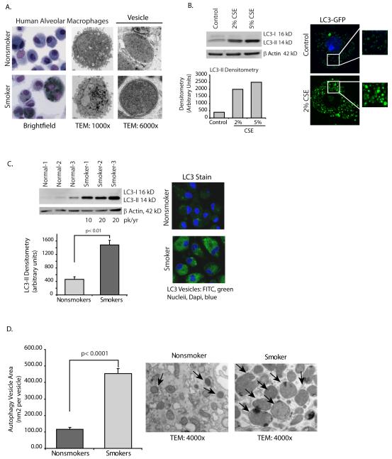
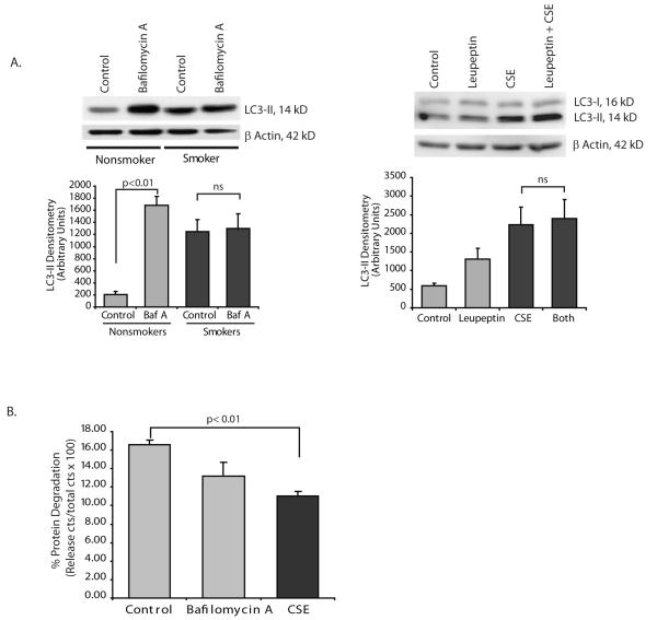
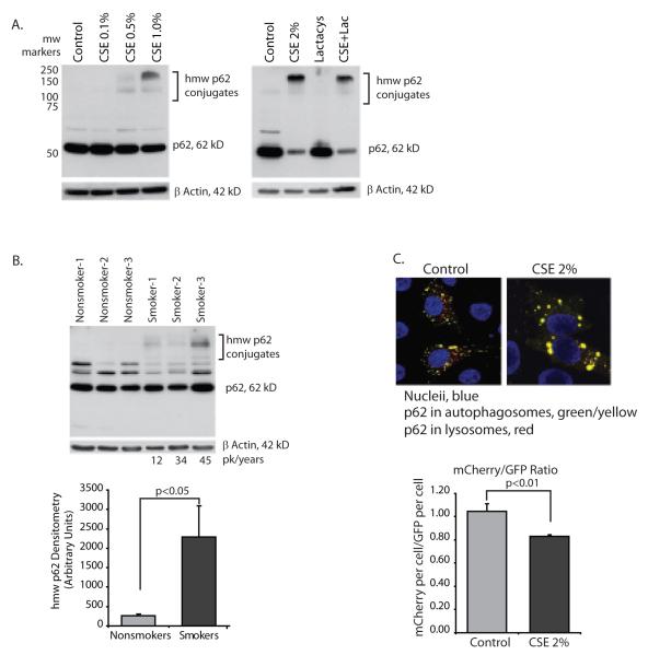
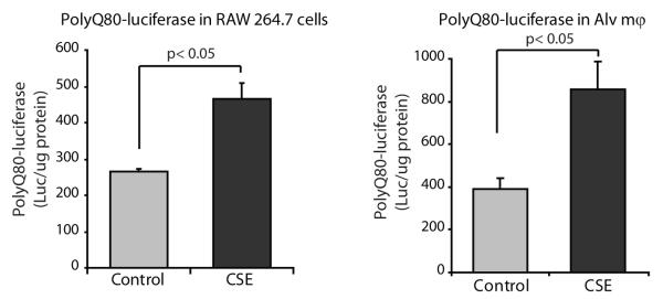
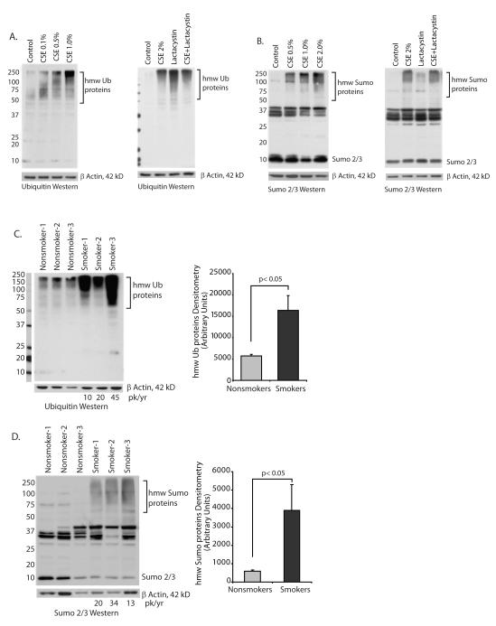
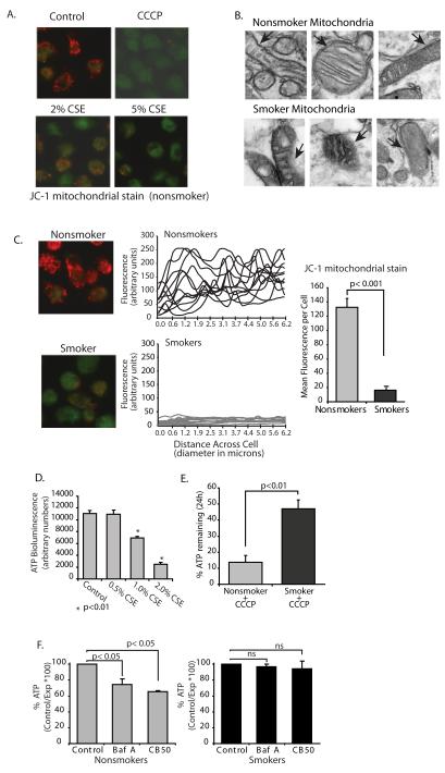
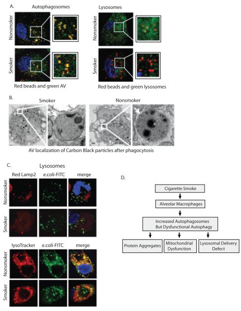
Alveolar macrophages are essential for clearing bacteria from the alveolar surface and preventing microbe-induced infections. It is well documented that smokers have an increased incidence of infections, in particular lung infections. Alveolar macrophages accumulate in smokers' lungs, but they have a functional immune deficit. In this study, we identify an autophagy defect in smokers' alveolar macrophages. Smokers' alveolar macrophages accumulate both autophagosomes and p62, a marker of autophagic flux. The decrease in the process of autophagy leads to impaired protein aggregate clearance, dysfunctional mitochondria, and defective delivery of bacteria to lysosomes. This study identifies the autophagy pathway as a potential target for interventions designed to decrease infection rates in smokers and possibly in individuals with high environmental particulate exposure.


Figures

Figure 1
Figure 1
Cigarette smoke exposure in vitro and in vivo causes an increase in numbers of autophagosomes. A. Alveolar macrophages were obtained from nonsmokers and smokers. Shown are representative TEM and Wright Giemsa stains. Higher magnification TEM image shows presence of double walled vesicles in smoker’s macrophages compared to lack of double walled vesicles in nonsmokers cells. Greater than 70% of vesicles in smoker cells had double membrane characteristic of autophagosomes. B. Alveolar macrophages from nonsmokers were exposed to 2% or 5% CSE for five hours. Whole cell lysates were obtained and Western analysis for LC3-II performed. Equal loading is demonstrated by probing the same blot for β actin. Image is representative of 4 separate experiments. On the right is a confocal image of human alveolar macrophages transfected with LC3-GFP. After overnight incubation, some of the cells were exposed to 2% CSE for 5 hours. Confocal analysis was performed to identify GFP positive vesicles. C. Alveolar macrophages from 3 nonsmokers and 3 smokers were analyzed for LC3-II by Western analysis of whole cell proteins. Below is a graph of densitometry results. Significance was determined using nonpaired Student’s t test. On the right is a representative image of fixed alveolar macrophages from a nonsmoker and from a smoker stained for LC3b. D. Alveolar macrophages from nonsmokers and smokers were fixed shortly after isolation. Transmission electron microscopy (TEM) samples were prepared and images obtained. Autophagosome size was determined by analyzing area (with Image J software) of double walled vesicles in cells with slices obtained through the middle of the cell (determined by nucleus). Measurements were obtained from 3 nonsmokers and 3 smokers (12 vesicles each cell, 4 cells each person) and area of vesicle obtained by drawing circles around vesicles and performing Image j analysis. Significance was determined using nonpaired Student’s t test.
Figure 2
Figure 2
Cigarette smoke exposure blocks autophagic flux. A. Nonsmoker alveolar macrophages were exposed to bafilomycin A (100 nM) for 5 hours. Whole cell lysates were obtained and LC3-II quantified by Western analysis. The densitometry graph represents 3 separate experiments. Identical experiments were performed using leupeptin ((50 ug/ml) to inhibit lysosome function. Western anlysis and densitometry from 3 separate experiments is shown. Significance was determined using nonpaired Student’s t test. B. Clearance of long lived proteins was determined by labeling nonsmoker alveolar macrophages with 14C leucine for 8 hours. Following a chase with cold leucine, cells were exposed to nothing (control), bafilomycin A or CSE 2%. Following an 8 hour incubation, cells and supernatants were saved. TCA precipitation isolated proteins from both samples. The percentage of long-lived proteins degraded in the culture period was determined using the formula a/a+b × 100 where a equals the TCA soluble fraction of the supernatant and b equals the TCA insoluble fraction of the cell lysate. Data was analyzed using nonpaired Student’s t test.
Figure 3
Figure 3
Cigarette smoke blocks delivery of ubiquitin chaperone protein, p62 to the lysosome. A. Nonsmoker alveolar macrophages were exposed to CSE at varying concentrations (0.1 to 1.0%) for 5 hours. Whole cell lysates were obtained and Western analysis performed for p62 protein. In the right panel, nonsmoker alveolar macrophages were exposed to lactacystin (10 uM) with and without CSE 2% for 5 hours. B. Alveolar macrophages from 3 nonsmokers and 3 smokers were analyzed for hmw p62 aggregates by Western analysis of whole cell proteins. Densitometry reflects the p62 positive bands between 100 and 250 kD. Significance was determined using nonpaired Student’s t test. C. Hela cells were transfected with mCherry-GFP-p62. Following an overnight incubation, cells ere exposed to 2% CSE for 5 hours. Confocal analysis was performed to identify yellow (green and red colocalization) vesicles versus red vesicles. 12 individual cells from each group were analyzed for red pixels versus green pixels (20 confocal slices per cell). Quantitation is shown in the accompanying graph. Significance was determined using nonpaired Student’s t test.
Figure 4
Figure 4
Cigarette smoke exposure results in cytosolic accumulation of protein aggregates. Raw 264.7 cells and nonsmoker alveolar macrophages were transfected with a plasmid encoding firefly luciferase fused to a N-terminal polyglutamine (80) repeats (polyQ80-luciferase). Following transfection, cells were incubated overnight to allow for an accumulation of polyQ-luciferase and then exposed to 2% CSE for 6 hours. Following CSE exposure, cells were lysed and luciferase assayed. Data represent mean ±SEM (n=3). Significance was determined using nonpaired Student’s t test.
Figure 5
Figure 5
Cigarette smoke exposure causes an accumulation of high molecular weight (hmw) ubiquitin and sumo-conjugated proteins. A. Ubiquitin analysis: The accumulation of hmw ubiquitin conjugates was assessed in alveolar macrophages from nonsmoker’s exposed to 0.1 to 1.0% CSE for 5 hours. Western analysis for ubiquitin was done on whole cell lysates. In the experiment shown on the right, nonsmoker alveolar macrophages were exposed to lactacystin (10 uM) with and without 2% CSE for 5 hours. Images are representative of three separate experiments. B. Sumo analysis: The accumulation of hmw sumo conjugates was assessed in alveolar macrophages from nonsmoker’s exposed to 0.1 to 1.0% CSE for 5 hours. Western analysis for sumo 2/3 was done on whole cell lysates. In the experiment shown on the right, nonsmoker alveolar macrophages were exposed to lactacystin (10 uM) with and without 2% CSE for 5 hours. Images are representative of three separate experiments. C. Ubiquitin analysis: Alveolar macrophages from 3 nonsmokers and 3 smokers were analyzed for hmw ubiquitin conjugates by Western analysis of whole cell proteins. Denstitometry reflects the ubiquitin positive bands between 75 and 250 kD. Significance was determined using nonpaired Student’s t test. D. Sumo analysis: Alveolar macrophages from 3 nonsmokers and 3 smokers were analyzed for hmw sumo conjugates by Western analysis of whole cell proteins. Densitometry reflects the sumo positive bands between 75 and 250 kD. Significance was determined using nonpaired Student’s t test.
Figure 6
Figure 6
Cigarette smoke exposure results in loss of mitochondrial electron transport chain function and accumulation of defective mitochondria. A. Human alveolar macrophages were cultured (1×10⁶/ml in 2 chamber microscope slides) with and without 2-5% CSE or the uncoupler (CCCP). At the end of the incubation period, the mitochondrial stain JC-1 was added as described in the methods. Red/orange stain denotes intact mitochondria with no disruption of membrane potential. Green staining denotes loss of mitΔΨ. B. TEM was performed on smoker and nonsmoker macrophages. Shown are mitochondria demonstrating damaged mitochondria in the smoker’s cells. Arrows in images point to intact (nonsmoker) and damaged (smoker) mitochondrial membranes. C. Nonsmoker and smoker macrophages were allowed to adhere to 2 chamber slides and stained with JC-1. Quantitation was performed as described in the methods and individual cell levels of red fluorescence from two separate experiments are shown in the line graphs and as a composite in the bar graph. D. Normal alveolar macrophages were cultured (96 well plate) with varying concentrations of CSE (0.5%-2.0%). ATP levels were measured using a chemiluminescence assay system. Data is presented as arbitrary luminescent units and is a composite of three experiments. Significance was determined using nonpaired Student’s t test. E. Nonsmoker and smoker alveolar macrophages were exposed the uncoupler, CCCP, for 24 hours. ATP levels with and without the uncoupler were obtained and percent remaining ATP calculated. Data is a composite of three smokers and three nonsmokers. Significance was determined using nonpaired Student’s t test. F. Nonsmoker alveolar macrophages were exposed to the specific inhibitor of vacuolar-type H+-ATPase, bafilomycin A (100 nM) or carbon black 50 nM nanoparticles for 5 hours. ATP levels were measured using a chemiluminescence assay system. Data is presented as arbitrary luminescent units and is a composite of three experiments. Significance was determined using nonpaired Student’s t test.
Figure 7
Figure 7
Cigarette smoke exposure impairs delivery of cargo to lysosomes. A. Alveolar macrophages from nonsmokers and smokers were placed in culture. Following a 3 hour stabilizing period, cells were exposed to 0.5 micron latex beads for 60 minutes. Nonphagocytosed beads were washed off and cells were incubated for a 2 hour chase period. Cells were then fixed and stained for either LC3b (autophagosomes) or Lamp2 (lysosomes). B. Alveolar macrophages from nonsmokers and smokers were placed in culture. Following a 3 hour stabilizing period, cells were exposed to 50 nM carbon black nanoparticles for 60 minutes. Nonphagocytosed nanoparticles were washed off and cells were incubated for a 2 hour chase period. Cells were then fixed and TEM analysis performed. Shown is a representative cell demonstrating carbon black particles in double walled vesicles. C. Alveolar macrophages from smokers and nonsmokers were cultured on a coverslip chamber slide. Cells were exposed to AlexaFlour 488-E.coli for 30 minutes, nonphagocytosed bacteria was washed off, a 1 hour chase incubation followed before staining the cells with Lysotracker Red DND-99 or fixing and staining for Lamp2 with an Alexa 568 secondary antibody. The images are representative of three separate experiments. The photomicrographs show red lysosomes, green E. coli and yellow merged image of E. coli in a lysosome. D. The diagram outlines the conclusions from this study. Prolonged cigarette smoke exposure leads to accumulation of nonfunctional autophagosomes by blocking lysosomal trafficking. The block in autophagic flux leads to decreased clearance of protein aggregates, mitochondrial dysfunction and reduced delivery of phagocytosed bacteria to the lysosome.

Similar articles

Cited by

References

    1. Niewoehner DE, Kleinerman J, Rice DB. Pathologic changes in the peripheral airways of young cigarette smokers. The New England journal of medicine. 1974;291:755–758. - PubMed
    1. Green GM. Cigarette smoke: protection of alveolar macrophages by glutathione and cysteine. Science (New York, N.Y. 1968;162:810–811. - PubMed
    1. Laurenzi GA, Guarneri JJ, Endriga RB, Carey JP. Clearance of Bacteria by the Lower Respiratory Tract. Science (New York, N.Y. 1963;142:1572–1573. - PubMed
    1. Klionsky DJ, Emr SD. Autophagy as a regulated pathway of cellular degradation. Science (New York, N.Y. 2000;290:1717–1721. - PMC - PubMed
    1. Levine B, Kroemer G. Autophagy in the pathogenesis of disease. Cell. 2008;132:27–42. - PMC - PubMed

Publication types