LRRK2 is involved in the IFN-gamma response and host response to pathogens
- PMID: 20921534
- PMCID: PMC3156100
- DOI: 10.4049/jimmunol.1000548
LRRK2 is involved in the IFN-gamma response and host response to pathogens
Abstract
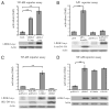
LRRK2 was previously identified as a defective gene in Parkinson's disease, and it is also located in a risk region for Crohn's disease. In this study, we aim to determine whether LRRK2 could be involved in immune responses. We show that LRRK2 expression is enriched in human immune cells. LRRK2 is an IFN-γ target gene, and its expression increased in intestinal tissues upon Crohn's disease inflammation. In inflamed intestinal tissues, LRRK2 is detected in the lamina propria macrophages, B-lymphocytes, and CD103-positive dendritic cells. Furthermore, LRRK2 expression enhances NF-κB-dependent transcription, suggesting its role in immune response signaling. Endogenous LRRK2 rapidly translocates near bacterial membranes, and knockdown of LRRK2 interferes with reactive oxygen species production during phagocytosis and bacterial killing. These observations indicate that LRRK2 is an IFN-γ target gene, and it might be involved in signaling pathways relevant to Crohn's disease pathogenesis.
Conflict of interest statement
The authors have no financial conflicts of interest.
Figures


Similar articles
-
Crohn's and Parkinson's Disease-Associated LRRK2 Mutations Alter Type II Interferon Responses in Human CD14+ Blood Monocytes Ex Vivo.J Neuroimmune Pharmacol. 2020 Dec;15(4):794-800. doi: 10.1007/s11481-020-09909-8. Epub 2020 Mar 16. J Neuroimmune Pharmacol. 2020. PMID: 32180132 Free PMC article.
-
Interferon-γ induces leucine-rich repeat kinase LRRK2 via extracellular signal-regulated kinase ERK5 in macrophages.J Neurochem. 2014 Jun;129(6):980-7. doi: 10.1111/jnc.12668. Epub 2014 Feb 24. J Neurochem. 2014. PMID: 24479685
-
An increase in LRRK2 suppresses autophagy and enhances Dectin-1-induced immunity in a mouse model of colitis.Sci Transl Med. 2018 Jun 6;10(444):eaan8162. doi: 10.1126/scitranslmed.aan8162. Sci Transl Med. 2018. PMID: 29875204 Free PMC article.
-
An emerging role for LRRK2 in the immune system.Biochem Soc Trans. 2012 Oct;40(5):1134-9. doi: 10.1042/BST20120119. Biochem Soc Trans. 2012. PMID: 22988878 Review.
-
LRRK2 in peripheral and central nervous system innate immunity: its link to Parkinson's disease.Biochem Soc Trans. 2017 Feb 8;45(1):131-139. doi: 10.1042/BST20160262. Biochem Soc Trans. 2017. PMID: 28202666 Free PMC article. Review.
Cited by
-
The G2019S LRRK2 mutation increases myeloid cell chemotactic responses and enhances LRRK2 binding to actin-regulatory proteins.Hum Mol Genet. 2015 Aug 1;24(15):4250-67. doi: 10.1093/hmg/ddv157. Epub 2015 Apr 29. Hum Mol Genet. 2015. PMID: 25926623 Free PMC article.
-
Parkinson's disease and immune system: is the culprit LRRKing in the periphery?J Neuroinflammation. 2012 Jul 9;9:94. doi: 10.1186/1742-2094-9-94. J Neuroinflammation. 2012. PMID: 22594666 Free PMC article. Review.
-
Human iPSC-derived microglia carrying the LRRK2-G2019S mutation show a Parkinson's disease related transcriptional profile and function.Sci Rep. 2023 Dec 13;13(1):22118. doi: 10.1038/s41598-023-49294-9. Sci Rep. 2023. PMID: 38092815 Free PMC article.
-
The Current State-of-the Art of LRRK2-Based Biomarker Assay Development in Parkinson's Disease.Front Neurosci. 2020 Aug 18;14:865. doi: 10.3389/fnins.2020.00865. eCollection 2020. Front Neurosci. 2020. PMID: 33013290 Free PMC article. Review.
-
Roco Proteins: GTPases with a Baroque Structure and Mechanism.Int J Mol Sci. 2019 Jan 3;20(1):147. doi: 10.3390/ijms20010147. Int J Mol Sci. 2019. PMID: 30609797 Free PMC article. Review.
References
-
- Xavier RJ, Podolsky DK. Unravelling the pathogenesis of inflammatory bowel disease. Nature. 2007;448:427–434. - PubMed
-
- Barrett JC, Hansoul S, Nicolae DL, Cho JH, Duerr RH, Rioux JD, Brant SR, Silverberg MS, Taylor KD, Barmada MM, et al. NIDDK IBD Genetics Consortium; Belgian-French IBD Consortium; Wellcome Trust Case Control Consortium. Genome-wide association defines more than 30 distinct susceptibility loci for Crohn’s disease. Nat Genet. 2008;40:955–962. - PMC - PubMed
-
- Paisán-Ruíz C, Jain S, Evans EW, Gilks WP, Simón J, van der Brug M, López de Munain A, Aparicio S, Gil AM, Khan N, et al. Cloning of the gene containing mutations that cause PARK8-linked Parkinson’s disease. Neuron. 2004;44:595–600. - PubMed
-
- Zimprich A, Biskup S, Leitner P, Lichtner P, Farrer M, Lincoln S, Kachergus J, Hulihan M, Uitti RJ, Calne DB, et al. Mutations in LRRK2 cause autosomal-dominant parkinsonism with pleomorphic pathology. Neuron. 2004;44:601–607. - PubMed
Publication types
MeSH terms
Substances
Grants and funding
LinkOut - more resources
Full Text Sources
Other Literature Sources
Medical
Research Materials

