. 2010 Nov;120(11):4007-20.
doi: 10.1172/JCI42447.

Distinct growth hormone receptor signaling modes regulate skeletal muscle development and insulin sensitivity in mice

Affiliations

Distinct growth hormone receptor signaling modes regulate skeletal muscle development and insulin sensitivity in mice

Mahendra D Mavalli et al. J Clin Invest. 2010 Nov.

Abstract

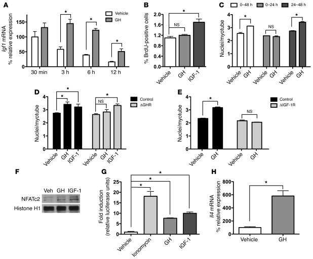
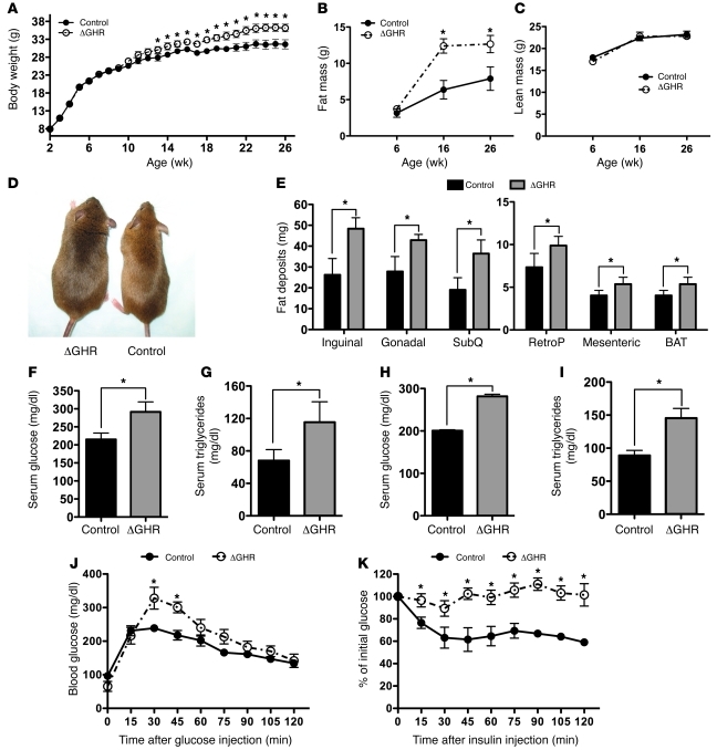
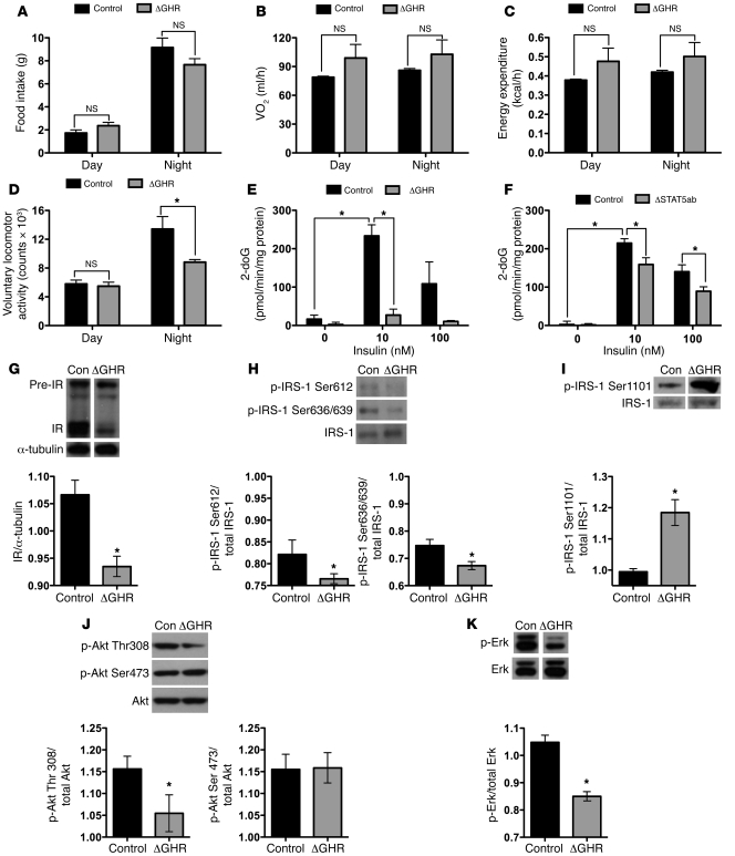
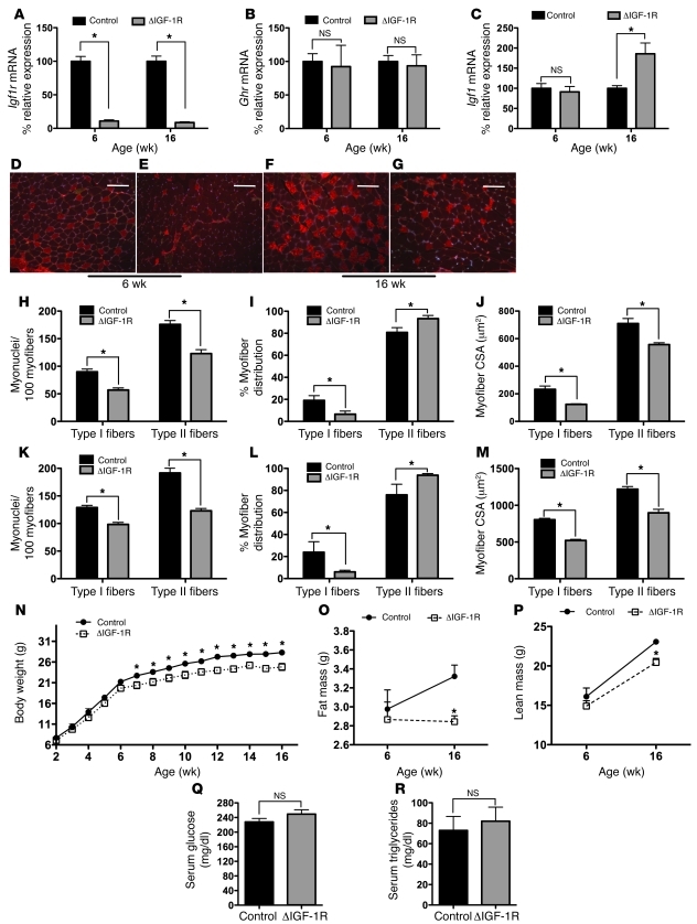
Skeletal muscle development, nutrient uptake, and nutrient utilization is largely coordinated by growth hormone (GH) and its downstream effectors, in particular, IGF-1. However, it is not clear which effects of GH on skeletal muscle are direct and which are secondary to GH-induced IGF-1 expression. Thus, we generated mice lacking either GH receptor (GHR) or IGF-1 receptor (IGF-1R) specifically in skeletal muscle. Both exhibited impaired skeletal muscle development characterized by reductions in myofiber number and area as well as accompanying deficiencies in functional performance. Defective skeletal muscle development, in both GHR and IGF-1R mutants, was attributable to diminished myoblast fusion and associated with compromised nuclear factor of activated T cells import and activity. Strikingly, mice lacking GHR developed metabolic features that were not observed in the IGF-1R mutants, including marked peripheral adiposity, insulin resistance, and glucose intolerance. Insulin resistance in GHR-deficient myotubes derived from reduced IR protein abundance and increased inhibitory phosphorylation of IRS-1 on Ser 1101. These results identify distinct signaling pathways through which GHR regulates skeletal muscle development and modulates nutrient metabolism.


Figures

Figure 1
Figure 1. GH-induced myoblast proliferation and fusion are mediated by IGF-1 production and intersect the NFATc2/IL-4 pathway.
(A) Primary mouse myoblasts were serum starved for 10 hours before stimulation with GH. Real-time PCR was performed using primers for IGF-1 at indicated times. (B) Myoblasts were serum starved for 10 hours before stimulation with GH or IGF-1 for 8 hours, with BrdU exposure for the full 8 hours of treatment. The percentage of BrdU-positive cells was assessed by flow cytometry. (C) Myoblasts were induced to differentiate and treated with GH for 0–48 hours, 0–24 hours, or 24–48 hours of differentiation. (D) GHR-floxed myoblasts were infected with adenoGFP (control) or adenoCre (ΔGHR) and differentiated in the presence of vehicle, GH, or IGF-1 for 48 hours. (E) IGF-1R–floxed myoblasts were infected with adenoGFP (control) or adenoCre (ΔIGF-1R) as treated in D. (F) Wild-type myoblasts were serum starved for 10 hours before stimulation with vehicle (Veh), GH, or IGF-1 for 1 hour and harvest of nuclear protein extracts. (G) Myoblasts were transfected with an NFAT luciferase reporter construct before induction of differentiation for 24 hours and treatment with vehicle, ionomycin, GH, or IGF-1 for 5 hours. (H) Myoblasts were serum starved for 10 hours before treatment with vehicle or GH for 5 hours. Real-time PCR was performed using primers for IL-4. All data shown is representative of at least 3 separate experiments performed from separate muscle cell preparations. Error bars indicate SEM. *P < 0.05.
Figure 2
Figure 2. GHR is required for normal myofiber specification, myonuclei accumulation, and muscle function.
mRNA was harvested from the gastrocnemius muscles of control and ΔGHR mice, and real-time PCR was performed using primers for (A) Ghr, (B) Igf1r, and (C) Igf1. (DG) Sections of medial gastrocnemius from control and ΔGHR mice were visualized using antibodies directed against MHC type I and laminin and counterstained with Hoechst 33258 to visualize myonuclei. Scale bars: 80 μm. (HM) Histomorphometric analyses of myonuclei per 100 myofibers, percentage myofiber distribution, and myofiber diameter (CSA) were performed on sections described above for (HJ) 6-week-old and (KM) 16-week-old control and ΔGHR mice. Muscle performance in 26-week-old control and ΔGHR mice was assessed by (N) grip strength and (O and P) rotarod testing, as described in Methods. For all studies shown, n = 6 for control and ΔGHR at all time points. Error bars indicate SEM. *P < 0.05.
Figure 3
Figure 3. Loss of GHR in skeletal muscle causes peripheral adiposity, glucose intolerance, and insulin resistance.
(A) Control (n = 9) and ΔGHR (n = 11) mice were weighed weekly from 2 to 26 weeks of age (image in D was taken at 26 weeks). qMR analysis of (B) fat and (C) lean mass in control and ΔGHR mice at 6, 16, and 26 weeks of age. (E) Fat pad mass at 26 weeks. SubQ, subcutaneous; RetroP, retroperitoneal; BAT, brown adipose tissue. (FI) Ad libitum serum glucose and triglyceride concentration at (F and G) 16 weeks and (H and I) 26 weeks of age. Control and ΔGHR mice were also subjected to (J) GTTs and (K) ITTs, as described in Methods. Unless otherwise indicated, n = 6 for control and ΔGHR at all time points. Error bars indicate SEM. *P < 0.05.
Figure 4
Figure 4. Metabolic phenotype of ΔGHR mice is associated with skeletal muscle insulin resistance.
(A) Food intake of 26-week-old control and ΔGHR mice. (B) Oxygen consumption (VO2) and (C) energy expenditure, as assessed by indirect calorimetry. (D) Voluntary locomotor activity assessed by computer-monitored infrared beam breaks (n = 4 control and ΔGHR mice for AD). (E and F) Myoblasts carrying (E) floxed GHR alleles or (F) floxed STAT5ab alleles were infected with adenoGFP (control) or adenoCre (ΔGHR or ΔSTAT5ab, respectively). Myoblasts were then serum starved for 12 hours, before a 1-hour pretreatment with vehicle or insulin, and then pulsed with 2-doG. Radiolabeled glucose uptake was determined by liquid scintillation counting. (GK) Protein lysates were harvested from differentiated control and ΔGHR primary myotubes and subjected to SDS-PAGE analysis. Membranes were immunoblotted with antibodies directed against the proteins indicated. Membranes were stripped and reprobed for the indicated loading control protein. In some cases, additional irrelevant lanes between those shown were removed (indicated by separating white space). Bands for the indicated proteins were quantified based on gray density, as described in the Methods, and normalized against respective loading control proteins. All in vitro data shown is representative of at least 3 separate experiments performed from separate muscle cell preparations. Error bars indicate SEM. *P < 0.05.
Figure 5
Figure 5. Mice lacking IGF-1R in skeletal muscle phenocopy the skeletal muscle defects of GHR mutants but are neither obese nor insulin resistant.
mRNA was harvested from the gastrocnemius muscles of control and ΔIGF-1R mice, and real-time PCR was performed using primers for (A) Igf1r, (B) Ghr, and (C) Igf1. (DG) Sections of gastrocnemius muscle from control and ΔIGF-1R mice were visualized using antibodies directed against MHC type I and laminin and counterstained with Hoechst 33258 to visualize myonuclei. Scale bars: 80 μm. (HM) Histomorphometric analyses of myonuclei per 100 myofibers, percentage myofiber distribution, and myofiber diameter (CSA) were performed on sections described above for (HJ) 6-week-old and (KM) 16-week-old control and ΔIGF-1R mice. (N) Control (n = 7) and ΔIGF-1R (n = 10) mice were weighed weekly from 2 to 16 weeks of age. qMR analysis of (O) fat and (P) lean mass in control and ΔIGF-1R mice at 6 and 16 weeks of age. Ad libitum (Q) serum glucose and (R) triglyceride levels were measured in control and ΔIGF-1R mice at 16 weeks of age. Unless otherwise indicated, n = 6 for control and ΔIGF-1R at all time points. Error bars indicate SEM. *P < 0.05.

Similar articles

Cited by

References

    1. Kablar B, et al. MyoD and Myf-5 define the specification of musculature of distinct embryonic origin. Biochem Cell Biol. 1998;76(6):1079–1091. doi: 10.1139/bcb-76-6-1079. - DOI - PubMed
    1. Hasty P, et al. Muscle deficiency and neonatal death in mice with a targeted mutation in the myogenin gene. Nature. 1993;364(6437):501–506. doi: 10.1038/364501a0. - DOI - PubMed
    1. Nabeshima Y, Hanaoka K, Hayasaka M, Esumi E, Li S, Nonaka I. Myogenin gene disruption results in perinatal lethality because of severe muscle defect. Nature. 1993;364(6437):532–535. doi: 10.1038/364532a0. - DOI - PubMed
    1. Horsley V, Pavlath GK. Forming a multinucleated cell: molecules that regulate myoblast fusion. Cells Tissues Organs. 2004;176(1–3):67–78. doi: 10.1159/000075028. - DOI - PubMed
    1. Horsley V, Friday BB, Matteson S, Kegley KM, Gephart J, Pavlath GK. Regulation of the growth of multinucleated muscle cells by an NFATC2-dependent pathway. J Cell Biol. 2001;153(2):329–338. doi: 10.1083/jcb.153.2.329. - DOI - PMC - PubMed

Publication types

MeSH terms