

doi: 10.4103/2153-3539.68332.
Digital images and the future of digital pathology
Affiliations
- PMID: 20922032
- PMCID: PMC2941968
- DOI: 10.4103/2153-3539.68332
Item in Clipboard
Digital images and the future of digital pathology
J Pathol Inform.
.
No abstract available
Figures
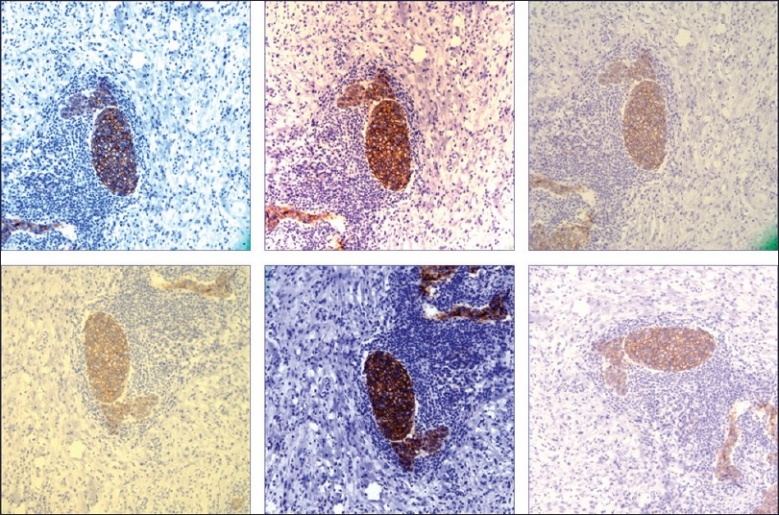
Different digital images of the same region on a glass slide photographed at the same magnification by six different pathologists, each using similar microscopes and the same attached digital cameras (HER-2/neu immunohistochemical stain)
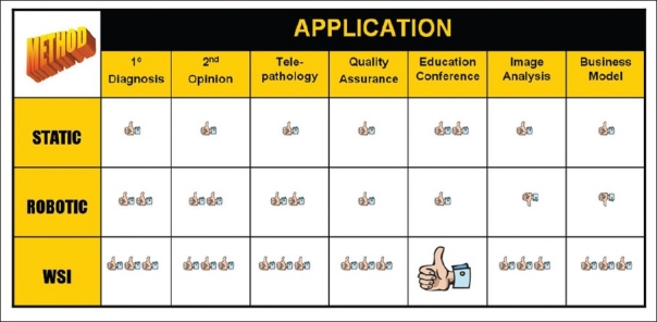
Table comparing the benefits of WSI to other modes of digital pathology. WSI gets more “thumbs up” for all applications compared to static images or live digital images viewed via robotic technology. WSI is a killer application for educational purposes
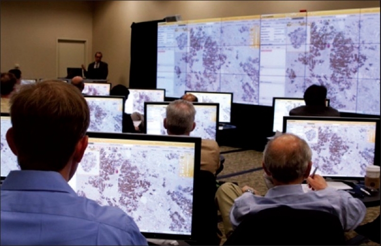
Whole slide images help create a “virtual multiheaded microscope” that supports interactive education (Image courtesy of BioImagene)
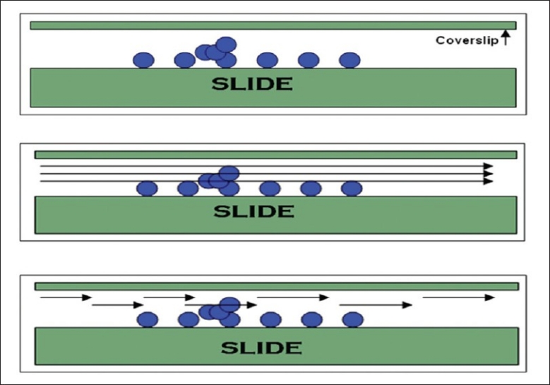
Cytology slides frequently contain 3D cell groups underneath the coverslip (top picture). The ability to view these groups in focus on a digital image can be achieved by multiplane scanning along multiple z axes (middle picture) or intercalation of scanned images along different focal points (bottom picture)
References
-
- May M. A better lens on disease. Sci Am. 2010;10:74–7. - PubMed
-
- Pinco J, Goulart RA, Otis CN, Garb J, Pantanowitz L. Impact of digital image manipulation in cytology. Arch Pathol Lab Med. 2009;133:57–61. - PubMed
-
- Weinstein RS, Graham AR, Richter LC, Barker GP, Krupinski EA, Lopez AM, et al. Overview of telepathology, virtual microscopy, and whole slide imaging: prospects for the future. Hum Pathol. 2009;40:1057–69. - PubMed
-
- Weaker FJ, Herbert DC. Transition of a dental histology course from light to virtual microscopy. J Dent Educ. 2009;73:1213–21. - PubMed
-
- Yamashiro K, Taira K, Matsubayashi S, Azuma M, Okuyama D, Nakajima M, et al. Comparison between a traditional single still image and a multiframe video image along the z-axis of the same microscopic field of interest in cytology: Which does contribute to telecytology? Diagn Cytopathol. 2009;37:727–31. - PubMed
LinkOut - more resources
Full Text Sources
Other Literature Sources
