

Retinal nerve fiber layer evaluation in multiple sclerosis with spectral domain optical coherence tomography
- PMID: 20922034
- PMCID: PMC2946989
- DOI: 10.2147/opth.s13278
Retinal nerve fiber layer evaluation in multiple sclerosis with spectral domain optical coherence tomography
Abstract
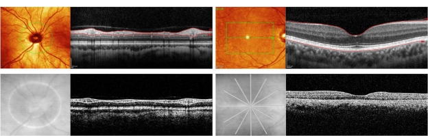
Purpose: Histopathologic studies have reported retinal nerve fiber layer (RNFL) thinning in various neurodegenerative diseases. Attempts to quantify this loss in vivo have relied on time-domain optical coherence tomography (TDOCT), which has low resolution and requires substantial interpolation of data for volume measurements. We hypothesized that the significantly higher resolution of spectral-domain optical coherence tomography (SDOCT) would better detect RNFL changes in patients with multiple sclerosis, and that RNFL thickness differences between eyes with and without optic neuritis might be identified more accurately.
Methods: In this retrospective case series, patients with multiple sclerosis were recruited from the Judith Jaffe Multiple Sclerosis Center at Weill Cornell Medical College in New York. Patients with a recent clinical diagnosis of optic neuritis (less than three months) were excluded. Eyes with a history of glaucoma, optic neuropathy (other than multiple sclerosis-related optic neuritis), age-related macular degeneration, or other relevant retinal and/or optic nerve disease were excluded. Both eyes of each patient were imaged with the Heidelberg Spectralis(®) HRA + OCT. RNFL and macular thickness were measured for each eye using the Heidelberg OCT software. These measurements were compared with validated published normal values, and were modeled as linear functions of duration of disease. The odds of an optic neuritis diagnosis as a function of RNFL and macular thickness were calculated.
Results: Ninety-four eyes were prospectively evaluated using OCT. Ages of patients ranged from 26 to 69 years, with an average age of 39 years. Peripapillary RNFL thinning was demonstrated in multiple sclerosis patients; mean RNFL thickness was 88.5 μm for individuals with multiple sclerosis compared with a reported normal value of 97 μm (P < 0.001). Eyes with a history of optic neuritis had more thinning compared with those without optic neuritis (83.0 μm versus 90.5 μm, respectively, P = 0.02). No significant differences were observed in macular thickness measurements between eyes with and without optic neuritis, nor were macular thickness measurements significantly different from normal values. As a function of multiple sclerosis duration and controlling for age, RNFL thickness was decreased in patients with a duration of multiple sclerosis greater than five years compared with those with a duration less than or equal to one year (P = 0.008).
Conclusions: Patients with a history of multiple sclerosis had RNFL thinning that was detectable on SDOCT. Decreasing RNFL thickness in eyes with optic neuritis was found, and the odds of having optic neuritis were increased significantly with decreasing RNFL thickness. Average RNFL thinning with increasing duration of disease was an excellent predictor of a reported history of optic neuritis. SDOCT retinal imaging may represent a high-resolution, objective, noninvasive, and easily quantifiable in vivo biomarker of the presence of optic neuritis and severity of multiple sclerosis.
Keywords: multiple sclerosis; nerve fiber layer; nerve fiber layer thickness; optic neuritis; optical coherence tomography; spectral-domain optical coherence tomography.
Figures
Similar articles
-
Peripapillary retinal nerve fiber layer thickness in sickle-cell hemoglobinopathies using spectral-domain optical coherence tomography.Am J Ophthalmol. 2013 Mar;155(3):456-464.e2. doi: 10.1016/j.ajo.2012.09.015. Epub 2012 Dec 4. Am J Ophthalmol. 2013. PMID: 23218697
-
[Retinal atrophy using optical coherence tomography (OCT) in 15 patients with multiple sclerosis and comparison with healthy subjects].Rev Neurol (Paris). 2008 Nov;164(11):927-34. doi: 10.1016/j.neurol.2008.03.008. Epub 2008 Jun 6. Rev Neurol (Paris). 2008. PMID: 18808761 French.
-
Reduced retinal nerve fiber layer and macular thickness in patients with multiple sclerosis with no history of optic neuritis identified by the use of spectral domain high-definition optical coherence tomography.J Clin Neurosci. 2011 Nov;18(11):1469-72. doi: 10.1016/j.jocn.2011.04.008. Epub 2011 Sep 13. J Clin Neurosci. 2011. PMID: 21917458
-
The Effects of Disease-Modifying Therapies on Optic Nerve Degeneration in Multiple Sclerosis.Eur J Neurol. 2025 Mar;32(3):e70081. doi: 10.1111/ene.70081. Eur J Neurol. 2025. PMID: 40047132 Free PMC article. Review.
-
Vision in multiple sclerosis: the story, structure-function correlations, and models for neuroprotection.J Neuroophthalmol. 2011 Dec;31(4):362-73. doi: 10.1097/WNO.0b013e318238937f. J Neuroophthalmol. 2011. PMID: 22089500 Free PMC article. Review.
Cited by
-
Optical coherence tomography in multiple sclerosis.Med Hypothesis Discov Innov Ophthalmol. 2024 Jan 31;12(4):187-193. doi: 10.51329/mehdiophthal1485. eCollection 2023 Winter. Med Hypothesis Discov Innov Ophthalmol. 2024. PMID: 38601055 Free PMC article.
-
Optical coherence tomography in multiple sclerosis: A 3-year prospective multicenter study.Ann Clin Transl Neurol. 2021 Dec;8(12):2235-2251. doi: 10.1002/acn3.51473. Epub 2021 Nov 18. Ann Clin Transl Neurol. 2021. PMID: 34792863 Free PMC article.
-
Optical coherence tomography in multiple sclerosis and neuromyelitis optica: an update.Mult Scler Int. 2011;2011:472790. doi: 10.1155/2011/472790. Epub 2011 Jun 2. Mult Scler Int. 2011. PMID: 22096638 Free PMC article.
-
Risk factors of internal carotid artery stenosis in patients with proliferative diabetic retinopathy: an analysis using optical coherence tomography and optical coherence tomography angiography.BMC Ophthalmol. 2024 Apr 9;24(1):156. doi: 10.1186/s12886-024-03391-z. BMC Ophthalmol. 2024. PMID: 38594643 Free PMC article.
-
Retinal Nerve Fiber Layer Thickness Correlates with Serum and Cerebrospinal Fluid Neurofilament Levels and is Associated with Current Disability in Multiple Sclerosis.Noro Psikiyatr Ars. 2021 Jan 16;58(1):34-40. doi: 10.29399/npa.27355. eCollection 2021 Mar. Noro Psikiyatr Ars. 2021. PMID: 33795950 Free PMC article.
References
-
- Frisén L, Hoyt WF. Insidious atrophy of the retinal nerve fibers in multiple sclerosis. Fundoscopic identification in patients with and without visual complaints. Arch Ophthalmol. 1974;92:91–97. - PubMed
-
- Kerrison JB, Flynn T, Green WR. Retinal pathologic changes in multiple sclerosis. Retina. 1994;14:445–451. - PubMed
-
- Gordon-Lipkin E, Chodkowski B, Reich DS, et al. Retinal nerve fiber layer is associated with brain atrophy in multiple sclerosis. Neurology. 2007;69:1603–1609. - PubMed
-
- Grazioli E, Zivadinov R, Weinstock-Guttman B, et al. Retinal nerve fiber layer thickness is associated with brain MRI outcomes in multiple sclerosis. J Neurol Sci. 2008;268:12–17. - PubMed
LinkOut - more resources
Full Text Sources
Research Materials
