

Swelling studies of camel and bovine corneal stroma
- PMID: 20922042
- PMCID: PMC2946997
- DOI: 10.2147/opth.s12576
Swelling studies of camel and bovine corneal stroma
Abstract
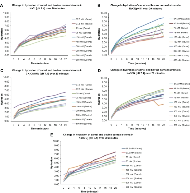
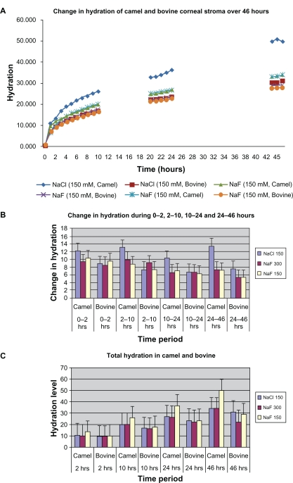
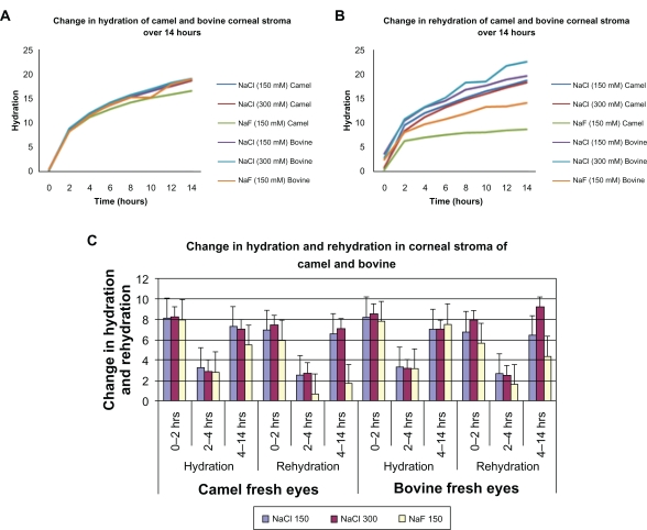
In the present study we investigated the swelling characteristics of fresh camel and bovine cornea in sodium salt solutions. Swelling studies were carried out at 20 minutes, 14 hours, and 46 hours on five fresh camel and 5 five fresh bovine corneas. During the 20-minute hydration of fresh corneal stroma was investigated using sodium chloride (NaCl), sodium bicarbonate (NaHCO(3)), sodium acetate (CH(3)COONa), sodium thiocyanate (NaSCN), and sodium floride (NaF) at 2-minute time intervals. During a 46-hour time period, the hydration study was carried out using NaCl (150, 300 mM) and NaF (150 mM) at random intervals. The 14-hour study was carried out to assess the rehydration of corneal stroma after 6 hours of drying. During the 20-minute swelling studies in the first 2 minutes the rate of hydration in both camel and bovine corneas was high but gradually reduced in the 2-20-minute period. The rates and levels of hydration of camel and bovine cornea were not significantly different from each other in all the strengths of solutions. During the 46-hour swelling studies, the initial rate of hydration (0-2 hours) of camel and bovine stroma, in all solutions was significantly higher (Z = 0.056) compared to hydration during later hours (2-46 hours). Camel stromal hydration (high) in 150 mM NaCl was significantly higher compared to bovine stromal hydration in the same solution during the 10-24, and 24-46-hour time periods. Rehydration in camel stroma was significantly lower than bovine in 150 mM NaF. The 20-minute study showed that there was no selective affinity for particular ions in camel or bovine corneal stroma. Initial swelling in both corneal and bovine stroma is faster and more prominant compared to later swelling. The swelling in camel cornea is more prominant compared to bovine corneal stroma. This could be due to higher negatively charged keratin sulfate-proteoglycans in the stroma. Lower rehydration in camel cornea suggests stronger leaching of proteoglycans from stroma in NaF.
Keywords: camel; proteoglycans; sodium bicarbonate; sodium thiocyanate; swelling.
Figures
Similar articles
-
Swelling of the collagen-keratocyte matrix of the bovine corneal stroma ex vivo in various solutions and its relationship to tissue thickness.Tissue Cell. 2000 Dec;32(6):478-93. doi: 10.1016/s0040-8166(00)80004-x. Tissue Cell. 2000. PMID: 11197230
-
The regulation of corneal hydration by a salt pump requiring the presence of sodium and bicarbonate ions.J Physiol. 1974 Jan;236(2):271-302. doi: 10.1113/jphysiol.1974.sp010435. J Physiol. 1974. PMID: 16992435 Free PMC article.
-
Effects of prior freezing or drying on the swelling behaviour of the bovine cornea.Chin Med J (Engl). 2009 Jan 20;122(2):212-8. Chin Med J (Engl). 2009. PMID: 19187649
-
Transparency, swelling and scarring in the corneal stroma.Eye (Lond). 2003 Nov;17(8):927-36. doi: 10.1038/sj.eye.6700574. Eye (Lond). 2003. PMID: 14631399 Review.
-
[Transplantation of corneal endothelial cells].Nippon Ganka Gakkai Zasshi. 2002 Dec;106(12):805-35; discussion 836. Nippon Ganka Gakkai Zasshi. 2002. PMID: 12610838 Review. Japanese.
References
-
- Maurice DM. The cornea and sclera. In: Davson H, editor. The Eye, Volume 1b, Vegetative Physiology and Biochemistry. 3rd ed. New York, NY: Academic Press; 1984. p. 509.
-
- Komai Y, Ushiki T. Three dimensional organization of collagen fibrils in human cornea and sclera. Invest Ophthalmol Vis Sci. 1991;32:2244–2258. - PubMed
-
- Michelacci YM. Collagens and proteoglycans of the corneal extracellular matrix. Braz J Med Biol Res. 2003;36:1037–1046. - PubMed
LinkOut - more resources
Full Text Sources
