

Metastasis from scapular Ewing's sarcoma presenting as sutural diastasis: An unusual presentation
- PMID: 20922089
- PMCID: PMC2940167
- DOI: 10.4103/0973-6042.68415
Metastasis from scapular Ewing's sarcoma presenting as sutural diastasis: An unusual presentation
Abstract
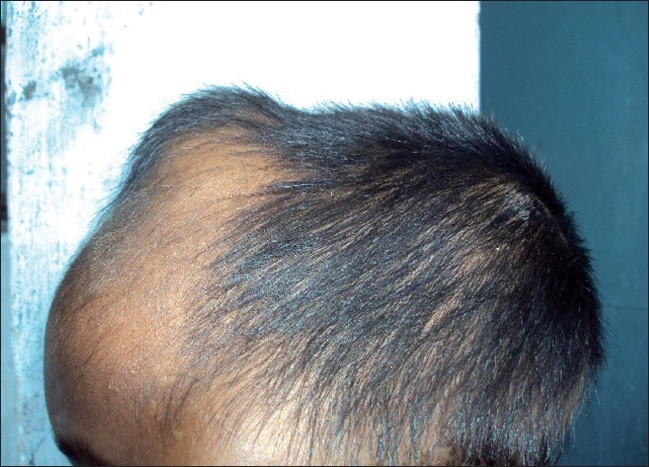
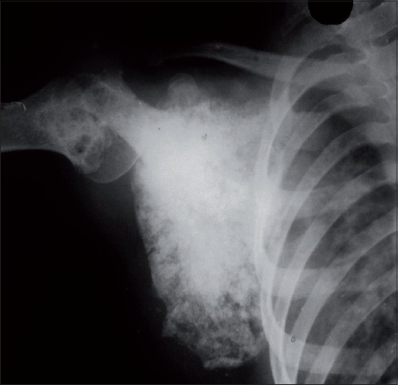
Ewing's sarcoma is a malignant non-osteogenic primary tumor of the bone. It is one of the most common primary malignant tumors of bone. Peak incidence is noticed in second decade of life with male preponderance of 1.6:1. It occurs most frequently in long bones and flat bones of pelvic girdles. In 30% cases, Ewing's sarcoma is multicentric in origin. In 14-50%, multiple metastases are present at the time of diagnosis. CNS spread is rare and isolated CNS involvement is not seen. Skull metastasis of Ewing's sarcoma is not rare compared to primary Ewing's sarcoma of the skull, but the actual frequency is unknown. We wish to report a case of "Primary Ewing's sarcoma of scapula with metastasis to Skull Vault in a Child resulting in sutural diastasis" diagnosed by clinicoradiological examination and confirmed by histopathology.
Keywords: Ewing’s sarcoma; non-osteogenic; skull vault; sutural diastasis.
Conflict of interest statement
Figures


References
-
- Dahlin DC, Unni KK. Bone Tumors. In: Thomas CC, Sprigfield IL, editors. General aspects and data on 8542 cases, No. 4. Illinois: Charles C Thomas Pub Ltd; 1986. pp. 269–305.
-
- Miller RW. Contrasting epidemiology of childhood osteosarcoma, Ewing’s tumor, and rhabdomyosarcoma. Natl Cancer Inst Monogr. 1981;56:9–15. - PubMed
-
- Ewing J. The Classic: Diffuse endothelioma of bone. Proceedings of the New York Pathological Society. 1921;12:17. Clin Orthop Relat Res. 2006;450:25–7. - PubMed
-
- Trigg ME, Glaubiger D, Nesbit ME., Jr The frequency of isolated CNS involvement in Ewing’s sarcoma. Cancer. 1982;49:2404–9. - PubMed
-
- Evans R, Nesbit M, Askin F, Burgert O, Cangir A, Foulkes M, et al. Local recurrence, rate and sites of metastases, and time to relapse as a function of treatment regimen, size of primary and surgical history in 62 patients presenting with non-metastatic ewing’s sarcoma of the pelvic bones. Int J Radiat Oncol Biol Phys. 1985;11:129–36. - PubMed
Publication types
LinkOut - more resources
Full Text Sources
