

An Assessment of the Relationship between the Maxillary Sinus Floor and the Maxillary Posterior Teeth Root Tips Using Dental Cone-beam Computerized Tomography
- PMID: 20922167
- PMCID: PMC2948741
An Assessment of the Relationship between the Maxillary Sinus Floor and the Maxillary Posterior Teeth Root Tips Using Dental Cone-beam Computerized Tomography
Abstract
Objectives: The aim of the present study was to assess the relationship between the maxillary sinus floor and the maxillary posterior teeth root tips using dental cone-beam CT.
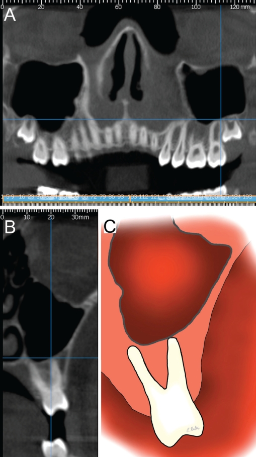
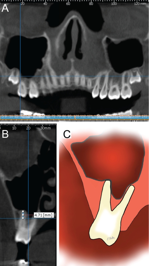
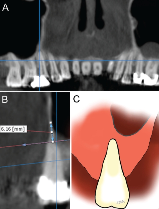
Methods: A total of 87 right and 89 left maxillary sinus regions from 92 patients were examined using dental cone-beam CT. Images were analyzed by a specialist in oral and maxillofacial radiology. Perpendicular lines were drawn on the cross-sectional images between the deepest point of the maxillary sinus floor and the root tips of the maxillary first and second premolars and first, second and third molars, and the distances were measured using built-in measurement tools. Means, standard deviations and minimum and maximum values were calculated for all right and left premolars and molars. T-tests were used to compare measurements between left and right sides and between female and male patients.
Results: The distance between sinus floor and root tip was longest for the first premolar root tip and shortest for the second molar buccodistal root tip for both right and left sides. No statistically significant differences were found between the right and left side measurements or between female and male patients (P>.05).
Conclusions: Knowledge of the anatomical relationship between the maxillary sinus floor and the maxillary posterior teeth root tips is important for the preoperative treatment planning of maxillary posterior teeth.
Keywords: Computerized tomography; Cone beam; Maxillary sinus; Posterior teeth.
Figures
References
-
- Misch CE. Contemporary implant dentistry. 2nd ed. St.Louis: CV Mosby Co; 1999. pp. 76–194.
-
- Mc Growan DA, Baxter PW, James J. The Maxillary Sinus and its Dental Implications. 1st ed. London: Wright Co; 1993. pp. 1–25.
-
- Hauman CH, Chandler NP, Tong DC. Endodontic implications of the maxillary sinus: a review. Int Endod J. 2002;35:127–141. - PubMed
-
- Waite DE. Maxillary sinus. Dent Clin North Am. 1971;15:349–368. - PubMed
-
- Tank PW. Grant’s Dissector. 13 ed. Philadelphia: Lippincott Williams & Wilkins; 2005. p. 198.
LinkOut - more resources
Full Text Sources