Comparative Study
. 2011 Feb;12(1):45-58.
doi: 10.1007/s10162-010-0238-z. Epub 2010 Oct 5.

Different cellular and genetic basis of noise-related endocochlear potential reduction in CBA/J and BALB/cJ mice

Affiliations
Comparative Study

Different cellular and genetic basis of noise-related endocochlear potential reduction in CBA/J and BALB/cJ mice

Kevin K Ohlemiller et al. J Assoc Res Otolaryngol. 2011 Feb.

Abstract

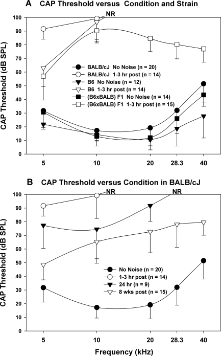
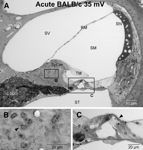
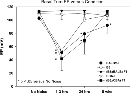
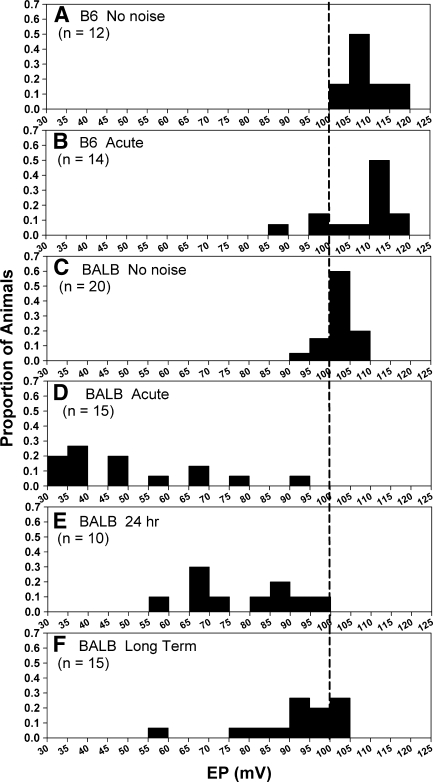
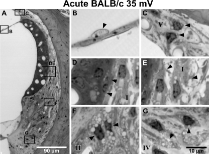
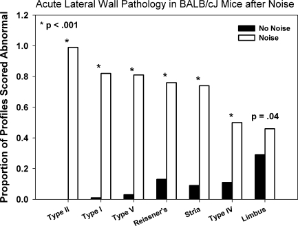
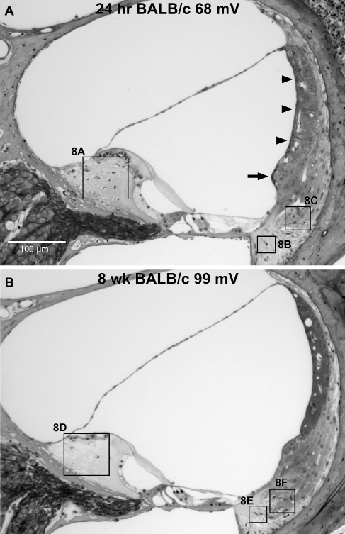
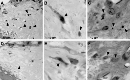
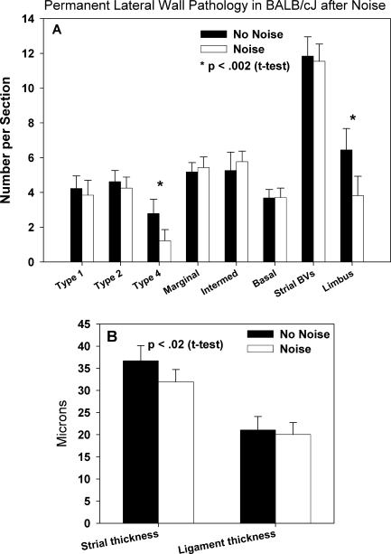
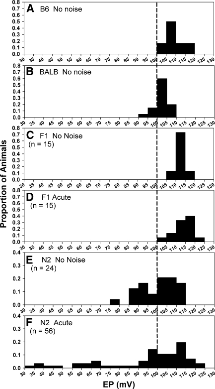
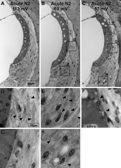
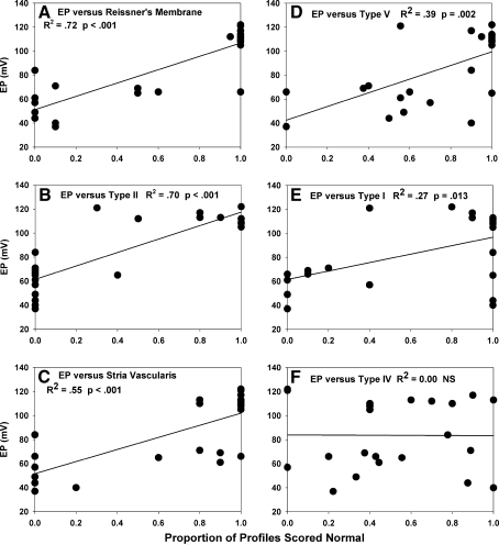
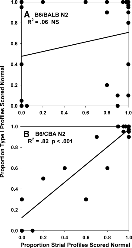
The acute and permanent effects of noise exposure on the endocochlear potential (EP) and cochlear lateral wall were evaluated in BALB/cJ (BALB) inbred mice, and compared with CBA/J (CBA) and C57BL/6 (B6) mice. Two-hour exposure to broadband noise (4-45 kHz) at 110 dB SPL leads to a approximately 50 mV reduction in the EP in BALB and CBA, but not B6. EP reduction in BALB and CBA is reliably associated with characteristic acute cellular pathology in stria vascularis and spiral ligament. By 8 weeks after exposure, the EP in CBA mice has returned to normal. In BALBs, however, the EP remains depressed by an average approximately 10 mV, so that permanent EP reduction contributes to permanent threshold shifts in these mice. We recently showed that the CBA noise phenotype in part reflects the influence of a large effect quantitative trait locus on Chr. 18, termed Nirep (Ohlemiller et al., Hear Res 260:47-53, 2010b). While CBA "EP susceptibility" alleles are dominant to those in B6, examination of (B6 × BALB) F1 hybrid mice and (F1 × BALB) N2 backcross mice revealed that noise-related EP reduction and associated cell pathology in BALBs are inherited in an autosomal recessive manner, and are dependent on multiple genes. Moreover, while N2 mice formed from B6 and CBA retain strong correspondence between acute EP reduction, ligament pathology, and strial pathology, N2s formed from B6 and BALB include subsets that dissociate pathology of ligament and stria. We conclude that the genes and cascades that govern the very similar EP susceptibility phenotypes in BALB and CBA mice need not be the same. BALBs appear to carry alleles that promote more pronounced long term effects of noise on the lateral wall. Separate loci in BALBs may preferentially impact stria versus ligament.


Figures

FIG. 1.
FIG. 1.
A Mean ± SD CAP thresholds for noise-exposed and unexposed control BALB, B6, and (B6 × BALB) F1 mice. B Mean ± SD CAP thresholds only for control BALB mice and BALB at various times after noise. Unexposed control and acute exposed mice (1–3 h after noise) are replotted from A to facilitate comparison. All noise exposures were 2 h, 4–45 kHz, 110 dB SPL. NR no response at highest sound level available.
FIG. 2.
FIG. 2.
A Typical cochlear duct of the upper basal turn in a BALB mouse 1–3 h after noise. B Expanded view of box centered on spiral limbus in (A). Stellate fibrocytes of limbus show subtle vacuolization (arrowhead). C Expanded view of organ of Corti. Pillars are bent and Nuel’s space is collapsed (arrowhead), but no breach of reticular lamina is apparent. SpL spiral ligament, StV stria vascularis, SV scala vestibuli, ST scala tympani, SM scala media, spLim spiral limbus, RM Reissner’s membrane, SpG spiral ganglion, TM tectorial membrane.
FIG. 3.
FIG. 3.
Mean ± SD basal turn EP in unexposed controls and at various times after noise exposure in inbred BALB, B6, and CBA/J mice, and in F1 hybrids formed from crossing BALB and CBA with B6. EP reduction by noise is dominant versus B6 in CBA; recessive in BALB. EP does not completely recover in BALBs. Asterisk Significantly different from unexposed controls of same strain by one-way ANOVA.
FIG. 4.
FIG. 4.
AF Distribution of basal turn EPs in B6 and BALB mice with and without noise exposure. Data are the same as those used to derive means and statistical results shown in Figure 3. BALBs are examined at various times after noise as shown. Vertical line marks 100 mV to aid comparisons. Data were derived by dividing the number of animals in each bin by sample size for each group, so that bin height indicates the proportion falling within a particular EP range.
FIG. 5.
FIG. 5.
A Cochlear upper basal turn lateral wall in example BALB mouse 1–3 h after noise. Animal is the same as shown in Figure 2. B Expanded view of box B in (A) showing enlarged pale nucleus of mesothelial cell of Reissner’s membrane (arrowhead). C Expanded view of box C in (A) showing vacuolated type V fibrocytes (arrowheads). DE Expanded view of boxes D and E in (A) showing vacuolated basal cells and possibly intermediate cells in stria with adjacent shrunken type I fibrocytes in spiral ligament (arrowheads). Shrinkage of fibrocytes is attended by increased clear space in ligament. f Expanded view of box F in (A) showing vacuolated type II fibrocytes (arrowheads). G Expanded view of box G in (A) showing vacuolated type IV fibrocytes (arrowheads). Roman numerals in (CG) denote major constituent fibrocyte types.
FIG. 6.
FIG. 6.
Quantitative analysis of acute (1–3 h post) noise-related pathology of lateral wall and spiral limbus in noise-exposed and control BALB mice. Sections were blindly scored “normal” or “abnormal” for each cell type or structure, and section totals were combined for each experimental group. P values indicate z test results.
FIG. 7.
FIG. 7.
A Example cochlear upper basal turn of BALB mouse taken for examination 24 h after noise exposure. Stria is swollen (arrowheads) and epithelial cells of spiral prominence are darkened (arrow). B Example cochlear upper basal turn of BALB mouse taken for examination 8 weeks after noise exposure. EPs are as shown. Boxes denoted 8A8F refer to expanded views in Figure 8.
FIG. 8.
FIG. 8.
Expanded views of boxed regions in Figure 7. AC At 24 h post-exposure, pyknotic nuclei and condensed cells of spiral limbus and in regions of ligament dominated by type IV fibrocytes and type II fibrocytes, respectively, indicate dying cells (arrowheads). DF By 8-week post-exposure, stellate fibrocytes of limbus are entirely missing in upper base and lower apex (arrowhead in (D)). Type IV fibrocytes are reduced in number. Arrowhead in (E) shows dying type IV. Type II fibrocytes (F, arrowheads) appear generally normal.
FIG. 9.
FIG. 9.
Quantitative analysis of permanent noise-related pathology (8-week post-exposure) in cochlear upper basal turn spiral limbus and lateral wall of control and exposed BALB mice. A Among metrics for strial and ligament cell types, as well as strial capillaries (blood vessel density (BVs)), significant differences were only found for type IV fibrocytes and stellate fibrocytes of limbus. B Significant thickness changes were found for stria, but not ligament. P values denote results of two-tailed t test.
FIG. 10.
FIG. 10.
AB Distribution of basal turn EPs in B6 and BALB mice without noise exposure (reproduced from Figure 4 to aid comparison). CD Distribution of EPs with and without noise in (B6 × BALB) F1 hybrid mice. EF Distribution of EPs in (F1 × BALB) N2 backcross mice with and without noise. Following noise, 25% (14/56) of N2 mice showed EPs below 80 mV. Note different y-axis range in (E) and (F) due to greater scatter. Vertical line marks 100 mV to facilitate comparisons. As in Figure 4, data were derived by dividing the number of animals in each bin by sample size for each group, so that bin height indicates the proportion falling within a particular EP range.
FIG. 11.
FIG. 11.
Examples of the three major patterns of cellular appearance observed in the cochlear upper basal turn 1–3 h after noise in B6/BALB N2 backcross mice. EPs are shown for each animal. A Normal EP attended by minimal pathology, as detected by light microscope. B Mouse with reduced EP combining minimal strial pathology with clear pathology of type I fibrocytes (expanded in (D), arrowheads) and type II fibrocytes (expanded in (E), arrowheads). C Mouse combining clear strial pathology (expanded in (F), arrowheads) with pathology of type II fibrocytes (expanded in (H), arrowheads), but showing minimal pathology of type I fibrocytes (expanded in (G)).
FIG. 12.
FIG. 12.
AF Correlation between basal turn EP and pathology of specific cells and structures in the upper basal turn of B6/BALB N2 backcross mice, observed 1–3 h after noise. The condition of Reissner’s membrane and type II fibrocytes of ligament showed the strongest correlation with EP (A, B), followed by progressively weaker correlation between EP and the appearance of the stria (C), type V fibrocytes (D), and type I fibrocytes (E). The appearance of type IV fibrocytes (F) and spiral limbus (not shown) was uncorrelated with EP.
FIG. 13.
FIG. 13.
Direct comparison the correlation between acute noise-related pathology of stria vascularis and type I fibrocytes of spiral ligament in B6/BALB N2 mice (A) and B6/CBA N2 mice (B). These were highly correlated in B6/CBA N2s, but completely uncorrelated in B6/BALB N2s.

Similar articles

Cited by

References

    1. Adams JC. Immunocytochemical traits of type IV fibrocytes and their possible relations to cochlear function and pathology. J Assoc Res Otolaryngol. 2009;10:369–382. doi: 10.1007/s10162-009-0165-z. - DOI - PMC - PubMed
    1. Ahmad M, Bohne BA, Harding GW. An in vivo tracer study of noise-induced damage to the reticular lamina. Hear Res. 2003;175:82–100. doi: 10.1016/S0378-5955(02)00713-X. - DOI - PubMed
    1. Beagley HA. Acoustic trauma in the guinea pig. I. Electrophysiology and histology. Acta Otolaryngol. 1965;60:437–451. doi: 10.3109/00016486509127027. - DOI - PubMed
    1. Bohne BA, Harding GW, Ou HC. Preparation and evaluation of the mouse temporal bone. In: Willott JF, editor. Handbook of mouse auditory research. Boca Raton, FL: CRC Press; 2001. pp. 171–187.
    1. Chang Q, Tang W, Ahmad S, Zhou B, Lin X. Gap junction mediated intercellular metabolite transfer in the cochlea in compromised in connexin 30 null mice. PLoS ONE. 2008;3:e4088. doi: 10.1371/journal.pone.0004088. - DOI - PMC - PubMed

Publication types

LinkOut - more resources