. 2010 Nov 5;9(11):6082-9.
doi: 10.1021/pr100924s. Epub 2010 Oct 15.

One-step sample concentration, purification, and albumin depletion method for urinary proteomics

Affiliations

One-step sample concentration, purification, and albumin depletion method for urinary proteomics

Ali R Vaezzadeh et al. J Proteome Res. .

Abstract

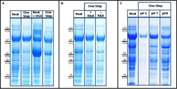
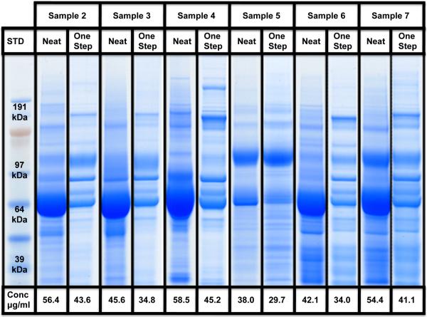
Workflows in urinary proteomics studies are often complex and require many steps to enrich, purify, deplete, and separate the complex mixture. Many of these methods are laborious, are time-consuming, and have the potential for error. Although individual steps of these methods have been previously studied, their downstream compatibilities with fractionation technologies such as off-gel electrophoresis have not been investigated. We developed a one-step sample preparation workflow that simultaneously (i) concentrates proteins, (ii) purifies by removing salts and other low molecular weight compounds, and (iii) depletes (albumin) from urine samples. This simple and robust workflow can be multiplexed and is compatible with a diverse range of downstream multidimensional separation technologies. Additionally, because of its high reproducibility and flexibility in processing samples with different volumes and concentrations, it has the potential to be used for standardization of urinary proteomics studies, as well as for studying other body fluids of similar complexity.


Figures

Figure 1
Figure 1. One-Step method workflow
The final workflow for preparation and analysis of urine samples. Urine is first spun down to remove debris and the pH is adjusted to neutral. Urine proteins are reduced and alkylated. This sample is then added to the Vivaspin 6 spin-filters. Anti-HSA resin is added. Samples are incubated and then spun down completing the protein concentration, purification and albumin depletion. Proteins are removed leaving the Anti-HSA resin behind. Proteins undergo in-solution digestion with trypsin. Samples could then be chemically labeled for quantitation and multiplexed*, prior to undergoing 1st dimension separation (Off-Gel electrophoresis is currently utilized**) and LC-MS/MS.
Figure 2
Figure 2. Efficiency, Recovery and pH sensitivity of the One-Step Method
A. The recovery and depletion efficiency of the One-Step enrichment, purification and depletion method applied to neat urine sample from a healthy individual with and without 20 μg spiked-in HSA. B. The effect of Reduction and Alkylation (R&A) on the depletion efficiency and recovery of the One-Step process. C. The effect of pH on the One-Step method recovery and depletion.
Figure 3
Figure 3. Reproducibility and efficiency of the One-Step method
The reproducibility and efficiency of the One-Step enrichment, purification and albumin depletion method is tested on urine samples 2 to 7. Protein concentrations before and after the One-Step method are presented.
Figure 4
Figure 4. One-Step method and Off-Gel Electrophoresis
The application of the One-Step sample preparation method to the Off-Gel technology. A. Demonstrates the average pI of the peptides identified in each fraction with their average standard deviations. B. Depicts the number of fractions each peptide has been identified in as a surrogate for the resolution. C. Shows the distribution of the peptides along the IPG strip for each sample.
Figure 5
Figure 5. Application of the One-Step method and Off-Gel Electrophoresis to different samples
The comparison of four urine sample (samples 2, 3, 5 and 7) prepared with the One-Step sample preparation method and analyzed using the Off-Gel technology. A. Demonstrates the average pI of the peptides identified in each fraction. B. Depicts the number of fractions each peptide has been identified in as a surrogate for the resolution. C. Shows the distribution of the peptides along the IPG strip for each sample.
Figure 6
Figure 6. Reproducibility of the One-Step method and Off-Gel Electrophoresis
Technical reproducibility of the One-Step urine sample preparation method in combination with Off-Gel separation and data-dependent LC-MS/MS. Blue bars represent the number of proteins identified only in technical replicate 1 and red bars indicate the number of proteins identified exclusively in technical replicate 2. Green bars show the number of proteins identified in both technical replicates.

References

    1. O'Riordan E, Gross SS, Goligorsky MS. Technology Insight: renal proteomics--at the crossroads between promise and problems. Nat Clin Pract Nephrol. 2006;2(8):445–58. - PubMed
    1. Lee RS, Monigatti F, Briscoe AC, Waldon Z, Freeman MR, Steen H. Optimizing sample handling for urinary proteomics. J Proteome Res. 2008;7(9):4022–30. - PMC - PubMed
    1. Orenes-Pinero E, Corton M, Gonzalez-Peramato P, Algaba F, Casal I, Serrano A, Sanchez-Carbayo M. Searching urinary tumor markers for bladder cancer using a two-dimensional differential gel electrophoresis (2DDIGE) approach. J Proteome Res. 2007;6(11):4440–8. - PubMed
    1. Lafitte D, Dussol B, Andersen S, Vazi A, Dupuy P, Jensen ON, Berland Y, Verdier JM. Optimized preparation of urine samples for two-dimensional electrophoresis and initial application to patient samples. Clin Biochem. 2002;35(8):581–9. - PubMed
    1. Lee RS, Monigatti F, Lutchman M, Patterson T, Budnik B, Steen JA, Freeman MR, Steen H. Temporal variations of the postnatal rat urinary proteome as a reflection of systemic maturation. Proteomics. 2008;8(5):1097–112. - PubMed

Publication types