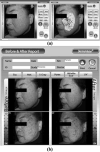
Dermatological feasibility of multimodal facial color imaging modality for cross-evaluation of facial actinic keratosis
- PMID: 20923462
- PMCID: PMC3021003
- DOI: 10.1111/j.1600-0846.2010.00464.x
Dermatological feasibility of multimodal facial color imaging modality for cross-evaluation of facial actinic keratosis
Abstract
Background/purpose: Digital color image analysis is currently considered as a routine procedure in dermatology. In our previous study, a multimodal facial color imaging modality (MFCIM), which provides a conventional, parallel- and cross-polarization, and a fluorescent color image, was introduced for objective evaluation of various facial skin lesions. This study introduces a commercial version of MFCIM, DermaVision-PRO, for routine clinical use in dermatology and demonstrates its dermatological feasibility for cross-evaluation of skin lesions.
Methods/results: Sample images of subjects with actinic keratosis or non-melanoma skin cancers were obtained at four different imaging modes. Various image analysis methods were applied to cross-evaluate the skin lesion and, finally, to extract valuable diagnostic information. DermaVision-PRO is potentially a useful tool as an objective macroscopic imaging modality for quick prescreening and cross-evaluation of facial skin lesions.
Conclusion: DermaVision-PRO may be utilized as a useful tool for the cross-evaluation of widely distributed facial skin lesions and as an efficient database management of patient information.
© 2010 John Wiley & Sons A/S.
Figures


Similar articles
-
Multimodal facial color imaging modality for objective analysis of skin lesions.J Biomed Opt. 2008 Nov-Dec;13(6):064007. doi: 10.1117/1.3006056. J Biomed Opt. 2008. PMID: 19123654 Free PMC article.
-
Digital photographic imaging system for the evaluation of various facial skin lesions.Annu Int Conf IEEE Eng Med Biol Soc. 2008;2008:4032-4. doi: 10.1109/IEMBS.2008.4650094. Annu Int Conf IEEE Eng Med Biol Soc. 2008. PMID: 19163597
-
Polarization color imaging system for on-line quantitative evaluation of facial skin lesions.Dermatol Surg. 2007 Nov;33(11):1350-6. doi: 10.1111/j.1524-4725.2007.33288.x. Dermatol Surg. 2007. PMID: 17958588
-
Role of In Vivo Reflectance Confocal Microscopy in the Analysis of Melanocytic Lesions.Acta Dermatovenerol Croat. 2018 Apr;26(1):64-67. Acta Dermatovenerol Croat. 2018. PMID: 29782304 Review.
-
Management of benign skin lesions commonly affecting the face: actinic keratosis, seborrheic keratosis, and rosacea.Curr Opin Otolaryngol Head Neck Surg. 2009 Aug;17(4):315-20. doi: 10.1097/MOO.0b013e32832d75e3. Curr Opin Otolaryngol Head Neck Surg. 2009. PMID: 19465852 Review.
Cited by
-
A multicenter pilot evaluation of the National Institutes of Health chronic graft-versus-host disease (cGVHD) therapeutic response measures: feasibility, interrater reliability, and minimum detectable change.Biol Blood Marrow Transplant. 2011 Nov;17(11):1619-29. doi: 10.1016/j.bbmt.2011.04.002. Epub 2011 Apr 12. Biol Blood Marrow Transplant. 2011. PMID: 21536143 Free PMC article.
-
Effects of Jae-Seng Acupuncture Treatment on the Improvement of Nasolabial Folds and Eye Wrinkles.Evid Based Complement Alternat Med. 2015;2015:273909. doi: 10.1155/2015/273909. Epub 2015 May 4. Evid Based Complement Alternat Med. 2015. PMID: 26064158 Free PMC article.
-
The mechanism and application of computer-assisted full facial skin imaging systems.Skin Health Dis. 2023 Dec 4;4(2):e320. doi: 10.1002/ski2.320. eCollection 2024 Apr. Skin Health Dis. 2023. PMID: 38577059 Free PMC article. Review.
References
-
- Fullerton A, Fischer T, Lahti A, et al. Guidelines for measurement of skin colour and erythema. A report from the Standardization Group of the European Society of Contact Dermatitis. Contact Dermatitis. 1996;35:1–10. - PubMed
-
- Jung B, Choi B, Shin Y, et al. Determination of optimal view angles for quantitative facial image analysis. J Biomed Opt. 2005;10:024002. - PubMed
-
- Jung B, Kim CS, Choi B, et al. Use of erythema index imaging for systematic analysis of port wine stain skin response to laser therapy. Lasers Surg Med. 2005;37:186–91. - PubMed
-
- Wallace VP, Crawford DC, Mortimer PS, et al. Spectrophotometric assessment of pigmented skin lesions: methods and feature selection for evaluation of diagnostic performance. Phys Med Biol. 2000;45:735–51. - PubMed
-
- Marghoob AA, Swindle LD, Moricz CZ, et al. Instruments and new technologies for the in vivo diagnosis of melanoma. J Am Acad Dermatol. 2003;49:777–97. quiz 98-9. - PubMed

