. 2010 Oct 6;99(7):2116-24.
doi: 10.1016/j.bpj.2010.06.035.

The N-terminus of the intrinsically disordered protein α-synuclein triggers membrane binding and helix folding

Affiliations

The N-terminus of the intrinsically disordered protein α-synuclein triggers membrane binding and helix folding

Tim Bartels et al. Biophys J. .

Abstract

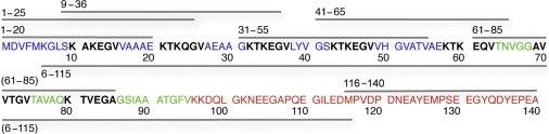
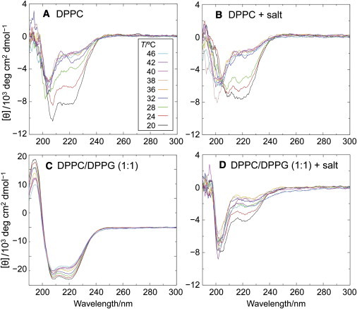
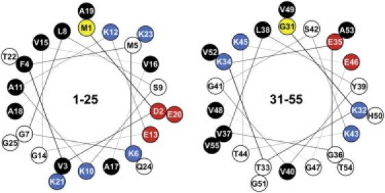
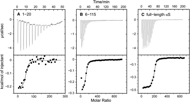
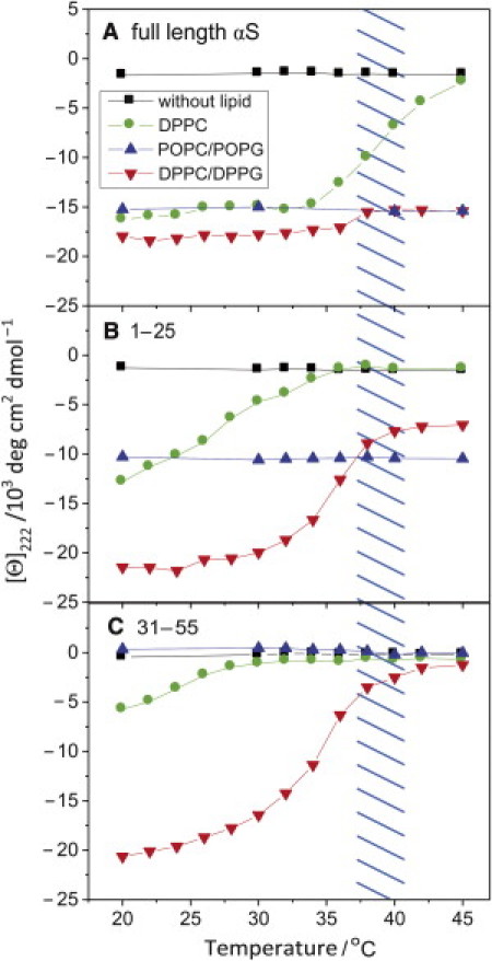
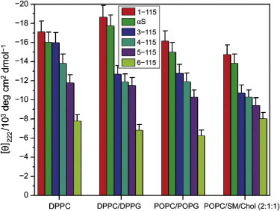
Alpha-synuclein (αS) is a 140-amino-acid protein that is involved in a number of neurodegenerative diseases. In Parkinson's disease, the protein is typically encountered in intracellular, high-molecular-weight aggregates. Although αS is abundant in the presynaptic terminals of the central nervous system, its physiological function is still unknown. There is strong evidence for the membrane affinity of the protein. One hypothesis is that lipid-induced binding and helix folding may modulate the fusion of synaptic vesicles with the presynaptic membrane and the ensuing transmitter release. Here we show that membrane recognition of the N-terminus is essential for the cooperative formation of helical domains in the protein. We used circular dichroism spectroscopy and isothermal titration calorimetry to investigate synthetic peptide fragments from different domains of the full-length αS protein. Site-specific truncation and partial cleavage of the full-length protein were employed to further characterize the structural motifs responsible for helix formation and lipid-protein interaction. Unilamellar vesicles of varying net charge and lipid compositions undergoing lateral phase separation or chain melting phase transitions in the vicinity of physiological temperatures served as model membranes. The results suggest that the membrane-induced helical folding of the first 25 residues may be driven simultaneously by electrostatic attraction and by a change in lipid ordering. Our findings highlight the significance of the αS N-terminus for folding nucleation, and provide a framework for elucidating the role of lipid-induced conformational transitions of the protein within its intracellular milieu.


Figures

Figure 1
Figure 1
Amino-acid sequence of α-synuclein. The N-terminal region is blue, the aggregation-prone (NAC) domain is in green, and the C-terminal region is in red. The seven imperfect repeats are indicated in bold type. Protein fragments are schematically shown as bars above (synthetic small peptides) or below (chemically truncated protein). For clarity, the N-terminal truncated mutants are omitted.
Figure 2
Figure 2
Temperature dependence of circular dichroism spectra reveals membrane binding and coil/helix transition of N-terminal peptide 1–20. Vesicle compositions: (A) DPPC; (B) DPPC, 140 mM NaCl; (C) DPPC/DPPG (1:1); and (D) DPPC/DPPG (1:1), 140 mM NaCl. All samples contained 10 mM Na phosphate, pH 7.0. Total phospholipid concentration was 5 mM and the peptide concentration was 30 μM in a cuvette of 0.2-cm pathlength. CD spectra in terms of decreasing ellipticity at 222 nm were recorded at temperatures of 46, 42, 40, 38, 36, 32, 28, 24, and 20°C. Control spectra acquired without peptide, but with the same buffer and phospholipid concentration, were subtracted for each temperature.
Figure 3
Figure 3
Helical-wheel representation of 1–25 and 31–55 peptides. View is from the N-terminus down the helix axis. Adjacent amino acids are offset by an angle of 100 deg. Residue designations are (yellow) first amino acid in the sequence; (red) negative amino acid charges; (blue) positive amino acid charges; (white) slightly polar, neutral amino acids; and (black) nonpolar, hydrophobic amino acids. Note the opposing lysine clusters, separated by 200 deg (K10, K21, and K12, K23 for the 1–25 peptide, and K32, K43 and K34, K45 for the 31–55 peptide, respectively).
Figure 4
Figure 4
Isothermal titration calorimetry yields percentage of vesicle-bound peptide or protein. SUV suspensions were titrated at 20°C into a 1.4-mL calorimeter cell containing the peptide or protein solutions. Single 7-μL injection volumes were typically employed. (A) Here, 30 μM of peptide 1–20 and (B) 8.5 μM peptide 6–115 in phosphate buffer, pH 7.0, titrated with DPPC/DPPG vesicles; molar ratio of the lipids 1:1, total lipid concentration in the syringe, 40 mM. (C) Here, 8.7 mM αS titrated with 45 mM DPPC/DPPG vesicles; other conditions as in panels A and B. The number of associated lipids and the binding constants Kb were obtained using the binding model for N independent sites provided with the software ORIGIN (MicroCal). (For details, see (19).)
Figure 5
Figure 5
Circular dichroism spectroscopy manifests helical folding due to local sequence properties and lipid composition of vesicle membranes. The helicity was obtained from mean-residue ellipticity values at 222 nm. Small unilamellar vesicles (phospholipid concentration 5 mM) and αS or αS-derived peptides in 100 mM KCl, 20 mM Na phosphate, pH 7.4, were mixed in a 0.2-cm pathlength CD cuvette, and incubated for 5 min to allow for temperature equilibration. The molar ratio of phospholipids in the binary mixtures was 1:1 and the lipid/protein (peptide) molar ratio was 167:1. Temperature, 20°C; typical errors ±6%. (Methods).
Figure 6
Figure 6
Temperature dependence of mean-residue ellipticity at 222 nm reveals varying affinities of N-terminal sequence domains to surface charges and membrane defects. (A) Full-length α-synuclein, (B) 1-25 peptide, and (C) 31-35 peptide. Small unilamellar vesicles (phospholipid concentration 5 mM) were mixed with 6 μM αS or 30 μM of the peptides in 100 mM KCl, 20 mM Na phosphate, pH 7.4, corresponding to phospholipid/protein molar ratios of 833:1 and 167:1. The molar ratio of phospholipids in the binary mixtures was 1:1. The temperature range of the phase transition for DPPC and DPPC/DPPG vesicles is indicated by the hatched area. Note the additive effect of electrostatic interaction and chain melting phase transition on the mean-residue ellipticity of the full-length protein and the 125 peptide.
Figure 7
Figure 7
Stepwise removal of N-terminal amino acids from α-synuclein yields a progressive decrease of membrane-induced helical folding. Mean-residue ellipticities of SUVs were determined in 20 mM sodium phosphate, pH 7.4, and 100 mM KCl. Total SUV lipid concentration was 5 mM; protein concentrations were 8.5 μM and 6 μM for the deletion and truncation mutants, corresponding to lipid/protein molar ratios of 588 and 833, respectively. The molar ratio of phospholipids in the binary mixtures was 1:1. Temperature, 20°C.

References

    1. Beyer K. Mechanistic aspects of Parkinson's disease: α-synuclein and the biomembrane. Cell Biochem. Biophys. 2007;47:285–299. - PubMed
    1. Haass C., Selkoe D.J. Soluble protein oligomers in neurodegeneration: lessons from the Alzheimer's amyloid β-peptide. Nat. Rev. Mol. Cell Biol. 2007;8:101–112. - PubMed
    1. Dev K.K., Hofele K., van der Putten H. Part II: α-synuclein and its molecular pathophysiological role in neurodegenerative disease. Neuropharmacology. 2003;45:14–44. - PubMed
    1. Moore D.J., West A.B., Dawson T.M. Molecular pathophysiology of Parkinson's disease. Annu. Rev. Neurosci. 2005;28:57–87. - PubMed
    1. Wakabayashi K., Yoshimoto M., Takahashi H. α-synuclein immunoreactivity in glial cytoplasmic inclusions in multiple system atrophy. Neurosci. Lett. 1998;249:180–182. - PubMed

Publication types