. 2010 Oct 19;107(42):18073-8.
doi: 10.1073/pnas.1008737107. Epub 2010 Oct 5.

In vivo two-photon imaging reveals monocyte-dependent neutrophil extravasation during pulmonary inflammation

Affiliations

In vivo two-photon imaging reveals monocyte-dependent neutrophil extravasation during pulmonary inflammation

Daniel Kreisel et al. Proc Natl Acad Sci U S A. .

Abstract

Immune-mediated pulmonary diseases are a significant public health concern. Analysis of leukocyte behavior in the lung is essential for understanding cellular mechanisms that contribute to normal and diseased states. Here, we used two-photon imaging to study neutrophil extravasation from pulmonary vessels and subsequent interstitial migration. We found that the lungs contained a significant pool of tissue-resident neutrophils in the steady state. In response to inflammation produced by bacterial challenge or transplant-mediated, ischemia-reperfusion injury, neutrophils were rapidly recruited from the circulation and patrolled the interstitium and airspaces of the lung. Motile neutrophils often aggregated in dynamic clusters that formed and dispersed over tens of minutes. These clusters were associated with CD115(+) F4/80(+) Ly6C(+) cells that had recently entered the lung. The depletion of blood monocytes with clodronate liposomes reduced neutrophil clustering in the lung, but acted by inhibiting neutrophil transendothelial migration upstream of interstitial migration. Our results suggest that a subset of monocytes serve as key regulators of neutrophil extravasation in the lung and may be an attractive target for the treatment of inflammatory pulmonary diseases.


Conflict of interest statement

The authors declare no conflict of interest.

Figures

Fig. 1.
Fig. 1.
Neutrophil and macrophage distribution in various tissues of LysM-GFP mice. Neutrophils (bright green) and macrophages (dim green) are easily distinguishable based on their different brightness levels and distinct morphological characteristics. Blood vessels (red) were labeled by i.v. injection of nontargeted 655-nm Q-dots and the laser-induced second harmonic generation signal appears blue. In addition to resident macrophages (white arrowheads), there is a large number of extravascular tissue-resident neutrophils (yellow arrowheads) seen in lungs in vivo (A) (Movie S1) and explanted lungs (B), and in lymph nodes (C). In heart tissue (D), resident macrophages were observed, but neutrophils were present only within blood vessels. (Scale bar: 80 μm.) Lower are zoomed views of Upper. (Scale bar: 20 μm.) Tissue-resident macrophages (white arrowheads) are found in other tissues, but neutrophils (yellow arrowheads) are detected primarily within the vasculature in brain (E), liver (F), kidney (G), small intestine (H), or hind footpad (I). (Scale bar: 15 μm.)
Fig. 2.
Fig. 2.
Time-lapse imaging of neutrophil behavior in lung tissue in vivo, ex vivo, and under inflammatory conditions. (A) Intravital 2P imaging of resident neutrophils (green) in the parenchyma of the lung in vivo. Images are individual frames from a continuous time-lapse movie (Movie S2). A rare motile cell (yellow arrowheads) is shown migrating through the interstitial tissue. (B) Individual cells were tracked and cell displacement squared (μm2) vs. time (min) shows a strong linear correlation indicative of random cell migration. Plots of average neutrophil track speed (C) and meandering index (MI), n = 20 (D). The MI was calculated by dividing the distance a cell traveled from its starting point by the track length. Values of >0.8 are commonly associated with chemotaxis, whereas values of <0.5 are consistent with random cell migration. (E) Neutrophils (green) migrating in explanted lung tissue (Movie S3). A representative neutrophil track is highlighted (yellow arrowhead). Neutrophil displacement squared vs. time plot (F), mean track speed (G), and MI (H) in lung explants, n = 20. (I) Neutrophil (green) behavior in vivo 5 min after intratracheal administration of bacteria (L. monocytogenes, EGD strain) (Movie S4). A representative neutrophil track is highlighted (yellow arrowhead). Neutrophil displacement squared vs. time plot (J), mean track speed (K), and MI (L) after bacterial challenge, n = 16. (Scale bars: 10 μm.) Relative time is displayed in min:sec.
Fig. 3.
Fig. 3.
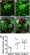
Neutrophils cluster after intratracheal bacterial challenge. Two-photon images of neutrophil (green) distribution 5 (A) and 30 min (B) after intratracheal administration of bacteria (Movie S6). Nontargeted Q-dots (red) were injected i.v. 30 min before bacterial challenge. Neutrophils in the clusters were often nonmotile. (Scale bar: 60 μm.) Two-photon images of neutrophil (green) distribution in lung grafts 2 h after transplant (C) and 2.5 h after transplant (D) (Movie S7). Time stamp is shown in min:sec. Yellow arrowheads show clusters that are forming or remain similar in size; white arrowheads show clusters that appear to dissociate. (E) The number of neutrophils per cluster in steady-state lungs (gray squares), lung explants (red triangles), and lungs after transplantation (blue diamonds) (*, 0.0371; **, 0.0088).
Fig. 4.
Fig. 4.
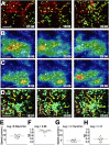
Leukocyte dynamics during transplant mediated ischemia reperfusion injury. (A) Blood vessels (red) were labeled by i.v. injection of nontargeted 655-nm Q-dots. A time-lapse 2P image sequence shows a Q-dot–positive cell leaving the pulmonary vasculature through a small branch of a medium-sized vessel (yellow track). Extravasation of the Q-dot–positive cell is associated with neutrophil extravasation and subsequent cluster formation (Movie S8). (Scale bar: 20 μm.) A heatmap visualization was generated to show the spatiotemporal changes in neutrophil velocity (B) and density (or integrated intensity) (C) (Movie S9). For speed, the color scale ranges from <2 μm/min (blue) to >10 μm/min (red). A local increase in cell velocity (B) (white arrowheads) precedes a 4-fold increase in cell density (yellow arrows) (C). (Scale bar: 60 μm.) (D) Neutrophil tracks (yellow arrows) show a symmetrical short-range migration bias toward the cluster. (Scale bar: 15 μm.) Time stamps in AD show relative time in min:sec. Neutrophils were divided in two groups based on distance from the center of the cluster, and the migration of each group was analyzed separately. Mean track speed (E) and MI (F) of neutrophils approaching within 50 μm of clusters, n = 13. Mean track speed (G) and MI (H) of neutrophils distal to clusters (>50 μm from clusters), n = 13.
Fig. 5.
Fig. 5.
Clodronate-liposome depletion impairs neutrophil transendothelial migration after transplantation. Pulmonary blood vessels (red) were labeled by i.v. injection of nontargeted 655-nm Q-dots. (A) Two-photon image of neutrophils (green) extravasating from a medium-size vessel (white lines) in a control lung graft at 2 h after transplantation. (B) Clodronate-liposome (CL) treatment of the transplant recipient results in neutrophil accumulation in medium-sized vessels and a reduced number of extravasated neutrophils. (C) The percentage of intravascular neutrophils observed at 2 h after engraftment in untreated recipients (control, <5%) and in clodronate-liposome treated recipients (CL, >90%). (Scale bar: 60 μm.)

References

    1. Tsai KS, Grayson MH. Pulmonary defense mechanisms against pneumonia and sepsis. Curr Opin Pulm Med. 2008;14:260–265. - PubMed
    1. Tate MD, et al. Neutrophils ameliorate lung injury and the development of severe disease during influenza infection. J Immunol. 2009;183:7441–7450. - PubMed
    1. Belperio JA, et al. CXCR2/CXCR2 ligand biology during lung transplant ischemia-reperfusion injury. J Immunol. 2005;175:6931–6939. - PubMed
    1. Zarbock A, Allegretti M, Ley K. Therapeutic inhibition of CXCR2 by Reparixin attenuates acute lung injury in mice. Br J Pharmacol. 2008;155:357–364. - PMC - PubMed
    1. Miller MJ, Wei SH, Parker I, Cahalan MD. Two-photon imaging of lymphocyte motility and antigen response in intact lymph node. Science. 2002;296:1869–1873. - PubMed

Publication types

LinkOut - more resources