

doi: 10.1534/genetics.110.123653.
Epub 2010 Oct 5.
Cdc14-dependent dephosphorylation of a kinetochore protein prior to anaphase in Saccharomyces cerevisiae
Affiliations
- PMID: 20923974
- PMCID: PMC2998326
- DOI: 10.1534/genetics.110.123653
Item in Clipboard
Cdc14-dependent dephosphorylation of a kinetochore protein prior to anaphase in Saccharomyces cerevisiae
Genetics.
2010 Dec.
Abstract
The budding yeast Cdc14 phosphatase reverses Cdk1 phosphorylation to promote mitotic exit. Although Cdc14 activity is thought to be restricted to anaphase, we found that dephosphorylation of the Dsn1 kinetochore protein in metaphase requires Cdc14. These data suggest that there is a nonnucleolar pool of active Cdc14 prior to anaphase.
Figures
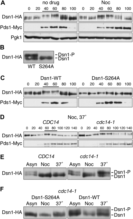
Cdc14 dephosphorylates Dsn1 prior to anaphase. (A) Dsn1–HA shows a cell-cycle dependent mobility shift. Cells containing Dsn1–HA and Pds1–Myc (SBY6079) were released from G1 into complete media in the presence or absence of 15 μg/ml nocodazole at room temperature, lysates were prepared, and Dsn1–HA was monitored for mobility shift by immunoblotting as previously described (Biggins et al. 1999). Decreased mobility shifts are indicated by lines above immunoblots. Pgk1 was used as a loading control, and Pds1–Myc was used to monitor cell cycle progression. Yeast strains used in this study are listed in Table 1. (B and C) Ser264 phosphorylation is largely responsible for the mobility shift of Dsn1–HA. Lysates were prepared from asynchronously growing cells (B) or G1-released cells (C) expressing Pds1–Myc and either Dsn1–WT–HA (SBY5866) or Dsn1–S264A–HA (SBY5770) and immunoblotted with anti-HA and anti-Myc antibodies. (D) Cdc14 dephosphorylates Dsn1 prior to anaphase. The experiment in A was repeated using CDC14 (SBY6079) or cdc14-1 (SBY6085) cells containing Dsn1–HA and Pds1–Myc that were released to 37°. (E) Cdc14 is required to maintain the dephosphorylated status of Dsn1 during a preanaphase arrest. CDC14 (SBY6079) or cdc14-1 (SBY6085) cells were arrested with nocodazole for 3 hr and then shifted to 37° for 30 min. (F) The phosphorylation restored during a nocodazole arrest largely depends on Ser264 phosphorylation. The experiment in E was repeated using cdc14-1 cells containing Dsn1–S264A–HA (SBY9188) or Dsn1–WT–HA (SBY9187).
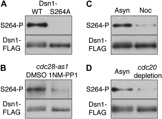
Dsn1–Ser264, a Cdk1 target site, is hypophosphorylated prior to anaphase. (A) A polyclonal phospho-specific antibody was custom-made by Pacific Immunology against phospho-Ser264 Dsn1 using peptide GGSTII(pS)PHKDIPEED. Dsn1–WT–FLAG (SBY7902) and Dsn1–S264A–FLAG (SBY8123) were immunoprecipitated using anti-FLAG antibodies as previously described (Akiyoshi et al. 2009), immunoblotted using the phospho-specific antibody at a 1:5000 dilution, and then reprobed with monoclonal FLAG antibodies. (B) Ser264 phosphorylation depends on Cdk1/Cdc28. Dsn1–FLAG proteins immunoprecipitated from cdc28–as1 mutant cells (SBY8042) (Bishop et al. 2000), treated with DMSO or 10 μm 1NM-PP1 for 30 min, were analyzed as in A. (C) Dsn1–FLAG proteins immunoprecipitated from asynchronous or preanaphase cells that were treated with 15 μg/ml nocodazole for 3 hr were analyzed as in A. (D) pGAL–CDC20 cells expressing Dsn1–FLAG (SBY8008) were asynchronously grown in galactose media and arrested in metaphase by adding glucose for 3 hr to deplete Cdc20. Dsn1–FLAG was immunoprecipitated and analyzed as in A.
References
-
- Bishop, A. C., J. A. Ubersax, D. T. Petsch, D. P. Matheos, N. S. Gray et al., 2000. A chemical switch for inhibitor-sensitive alleles of any protein kinase. Nature 407 395–401. - PubMed
Publication types
MeSH terms
Substances
Grants and funding
LinkOut - more resources
Full Text Sources
Molecular Biology Databases
Miscellaneous
