. 2010 Oct 15;70(20):8179-86.
doi: 10.1158/0008-5472.CAN-10-1871. Epub 2010 Oct 5.

Impact of stromal sensitivity on radiation response of tumors implanted in SCID hosts revisited

Affiliations

Impact of stromal sensitivity on radiation response of tumors implanted in SCID hosts revisited

Mónica García-Barros et al. Cancer Res. .

Abstract

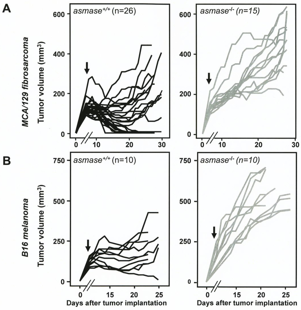
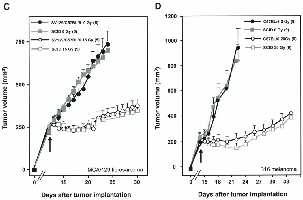
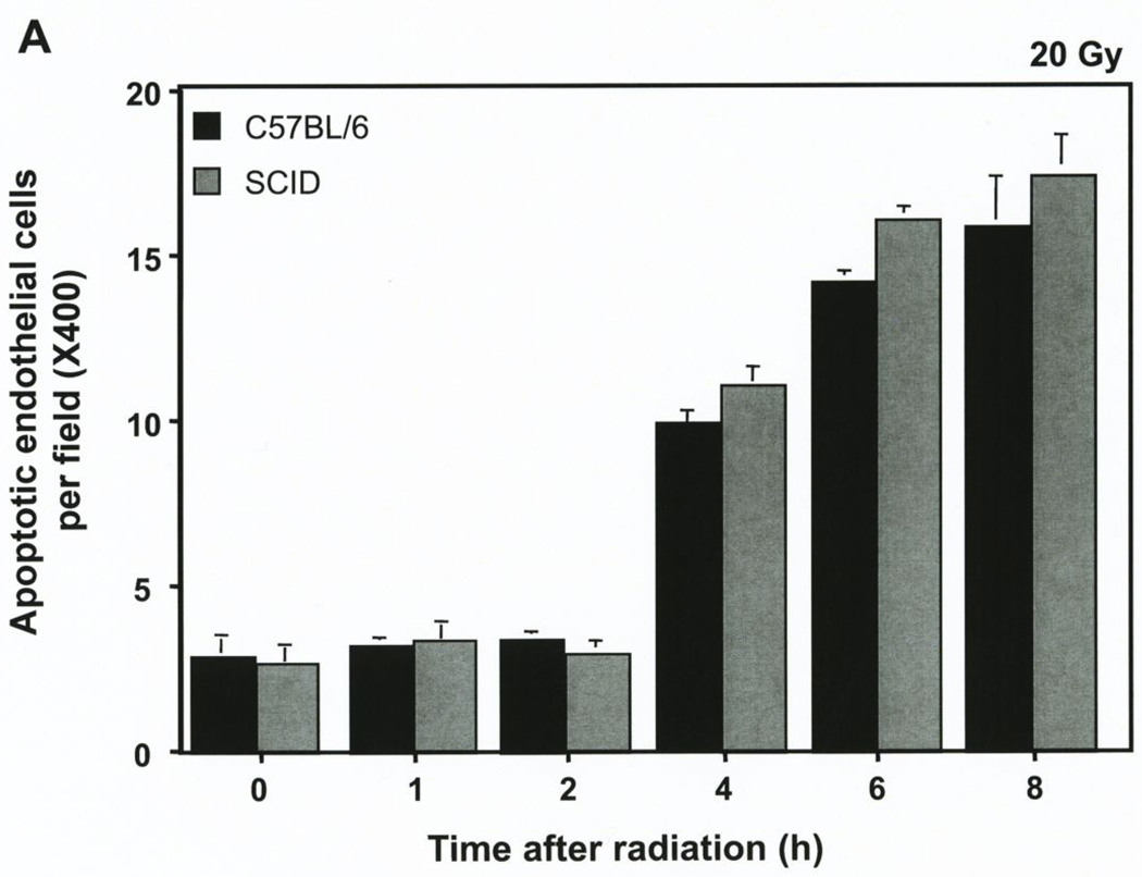
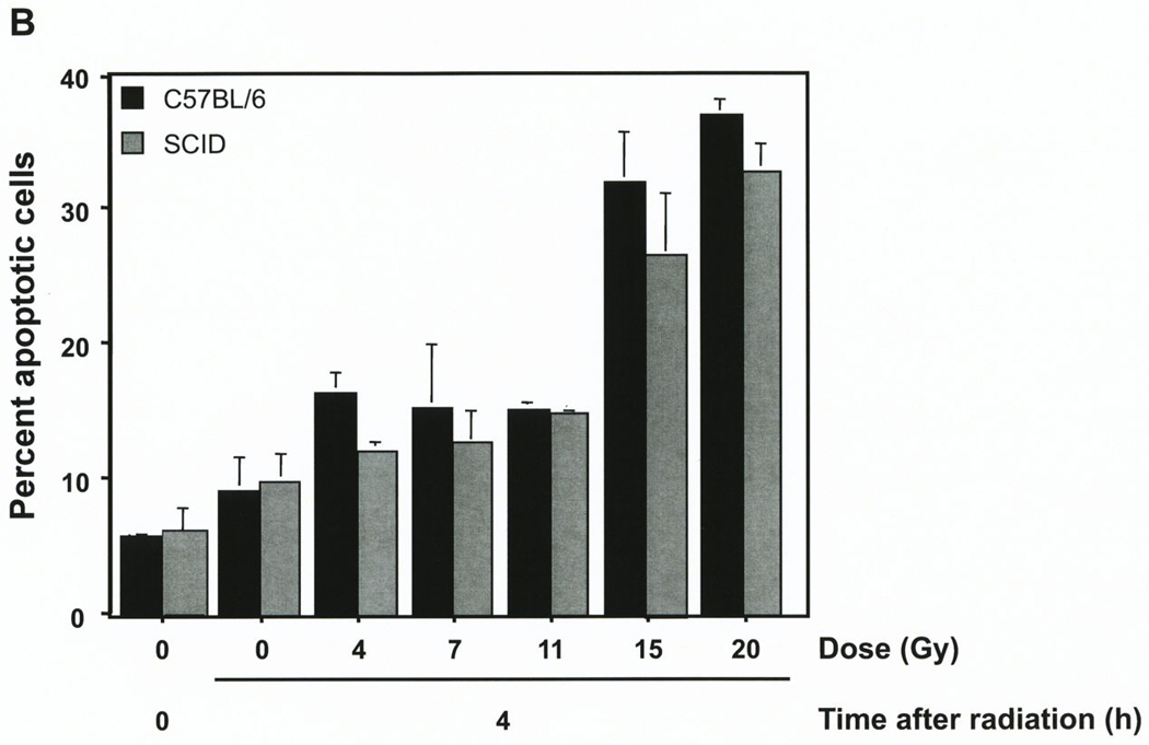
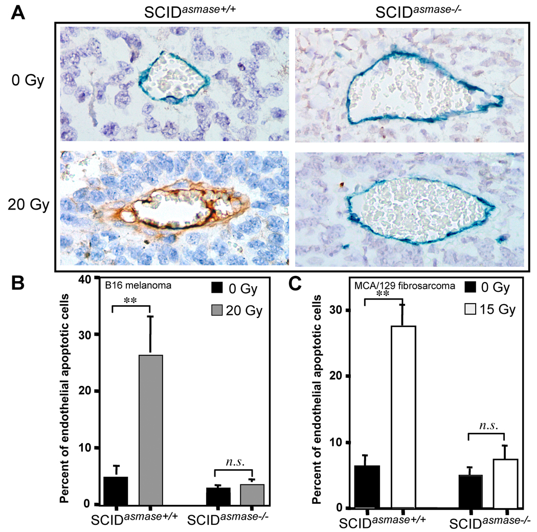
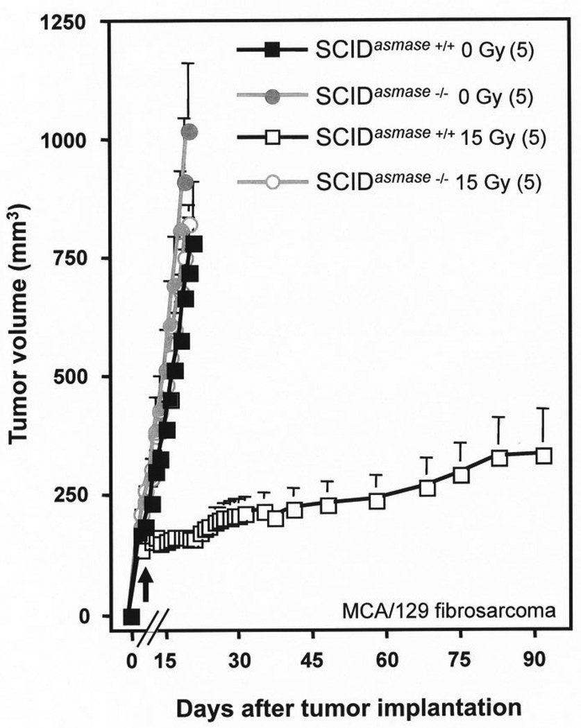
Severe combined immunodeficient (SCID) mice carry a germ-line mutation in DNA-PK, associated with deficiency in recognition and repair DNA double-strand breaks. Thus, SCID cells and tissues display increased sensitivity to radiation-induced postmitotic (clonogenic) cell death. Nonetheless, the single-radiation doses required for 50% permanent local control (TCD(50)) of tumors implanted in SCID mice are not significantly different from the TCD(50) values of the same tumors in wild-type hosts. Whereas the tumor stroma is derived from the host, the observation that tumors implanted in SCID mice do not exhibit hypersensitivity to radiation might imply that stromal endothelial elements do not contribute substantially to tumor cure by ionizing radiation. Here, we challenge this notion, testing the hypothesis that ASMase-mediated endothelial apoptosis, which results from plasma membrane alterations, not DNA damage, is a crucial element in the cure of tumors in SCID mice by single-dose radiotherapy (SDRT). We show that the endothelium in MCA/129 fibrosarcomas and B16 melanomas exhibits a wild-type apoptotic phenotype in SCID hosts, abrogated in tumors in SCID(asmase-/-) littermates, which also acquire resistance to SDRT. Conversion into a radioresistant tumor phenotype when implanted in SCID(asmase-/-) hosts provides compelling evidence that cell membrane ASMase-mediated microvascular dysfunction, rather than DNA damage-mediated endothelial clonogenic lethality, plays a mandatory role in the complex pathophysiologic mechanism of tumor cure by SDRT, and provides an explanation for the wild-type SDRT responses reported in tumors implanted in SCID mice.


Conflict of interest statement

The authors have declared that no conflicts of interest exist.

Figures

Figure 1
Figure 1. Tumor radiosensitivity depends on host asmase genotype
A, B. Effect of radiation on MCA/129 fibrosarcomas (A) or B16 melanomas (B) implanted in the right hind limb of asmase+/+ and asmase−/− hosts and irradiated at the post-implantation day indicated by the arrows. Lines indicate change in tumor volume over time for individual tumors measured daily. n indicates number of tumors treated per group. C, D. Effect of radiation on MCA/129 fibrosarcoma (C) or B16 melanoma (D) implanted in the right hind limb of wild-type strains (as indicated) or SCID hosts and irradiated at the post-implantation day indicated by the arrows. Value at each data point is the mean±SEM. Number of mice in each group is in parentheses.
Figure 2
Figure 2. The SCID mutation does not impact radiation-induced apoptosis of B16 melanoma endothelium
A. B16 melanomas were removed before and at the indicated times after exposure to 20 Gy, and 5 µm histologic sections were immunostained for apoptosis by TUNEL and for the endothelial cell surface marker MECA-32. Blinded scoring of apoptotic endothelial (TUNEL-MECA-32 double stained) cells was carried out manually. Data (means±SD) were compiled from 20 random hpfs (400X magnification) for each time point and collated from three independent experiments. B. A homogenous population of endothelial cells (~95% purity) was isolated by negative and positive selection from B16 melanoma tumors growing in either SCID or C57BL/6 mice. Immediately after isolation these cells were treated in single cell suspension with escalating radiation doses (0–20 Gy). A minimum of 500 cells, stained with bis-benzimide, was scored for apoptosis 4 hours post irradiation. Data (mean±SEM) were collated from three independent experiments.
Figure 3
Figure 3. Host asmase−/− but not the SCID mutation impacts radiation-induced apoptosis in implanted tumor endothelium
A. Representative 5 µm histologic B16 melanoma sections of tumor obtained at 6 hours after exposure to 20 Gy and doubly-immunostained for TUNEL and MECA-32. Apoptotic endothelium manifest a red-brown TUNEL-positive nuclear signal surrounded by dark blue plasma membrane signal of MECA-32. B, C. Incidence of apoptotic endothelial nuclei in tissue sections of control and 20 Gy-irradiated B16 melanoma (B) or 15 Gy-irradiated MCA/129 fibrosarcoma (C). Data (means±SD) were compiled from 20 random hpfs (400X magnification) and collated from three independent experiments.
Figure 4
Figure 4. Effect of the asmase−/− mutation on the radiation response of MCA/129 fibrosarcomas implanted in SCID mice
Tumors were implanted in the right hind limb of SCIDasmase+/+ or SCIDasmase−/− hosts and irradiated (15 Gy) at the post-implantation day indicated by the arrow. Value at each data point is the mean±SEM. Number of mice in each group is in parentheses.

References

    1. Brown M, Bristow R, Glazer P, et al. Comment on "Tumor response to radiotherapy regulated by endothelial cell apoptosis" (II) Science. 2003;302:1894. author reply. - PubMed
    1. Budach W, Taghian A, Freeman J, Gioioso D, Suit HD. Impact of stromal sensitivity on radiation response of tumors. J Natl Cancer Inst. 1993;85:988–993. - PubMed
    1. Fuks Z, Kolesnick R. Engaging the vascular component of the tumor response. Cancer Cell. 2005;8:89–91. - PubMed
    1. Garcia-Barros M, Paris F, Cordon-Cardo C, et al. Tumor response to radiotherapy regulated by endothelial cell apoptosis. Science. 2003;300:1155–1159. - PubMed
    1. Gerweck LE, Vijayappa S, Kurimasa A, Ogawa K, Chen DJ. Tumor cell radiosensitivity is a major determinant of tumor response to radiation. Cancer Res. 2006;66:8352–8355. - PubMed

Publication types