. 2010 Oct;30(9):1441-54.
doi: 10.1097/IAE.0b013e3181ee5ce8.

Drusen characterization with multimodal imaging

Affiliations

Drusen characterization with multimodal imaging

Richard F Spaide et al. Retina. 2010 Oct.

Abstract

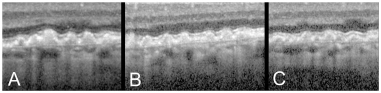
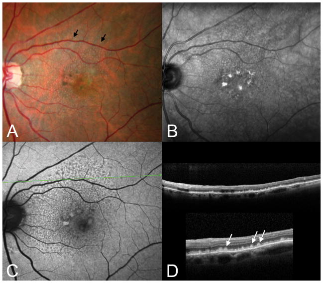
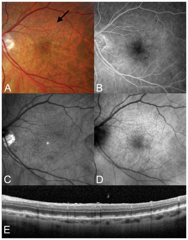
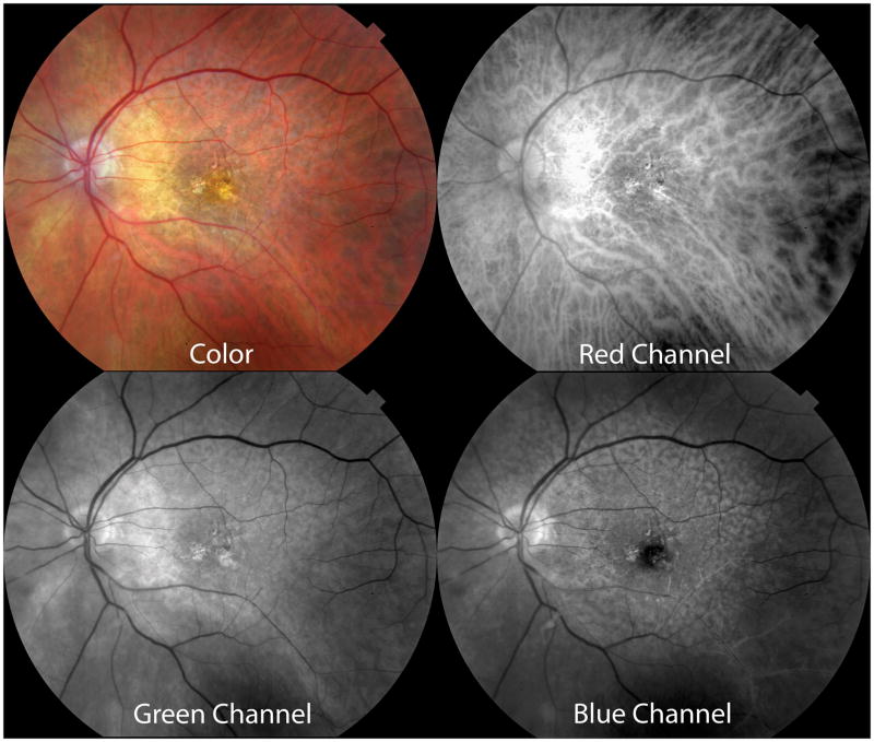
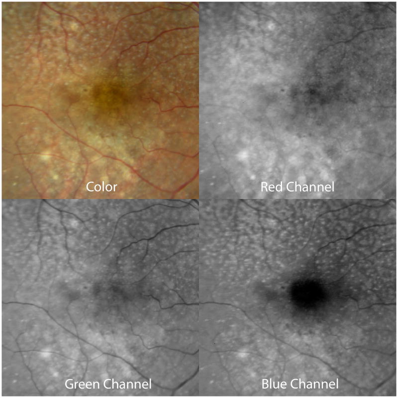
Purpose: To characterize the known appearance of cuticular drusen, subretinal drusenoid deposits (reticular pseudodrusen), and soft drusen as revealed by multimodal fundus imaging and to create an explanatory model that accounts for these observations.

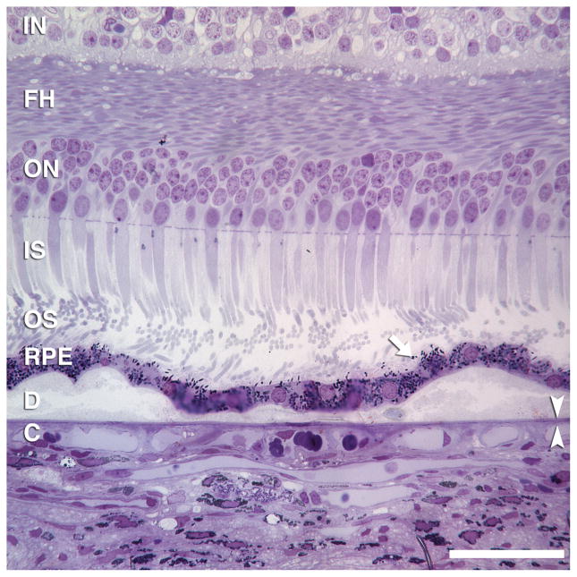
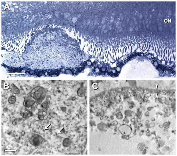
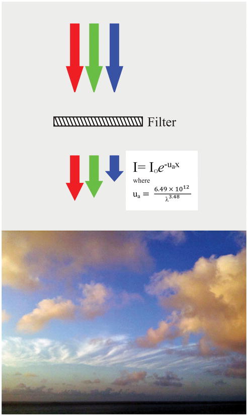
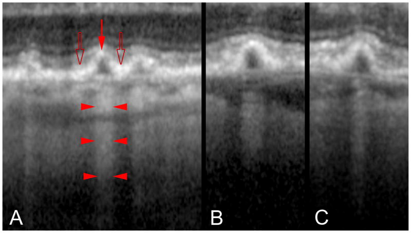
Methods: Reported color, fluorescein angiographic, autofluorescence, and spectral domain optical coherence tomography images of patients with cuticular drusen, soft drusen, and subretinal drusenoid deposits were reviewed, as were actual images from affected eyes. Representative histological sections were examined. The geometry, location, and imaging characteristics of these lesions were evaluated. A hypothesis based on the Beer-Lambert law of light absorption was generated to fit these observations.

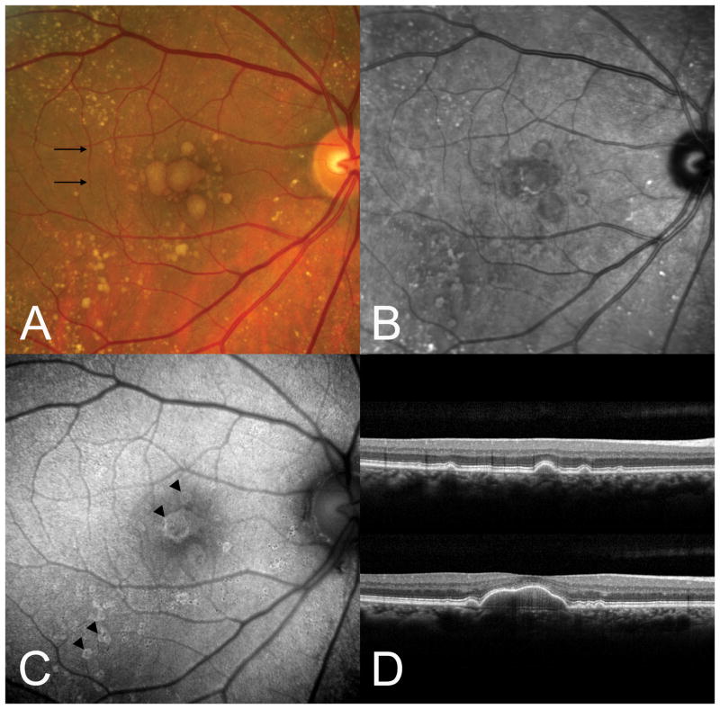
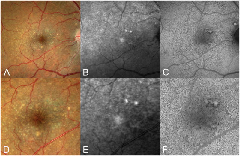
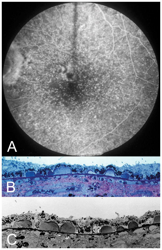
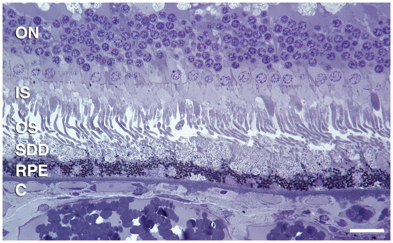
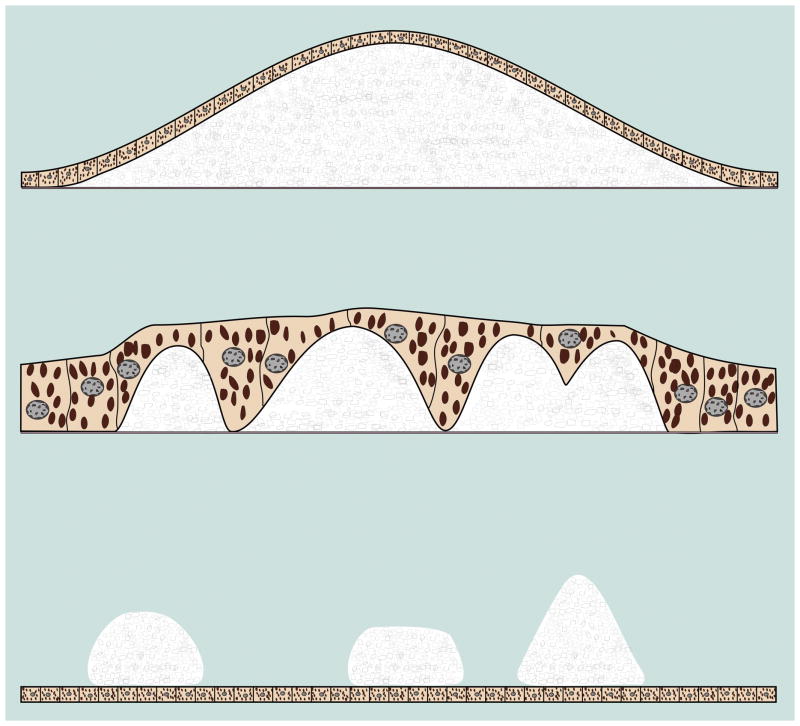
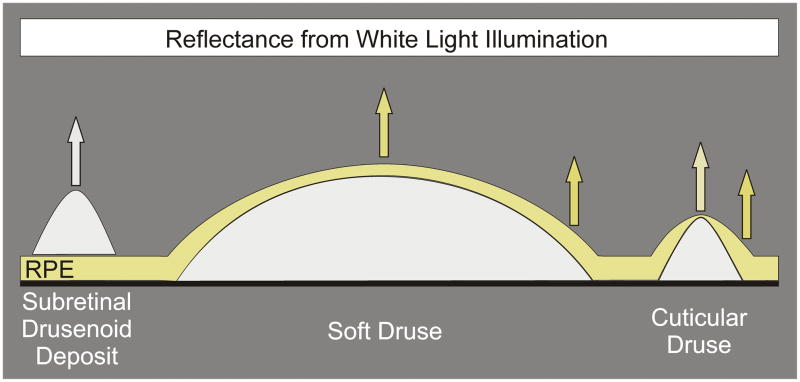
Results: Cuticular drusen appear as numerous, uniform, round, yellow-white punctate accumulations under the retinal pigment epithelium (RPE). Soft drusen are larger, yellow-white dome-shaped mounds of deposit under the RPE. Subretinal drusenoid deposits are polymorphous light-gray interconnected accumulations above the RPE. Based on the model, both cuticular and soft drusen appear yellow because of the removal of shorter wavelength light by a double pass through the RPE. Subretinal drusenoid deposits, which are located on the RPE, are not subjected to short-wavelength attenuation and therefore are more prominent when viewed with blue light. The location and morphology of extracellular material in relationship to the RPE, and associated changes to RPE morphology and pigmentation, appeared to be the primary determinants of druse appearance in different imaging modalities.

Conclusion: Although cuticular drusen, subretinal drusenoid deposits, and soft drusen are composed of common components, they are distinguishable by multimodal imaging because of differences in location, morphology, and optical filtering effects by drusenoid material and the RPE.


Conflict of interest statement

The authors have no financial interests.

Figures

Figure 1
Figure 1
Large soft drusen. A. Color photograph. The black arrows indicate the scan lines for the optical coherence tomographic (OCT) sections. B. The near-infrared reflectance scanning laser ophthalmoscopic (SLO) picture shows decreased brightness in the region of the soft drusen. C. There is a subtle increase in autofluorescence at the outer edges of several drusen. D. Representative OCT scans showing the deposition of material under the retinal pigment epithelium (RPE). Note that drusen color in A is not related to the thickness of sub-RPE material seen in D.
Figure 2
Figure 2
Two small soft drusen connected by basal linear deposit. Drusen contain membranous debris (lipoprotein-derived debris) and neutral lipid pools. RPE morphology and pigmentation is minimally affected. Basal linear deposit has also been called diffuse drusen. 1-μm thick, toluidine-blue stained section of eye post-fixed in osmium. Scale bar, 50 μm. Retinal layers: IN, inner nuclear; FH, Fibers of Henle in the outer plexiform layer; ON, outer nuclear; IS, inner segments; OS, outer segments; RPE, retinal pigment epithelium; C, choriocapillaris. Arrow indicates individual melanin granules in RPE apical processes. Arrowheads bracket Bruch’s membrane.
Figure 3
Figure 3
Cuticular drusen. A. Color photograph; B, near-infrared SLO image, C, autofluorescence image. D-F show 200% magnifications of the central portion of A–C, respectively. Note that the numerous small drusen in the color photograph are much more difficult to see in the near infrared images. The autofluorescence image shows innumerable small hypo-autofluorescent dots. These may represent the numerical summation of both thinning of the overlying RPE and focal areas of RPE degeneration.
Figure 4
Figure 4
Cuticular drusen, clinical and histologic images. A. Fluorescein angiography shows a starry-sky pattern of many punctate dots of hyperfluorescence. Light microscopic (B) and electron microscopic (C) of this patient at post-mortem shows numerous small ovoid accumulations protruding into the RPE monolayer. Note the thinning of the RPE over the apex, and the thickening of RPE at the base, of each druse. (Courtesy of John Sarks, MD.)
Figure 5
Figure 5
OCT findings of cuticular drusen. A, B, C. OCT images from 3 different patients show closely packed blunted triangles, the bases of which sit on Bruch’s membrane with apices towards the retina.
Figure 6
Figure 6
Subretinal drusenoid deposits, conical. A. The color photograph shows numerous pinpoint drusen-like structures (arrows) that superficially resemble cuticular drusen. B. The near-infrared SLO image shows dark spots corresponding to the small drusenoid deposits seen in A. C. The autofluorescence image shows hypo-autofluorescent spots corresponding to the drusenoid deposits, which are seen in cross-section in the OCT (D, top). The inset in lower D shows a 200% enlargement. Note the numerous conical deposits above the RPE.
Figure 7
Figure 7
Subretinal drusenoid deposits, flat. A. Subtle subretinal drusenoid deposits are visible as grayish spots in the superior macula. B. There are no corresponding findings in the fluorescein angiogram. C. The SD-OCT section shows relatively flat aggregates between the boundary between the inner and outer segments and the underlying RPE.
Figure 8
Figure 8
Disposition and ultrastructure of subretinal drusenoid debris and soft drusen. New sections from Case 1 were post-fixed by the osmium-tannic acid-paraphenylenediamine method for neutral lipid and sectioned at 1 μm for staining with toluidine blue (A) or sectioned for transmission electron microscopy (B, C). A. Adjacent sub-retinal drusenoid deposit (at left) and soft druse (at right) that is attached to the RPE. The subretinal drusenoid deposit is well-formed, with overlying photoreceptors that are deflected or shortened. It includes an apical cap of material, distinct from outer segments in both staining density and size, and possibly corresponding to an acetone-resistant cap previously described. The soft druse has partial contents that bind little stain. Both RPE underlying the deposit and overlying the druse is minimally disturbed. Bar, 25 μm. ON, outer nuclear layer. B, C. The interior of the subretinal drusenoid deposit has complex membranous whorls with neutral lipid interiors dispersed throughout a ground substance with globular proteins (arrows). In contrast, the soft druse has membranous debris of simpler shapes, mostly spheres, and heterogeneous sizes. Much of the druse contents are missing, consistent with the known physical fragility of these lesions [40]. Globular proteins present in B are absent in C. Other ultrastructural descriptions using osmium post-fixation indicate that subretinal drusenoid debris and soft drusen both contain abundant membranous debris [26]. Bar, 1 μm. Bracketing arrowheads indicate RPE basal lamina. (Prepared by Jeffrey D. Messinger, DC).
Figure 9
Figure 9
Flattened subretinal drusenoid debris. Histological section was prepared as described in Figure 8A. Layers are labeled as in Figure 2. Bar, 20 μm.
Figure 10
Figure 10
Schematic of lesion sizes, shapes, and relations with RPE. Soft drusen are formed by mounds of deposit under the RPE. Soft drusen generally range in size from 63 to greater than 1000 microns in diameter. There may be some attenuation of the RPE over the apex of the druse. Cuticular drusen are 50 to 75 microns in diameter and jut up through the thickness of the overlying RPE. Subretinal drusenoid deposits show a range of sizes larger than that of soft drusen; they can be confluent and consequently be quite large but also can have refractile elements that emulate the appearance of cuticular drusen.
Figure 11
Figure 11
Spectral filtering of light by RPE and atmosphere. Left, The RPE attenuates light, particularly for shorter wavelengths of light as shown by the varying nature of the extinction coefficient ua. If white light, illustrated as red, green, and blue arrows of equivalent lengths to represent equivalent intensities, pass through a filter with the optical qualities of the RPE, the blue light is preferentially absorbed. The resultant spectral profile is similar to that of sunlight. Right, When the sun is low in the sky, shorter wavelengths are increasingly filtered from sunlight, due to the greater path length through blue-scattering atmosphere. Clouds are therefore different colors due to the amount of blue-filtering atmosphere that sunlight passes through, i.e., less for high white cirrus clouds, more for low yellow cumulus clouds. By analogy, the material in soft drusen appears yellow by ophthalmoscopy not because druse contents are yellow, but because of optical filtering by the overlying RPE.
Figure 12
Figure 12
The RPE absorbs and attenuates blue light. Subretinal drusen deposits are on top of the RPE and therefore do not have any blue light attenuation. Soft and cuticular drusen, in contrast, are under the RPE, which attenuates blue light in their reflective spectral profiles. Local thickness changes in the overlying RPE are more acute for cuticular drusen than for soft drusen. RPE thickness at the apex of a cuticular druse is less than at the druse edge. This positions less pigment to block the excitation light used in fluorescein angiography and may account for the pinpoint hyperfluorescence seen with these drusen.
Figure 13
Figure 13
Contrast between subretinal drusenoid deposits and the background. Color photograph showing subretinal drusenoid deposits and the composite color channels in the color photograph. The red channel offers little contrast between the subretinal drusenoid deposits and the underlying choroid, in part because of the high penetration of red light through the RPE and the large proportion of red light reflected by the choroidal blood vessels. The contrast between the subretinal drusenoid deposits is increased somewhat in the green channel and is most evident in the blue channel. The RPE preferentially absorbs blue light and thus offers a darker background. Note the dark central macula in the blue channel because of light absorption by the macular pigment.
Figure 14
Figure 14
Subretinal drusenoid deposits and soft drusen are distinct in different color channels. In the red channel of the color photograph, soft drusen (in superior macula) are brighter and partially reduce the visibility of the underlying choroidal vessels. In the green and blue channels, the subretinal drusenoid deposits (in inferior macula) are brighter. In the blue channel, the soft drusen are much less evident.
Figure 15
Figure 15
Penetration of light through cuticular drusen revealed by OCT. A. Each druse shows thinning of the overlying RPE at its apex (red arrow) and RPE thickening at its base (open arrows). Penetration of light into the underlying choroid is blocked at sites adjacent to the drusen (bodies of the arrowheads) while transmission of light is increased in the center of each druse (at the points of the arrowhead). B, C. The transmission of light into the deeper layers thus varies not according to the thickness of the druse material, but by the thickness of the RPE.

References

    1. Bressler SB, Bressler NM, Sarks SH, Sarks JP. Age-related macular degeneration: nonneovascular early AMD, intermediate AMD, and geographic atrophy. In: Ryan SJ, editor. Retina. Mosby; 2006. pp. 1041–1074.
    1. Klein R, Klein BEK, Linton KLP. Prevalence of age-related maculopathy. Ophthalmol. 1992;99:933–943. - PubMed
    1. van der Schaft TL, Mooy CM, de Bruijn WC, et al. Histologic features of the early stages of age-related macular degeneration. Ophthalmol. 1992;99:278–286. - PubMed
    1. Mullins RF, Hageman GS. Human ocular drusen possess novel core domains with a distinct carbohydrate composition. J Histochem Cytochem. 1999;47:1533–1539. - PubMed
    1. Mullins RF, Russell SR, Anderson DH, Hageman GS. Drusen associated with aging and age-related macular degeneration contain proteins common to extracellular deposits associated with atherosclerosis, elastosis, amyloidosis, and dense deposit disease. FASEB J. 2000;14:835–846. - PubMed

Publication types