. 2010 Nov 17;29(22):3773-86.
doi: 10.1038/emboj.2010.245. Epub 2010 Oct 5.

Redox regulation of the stability of the SUMO protease SENP3 via interactions with CHIP and Hsp90

Affiliations

Redox regulation of the stability of the SUMO protease SENP3 via interactions with CHIP and Hsp90

Shan Yan et al. EMBO J. .

Abstract

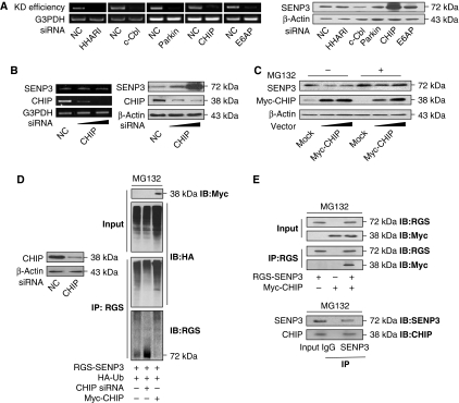
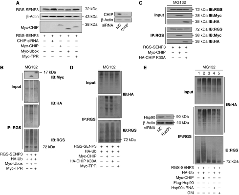
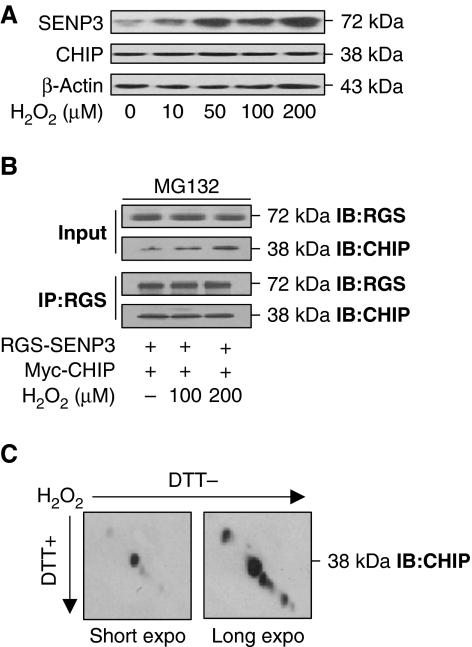
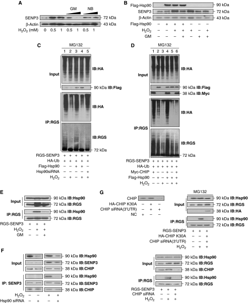
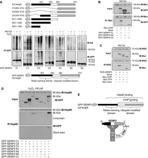
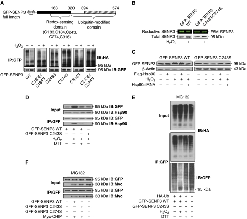
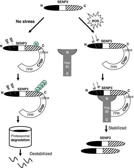
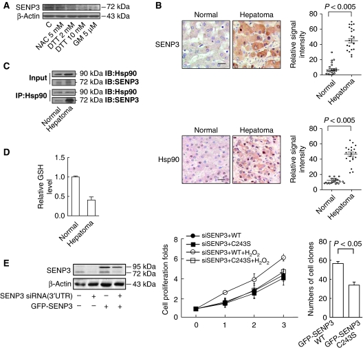
The molecular chaperone heat shock protein 90 (Hsp90) and the co-chaperone/ubiquitin ligase carboxyl terminus of Hsc70-interacting protein (CHIP) control the turnover of client proteins. How this system decides to stabilize or degrade the client proteins under particular physiological or pathological conditions is unclear. We report here a novel client protein, the SUMO2/3 protease SENP3, that is sophisticatedly regulated by CHIP and Hsp90. SENP3 is maintained at a low basal level under non-stress condition due to Hsp90-independent CHIP-mediated ubiquitination. Upon mild oxidative stress, SENP3 undergoes thiol modification, which recruits Hsp90. Hsp90/SENP3 association protects SENP3 from CHIP-mediated ubiquitination and subsequent degradation, but this effect of Hsp90 requires the presence of CHIP. Our data demonstrate for the first time that CHIP and Hsp90 interplay with a client alternately under non-stress and stress conditions, and the choice between stabilization and degradation is made by the redox state of the client. In addition, enhanced SENP3/Hsp90 association is found in cancer. These findings provide new mechanistic insight into how cells regulate the SUMO protease in response to oxidative stress.


Conflict of interest statement

The authors declare that they have no conflict of interest.

Figures

Figure 1
Figure 1
SENP3 stability is regulated by CHIP. (A) HeLa cells were transfected with 50 nM non-specific siRNA (NC) or specific siRNA for five genes, respectively, for 48 h. The efficiency of knockdown (KD efficiency) was determined by RT–PCR (left). SENP3 protein level was evaluated by IB using anti-SENP3 (right). (B) HeLa cells were transfected with 50 nM NC or CHIP siRNA (20 or 50 nM) for 48 h. SENP3 level and the efficiency of siRNA were determined by RT–PCR (left). SENP3 abundance was examined by IB after cells were transfected with 50 nM NC or CHIP siRNA (20 or 50 nM) for 48 h. The expression of CHIP protein was monitored by anti-CHIP (right). (C) SENP3 abundance was examined by IB after HeLa cells were transfected with mock DNA (40 ng) or Myc-CHIP (20 or 40 ng) for 48 h in the absence/presence of MG132 (10 μM) for the last 12 h. The expression of CHIP protein was monitored by anti-Myc. (D) The efficiency of CHIP knockdown was determined by IB after HEK293T cells were transfected with 50 nM NC and CHIP siRNA for 48 h (left). Cells were co-transfected with CHIP siRNA, Myc-CHIP, and HA-ubiquitin (HA-Ub) for 48 h, and MG132 (10 μM) was added for the later 12 h. Co-IP using anti-RGS and IB using the indicated antibodies were performed to determine the ubiquitin conjugation of SENP3 (right). (E) Cells were co-transfected with Myc-CHIP and RGS-SENP3 for 48 h and incubated with MG132 (10 μM) for the later 12 h. The exogenous proteins were co-immunoprecipitated by anti-RGS and immunoblotted using the indicated antibodies (upper). Cells were incubated with MG132 (10 μM) for 12 h. Co-IP using IgG or anti-SENP3 antibodies were performed, and precipitation of endogenous CHIP was determined by IB with anti-CHIP (lower).
Figure 2
Figure 2
SENP3 is degraded by CHIP in an Hsp90-independent manner. (A) HEK293T cells were co-transfected with CHIP siRNA, Myc-CHIP, or its domains (Myc-TPR or Myc-Ubox) and RGS-SENP3 for 48 h. The abundance of SENP3 was examined, and the efficiencies for Myc-CHIPs transfection and endogenous CHIP knockdown were monitored by IB. (B) Cells were co-transfected with TPR and Ubox domains of CHIP (Myc-TPR or Myc-Ubox), RGS-SENP3, and HA-ubiquitin (HA-Ub) for 48 h, and MG132 (10 μM) was added for the later 12 h. Co-IP using anti-RGS and IB using the indicated antibodies were carried out to determine the ubiquitination of SENP3. (C) Cells were co-transfected with Myc-CHIP, HA-CHIP (K30A), and RGS-SENP3 for 48 h and incubated with MG132 (10 μM) for the later 12 h. The exogenous proteins were co-immunoprecipitated by anti-RGS and immunoblotted using the indicated antibodies. (D) Cells were co-transfected with Myc-CHIP, HA-CHIP (K30A), TPR domain of CHIP (Myc-TPR), RGS-SENP3, and HA-Ub for 48 h, and MG132 (10 μM) was added for the later 12 h. Co-IP using anti-RGS and IB using the indicated antibodies were carried out to determine the ubiquitination of SENP3. (E) Cells were co-transfected with 50 nM NC or Hsp90 siRNA for 48 h, and the efficiency of the Hsp90 knockdown was determined by IB (left). Cells were co-transfected with RGS-SENP3 and HA-Ub with or without Myc-CHIP, Flag-Hsp90, or Hsp90siRNA for 48 h and MG132 was added for the later 12 h. Geldanamycin (GM, 5 μM) was added as indicated for the last 1 h. Co-IP using anti-RGS and IB using the indicated antibodies were performed to determine the ubiquitination (right).
Figure 3
Figure 3
The protein level of CHIP, its association with SENP3, and its redox state remain unchanged upon oxidative stress. (A) HeLa cells were treated with the indicated concentrations of H2O2 for 1 h. SENP3 and CHIP protein levels were evaluated, respectively, by IB. (B) Myc-CHIP and RGS-SENP3 were co-transfected into HEK293T cells for 48 h with MG132 (10 μM) incubation for the later 12 h. H2O2 was added at the indicated concentrations for the last 1 h. Exogenous proteins were co-immunoprecipitated using anti-RGS and immunoblotted using the indicated antibodies. (C) HeLa cells were treated with 100 μM H2O2 for 1 h. CHIP protein was examined by redox diagonal electrophoresis and IB using CHIP antibody. The blots with short- and long-time exposure were displayed to show the position of CHIP relative to the diagonal line.
Figure 4
Figure 4
Hsp90 repress SENP3 degradation upon oxidative stress. (A) HeLa cells were pre-treated with geldanamycin (GM, 1 or 5 μM) for 1 h or novobiocin (NB, 0.1 or 0.5 mM) for 12 h. H2O2 was added at the indicated concentrations for the last 1 h. SENP3 protein levels were evaluated by IB. (B) HEK293T cells were transfected with Flag-Hsp90 for 48 h and exposed to 100 μM H2O2 or/and 5 μM GM as indicated for the last 1 h. SENP3 protein levels were evaluated and Hsp90 transfection efficiency was monitored by IB. (C) HEK293T cells were co-transfected with RGS-SENP3 and HA-Ub with or without Flag-Hsp90 or Hsp90siRNA (50 nM) for 48 h, and MG132 was then added for the later 12 h. H2O2 was added as indicated for the last 1 h. Co-IP using anti-RGS and IB using the indicated antibodies were performed to determine the ubiquitination of SENP3. (D) HEK293T cells were co-transfected with RGS-SENP3 and HA-Ub with or without Myc-CHIP or/and Flag-Hsp90 for 48 h, and MG132 was added for the later 12 h. H2O2 was added as indicated for the last 1 h. Co-IP using anti-RGS and IB using the indicated antibodies were performed to determine the ubiquitination of SENP3. (E) HEK293T cells were transfected with RGS-SENP3 for 48 h and exposed to 100 μM H2O2 or/and 5 μM GM for the last 1 h. The proteins were co-immunoprecipitated using anti-RGS, and binding of endogenous Hsp90 with RGS-SENP3 was determined by IB using the indicated antibodies. (F) HEK293T cells were transfected with Hsp90siRNA (50 nM) for 48 h and exposed to 100 μM H2O2 as indicated for the last 1 h. The endogenous proteins were co-immunoprecipitated using anti-SENP3, and binding of endogenous CHIP or Hsp90 with SENP3 was determined, respectively, by IB using the indicated antibodies for re-blotting. (G) HEK293T cells were co-transfected with HA-CHIP K30A and non-specific siRNA or siRNA (3′UTR) that specifically targeted to endogenous CHIP, respectively. CHIP protein level was evaluated by IB using anti-CHIP to determine the efficiency and specificity of the CHIP siRNA (upper panel, left). RGS-SENP3 with or without CHIP siRNA (3′UTR) plus HA-CHIP K30A were co-transfected into HEK293T cells for 48 h, and MG132 was added for the later 12 h. H2O2 (100 μM) was added as indicated for the last 1 h. The proteins were co-immunoprecipitated using anti-RGS and immunoblotted using the indicated antibodies to determine the binding of endogenous Hsp90 with SENP3 in the presence of wild-type or mutant CHIP (upper, right). HEK293T cells were co-transfected with RGS-SENP3 or/and CHIP siRNA for 48 h. H2O2 was added for the last 1 h. The proteins were co-immunoprecipitated using anti-RGS and immunoblotted using the indicated antibodies to determine the binding of endogenous Hsp90 with SENP3 in the presence/absence of endogenous CHIP (bottom).
Figure 5
Figure 5
SENP3 interplays with CHIP and Hsp90 in a sophisticated way. (A) SENP3 truncates were illustrated (top). HEK293T cells were co-transfected with HA-Ub and GFP-SENP3 full length (FL) or truncates for 48 h. Cells were treated with MG132 (10 μM) for the later 12 h and H2O2 (100 μM) for the last 1 h. Co-IP using anti-GFP and IB using anti-HA were performed to determine the ubiquitination of SENP3, and GFP-SENP3 was detected with anti-GFP (middle). The domains of SENP3 were identified (bottom). (B) Myc-CHIP was co-transfected into HEK293T cells with FL or two truncates of GFP-SENP3 (N2 and C2) for 48 h, respectively. Cells were cultured in the presence of MG132 (10 μM) for the later 12 h. The proteins were co-immunoprecipitated using anti-Myc and immunoblotted using the indicated antibodies. (C) RGS-SENP3 was co-transfected into HEK293T cells with FL or two domains of Myc-CHIP (TPR and Ubox) for 48 h, respectively. Cells were cultured in the presence of MG132 (10 μM) for the later 12 h. The proteins were co-immunoprecipitated using anti-RGS and immunoblotted using the indicated antibodies. (D) HEK293T cells were transfected with GFP-SENP3 FL and truncates (N1, N2, N3, C1, C2, or C3), respectively, for 48 h. H2O2 was added for the last 1 h. Co-IP using anti-Hsp90 and IB using the indicated antibodies were performed to determine the binding of endogenous Hsp90 with SENP3 and its truncates. (E) A speculative model depicting the interplay among SENP3, Hsp90, and CHIP upon oxidative stress.
Figure 6
Figure 6
Blockade of SENP3 ubiquitination is triggered by oxidative modification of cysteines on SENP3, which recruits Hsp90. (A) The identified domains of SENP3 and the sites for mutagenesis with cysteines replaced by serines in the redox-sensing domain (upper). HEK293T cells were co-transfected with HA-Ub and GFP-SENP3 wild-type (WT) or C/S mutants for 48 h. Cells were treated with MG132 (10 μM) for the later 12 h and H2O2 (100 μM) for the last 1 h. Co-IP using anti-GFP and IB using the anti-HA were performed to determine the ubiquitination of SENP3, and GFP-SENP3 was detected with anti-GFP (bottom). (B) HEK293T cells were transfected with GFP-SENP3 WT or C243/274S mutant for 48 h. Cells were treated with H2O2 (100 μM) for 30 min before incubation with F5M for another 30 min. IP with anti-GFP was performed. Total GFP-SENP3 protein levels were evaluated by IB with anti-GFP. Reductive GFP-SENP3 with F5M fluorescence in the same bands were visualized and photographed. (C) HEK293T cells were transfected with GFP-SENP3 WT or mutant C243S with or without Flag-Hsp90 or Hsp90 siRNA for 48 h, and 100 μM H2O2 was added for the last 1 h. SENP3 protein levels were evaluated by IB. (D) HEK293T cells were transfected with GFP-SENP3 WT or mutant C243S for 48 h. A measure of 100 μM H2O2 and 5 mM DTT were added for the last 1 h. The proteins were co-immunoprecipitated using anti-GFP and immunoblotted using the indicated antibodies to determine the binding of endogenous Hsp90 with WT or mutant SENP3. (E) HEK293T cells were co-transfected with HA-Ub and GFP-SENP3 WT or mutant C243S for 48 h and MG132 was added for the later 12 h. H2O2 and DTT were added for the last 1 h. The proteins were co-immunoprecipitated using anti-GFP and immunoblotted using the indicated antibodies to determine the ubiquitination of SENP3. (F) HEK293T cells were co-transfected with GFP-SENP3 WT or mutants C243S and C274S, and Myc-CHIP for 48 h. MG132 was added for the later 12 h. The proteins were co-immunoprecipitated using anti-GFP and immunoblotted using the indicated antibodies to determine the binding of Myc-CHIP with SENP3 WT or mutants.
Figure 7
Figure 7
SENP3 interacts with CHIP and Hsp90 in differential modes under non-stress and oxidative stress conditions. A model for the mechanisms underlying stabilization and degradation of SENP3.
Figure 8
Figure 8
Accumulation of SENP3 in cancer cells correlates with enhanced Hsp90–SENP3 association and increased ROS. (A) HepG2 cells were pre-treated with NAC for 4 h, DTT or GM for 1 h. SENP3 protein level was evaluated by IB using anti-SENP3. (B) Immunohistochemistry for SENP3 and Hsp90 were performed in 20 hepatoma and adjacent normal tissues. Scale bar=10 μM (left). The relative signal intensity was analysed (right). (C) IP was performed in 100 mg of tissues evenly derived from two pairs of fresh hepatoma and adjacent normal tissues using anti-Hsp90, and IB was performed with 1/2 loading of hepatoma sample. (D) The GSH level was assessed in the tissues similar to (C). Quantification showed the means±s.d. of the relative GSH levels. (E) HeLa cells were co-transfected with NC or SENP3 3′UTR siRNA and the construct of GFP-SENP3 WT for 72 h. The endogenous SENP3 and the exogenous GFP-SENP3 were determined with anti-SENP3 and anti-GFP (left). HeLa cells were co-transfected with SENP3 3′UTR siRNA and the constructs of GFP-SENP3 WT or C243S. After 48 h, cells were re-seeded and treated with 100 μM H2O2 for 1 h once a day from the second day. Cell proliferation was assessed and indicated by a fold increase. The values are expressed as the means±s.d. of two independent experiments (middle). HepG2 cells were stably transfected with GFP-SENP3 WT or C243S and retroviral vector expressing shRNA for SENP3. Colony formation assay was performed and indicated by the numbers of colonies in soft agar per 1000 seeded cells (n=3 dishes) (right).

References

    1. Broadley SA, Hartl FU (2009) The role of molecular chaperones in human misfolding diseases. FEBS Lett 583: 2647–2653 - PubMed
    1. Cai J, Niu X, Chen Y, Hu Q, Shi G, Wu H, Wang J, Yi J (2008) Emodin-induced generation of reactive oxygen species inhibits RhoA activation to sensitize gastric carcinoma cells to anoikis. Neoplasia 10: 41–51 - PMC - PubMed
    1. Chiosis G, Vilenchik M, Kim J, Solit D (2004) Hsp90: the vulnerable chaperone. Drug Discov Today 9: 881–888 - PubMed
    1. Cowen LE (2009) Hsp90 orchestrates stress response signaling governing fungal drug resistance. PLoS Pathog 5: e1000471. - PMC - PubMed
    1. Cyr DM, Hohfeld J, Patterson C (2002) Protein quality control: U-box-containing E3 ubiquitin ligases join the fold. Trends Biochem Sci 27: 368–375 - PubMed

Publication types

LinkOut - more resources