. 2010 Nov;6(11):837-43.
doi: 10.1038/nchembio.451. Epub 2010 Oct 10.

Coupling of receptor conformation and ligand orientation determine graded activity

Affiliations

Coupling of receptor conformation and ligand orientation determine graded activity

John B Bruning et al. Nat Chem Biol. 2010 Nov.

Abstract

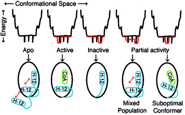
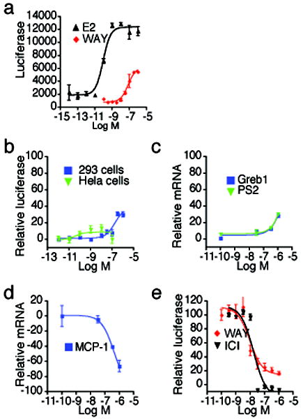
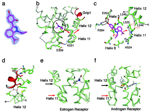
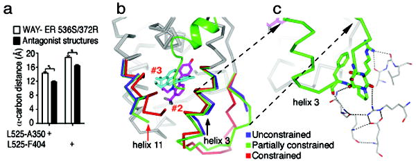
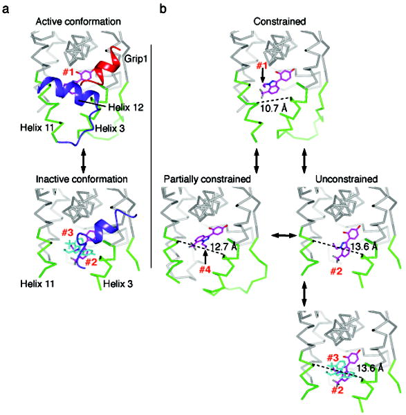
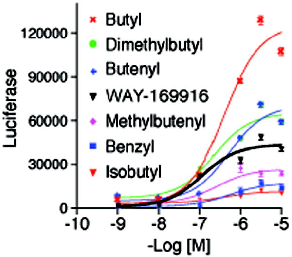
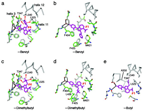
Small molecules stabilize specific protein conformations from a larger ensemble, enabling molecular switches that control diverse cellular functions. We show here that the converse also holds true: the conformational state of the estrogen receptor can direct distinct orientations of the bound ligand. 'Gain-of-allostery' mutations that mimic the effects of ligand in driving protein conformation allowed crystallization of the partial agonist ligand WAY-169916 with both the canonical active and inactive conformations of the estrogen receptor. The intermediate transcriptional activity induced by WAY-169916 is associated with the ligand binding differently to the active and inactive conformations of the receptor. Analyses of a series of chemical derivatives demonstrated that altering the ensemble of ligand binding orientations changes signaling output. The coupling of different ligand binding orientations to distinct active and inactive protein conformations defines a new mechanism for titrating allosteric signaling activity.


Conflict of interest statement

Competing financial interests The authors declare no competing financial interests.

Figures

Figure 1
Figure 1. An Energy Landscape Model of Ligand Binding
The relationship between protein conformations and energy is depicted as an energy well, with folded proteins populating the bottom regions of the well. In the statistical ensemble model of ligand binding, the relative population of different protein conformers is determined by the statistical sum of the thermodynamic energy of each protein-ligand substate. In the absence of ligand, nuclear receptors are thought to be conformationally dynamic, shown as black spikes at the bottom of the energy well. Full agonists or antagonists productively interact with a subset of lower energy conformations, shown in red. For nuclear receptors, agonist or antagonist ligands stabilize specific conformations of helix 12 (H-12). Partial agonists could either stabilize a unique conformer with suboptimal coactivator binding, or induce a mixed population of active and inactive conformers.
Figure 2
Figure 2. WAY-169916 is an ERα partial agonist
(a) Estrogen responsive luciferase assays were performed by transfection of a 3xERE-luc reporter in ER-positive MCF-7 cells. After 24 hr, cells were treated with the indicated doses of ligands overnight, and then assessed for luciferase activity. (b) The indicated cells were transfected as in (a), but with the addition of an ERα expression plasmid. Shown are dose responses to WAY-169916, relative to estradiol treatment. (c) Estrogen responsive gene expression was assayed in MCF-7 cells treated for 2 hr with the indicated doses of WAY-169916. Cells were then processed for qRT-PCR analysis of mRNA, which was normalized to 18S rRNA levels. (d) The NFκB responsive gene, MCP-1 was induced by treating MCF-7 cells for 2 hr with 15 ng/ml of TNFα, and increasing doses of WAY-169916, and then processed for qRT-PCR analysis of mRNA levels. (e) A mammalian two-hybrid assay was used to define the ligand dependent interaction of the Gal4-Grip1 coactivator and VP16-ERα LBD, using a Gal4 response element luciferase reporter. Data represent mean + SEM for (a-e), and are plotted as a percentage of the maximal response seen with estradiol for (b-e). ICI = ICI182,780 (faslodex, Fulvestrant)
Figure 3
Figure 3. The structure of WAY-169916 bound to the active conformation of ERα
(a) A 2Fo-Fc map shows the electron density for the ligand, and is contoured to 2.0 σ. (b) A portion of the structure of WAY-169916 bound to ERα in the active conformation (green) is shown superimposed with the structure of ERα bound to a full agonist (gray) . The ligand is oriented in the Z-axis of the image and is circled. The Grip1 coactivator peptide is colored red. The red arrow indicates the shift in the positioning of helix 11, and the associated shift in the C-terminus of helix 12 in the WAY-169916 structure. (c) Selected residues in the ligand-binding pocket are shown, illustrating how the positioning of WAY-169916 (magenta) induces a shift in helix 11 (green), relative to the full agonist structure (gray). The red dashed lines show clashes between the WAY-169916 induced position of helix 11 and helix 12 in the superimposed full agonist structure. (d) The WAY-169916 (green) and full agonist structure (gray) were superimposed on the coactivator binding site, including helices 3-5 and 12. Shown are the amino acids that directly contact the Grip1 peptide, which is colored red in the WAY-169916 structure. (e) The WAY-169916 structure and 6 published partial agonist structures (green) , were superimposed with 7 full agonist structures (gray) ,, and shown as c-α traces. (f) A series of 17 androgen receptor structures were superimposed, and colored as in (e). Here, partial agonists distort the helix 12 portion of the coactivator binding cleft.
Figure 4
Figure 4. An unconstrained inactive conformation of ERα shows two non-overlapping orientations of WAY-169916
(a) The width of the ligand-binding pocket was measured between the main chain carbon of the indicated amino acids. Shown is the distance for the 4 chains of the WAY-169916 inactive conformation structure compared with 11 published ERα structures bound to different antagonists. (b) The active conformer structure (red) was superimposed with the two chains of the dimer in the inactive conformation structure. Both of these chains (blue and green) show similar large shifts in helix 11 (red arrow), but differential shifts in helix 3 (black arrow). Two molecules of bound WAY-169916 are colored magenta and cyan, defining ligand orientations #2 and #3, respectively. (c) The chain with a partially constrained conformer of helix 3 shows stabilization of the preceding loop by crystal packing. The symmetry related model is show as thin sticks, and the dashed lines represent hydrogen bonds. The partially constrained conformer is shows as c-alpha trace, with helix 3 and the preceding loop colored green, along with selected amino acids that participate in crystal packing.
Figure 5
Figure 5. Protein substates define a pathway of ligand dynamics
(a) A comparison of the active and inactive conformation structures, with helix 12 colored blue and the Grip1 coactivator peptide is colored red. In the active conformation, helix 12 packs against helices 3 and 11, constraining their movement. The ligand is bound in orientation #1, and is colored magenta. As the strained active conformation results in the relocation of helix 12 into the inactive conformation, the pocket widens, allowing binding of multiple ligands in orientations #2 and #3, colored blue. (b) A model of ligand dynamics associated with different protein substates. Helix 12 is not shown to allow visualization of the different ligand orientations associated with distinct protein conformers. The width of the ligand binding pocket supports distinct ligand binding orientations, #1-4.
Figure 6
Figure 6. Activity profiles of WAY-169916 derivatives
Estrogen responsive luciferase assays were performed by transfection of a 3xERE-luc reporter in ERα-positive MCF-7 cells. After 24 hr, cells were treated with the indicated doses of ligands overnight, and then assessed for luciferase activity.
Figure 7
Figure 7. Ligand binding ensembles direct ERα activity profiles
(a) The structure of the benzyl derivative with ERα in the inactive conformation has one molecule in the dimer with an unconstrained conformer, and one molecule with a partially constrained conformer. The unconstrained conformer is shown in green, bound to the benzyl derivative, which is colored magenta. The ligand adopts an orientation identical to orientation #4 of WAY-169916. The superimposed structure of WAY-169916 in the active conformation is colored gray. The red arrows indicate clashes with the superimposed active conformation structure, and widening of the pocket to accommodate this ligand orientation. (b) Same as (a), but with the partially constrained inactive conformer of ERα. The ligand adopts an orientation identical to orientation #1 of WAY-169916. Here, there are no clashes with the superimposed active conformation. (c) The dimethylbutyl substitution was modeled onto the structure of the benzyl derivative in the unconstrained conformer in orientation #4, and shows similar clashes with superimposed active conformation structure. (d) The dimethylbutyl derivative was modeled onto ligand orientation #1, in the partially constrained conformer. As with the benzyl derivative, there are no clashes with the active conformation. However, the dimethylbutyl shows additional favorable VDW contacts with L428 and F425, suggesting that this orientation is more highly populated in solution. (e) The butyl derivative was modeled into ligand orientation #4 on the active conformation structure. The butyl group forms favorable VDW interactions with the indicated amino acids.

References

    1. Onuchic JN, Luthey-Schulten Z, Wolynes PG. Theory of protein folding: the energy landscape perspective. Annu Rev Phys Chem. 1997;48:545–600. doi: 10.1146/annurev.physchem.48.1.545. - DOI - PubMed
    1. Boehr DD, Dyson HJ, Wright PE. An NMR perspective on enzyme dynamics. Chem Rev. 2006;106:3055–3079. doi: 10.1021/cr050312q. - DOI - PubMed
    1. Popovych N, Sun S, Ebright RH, Kalodimos CG. Dynamically driven protein allostery. Nat Struct Mol Biol. 2006;13:831–838. - PMC - PubMed
    1. Eisenmesser EZ, et al. Intrinsic dynamics of an enzyme underlies catalysis. Nature. 2005;438:117–121. doi: 10.1038/nature04105. - DOI - PubMed
    1. Volkman BF, Lipson D, Wemmer DE, Kern D. Two-state allosteric behavior in a single-domain signaling protein. Science. 2001;291:2429–2433. doi: 10.1126/science.291.5512.2429. - DOI - PubMed

Publication types