. 2010 Nov;239(11):2980-8.
doi: 10.1002/dvdy.22431.

Sox17 and chordin are required for formation of Kupffer's vesicle and left-right asymmetry determination in zebrafish

Affiliations

Sox17 and chordin are required for formation of Kupffer's vesicle and left-right asymmetry determination in zebrafish

Emil Aamar et al. Dev Dyn. 2010 Nov.

Abstract

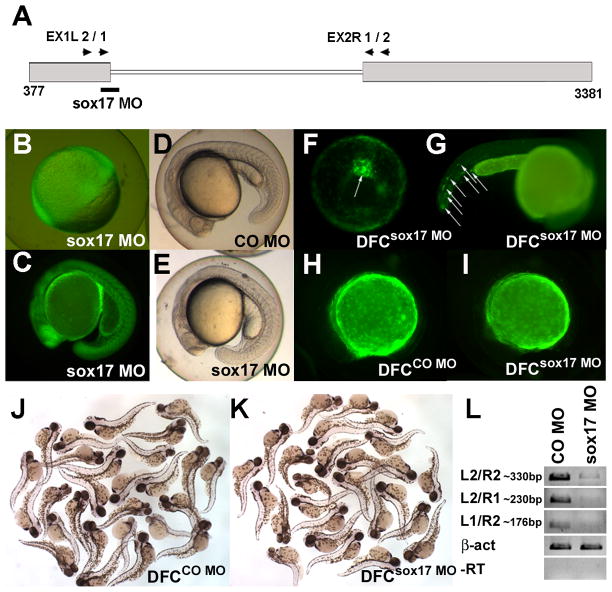
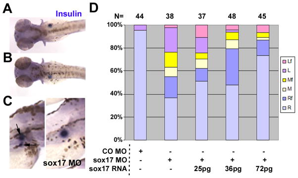
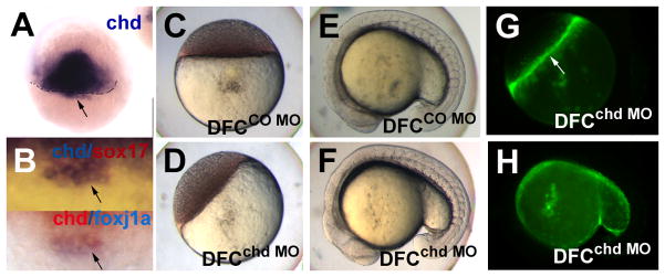
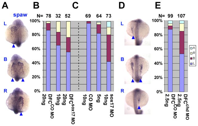
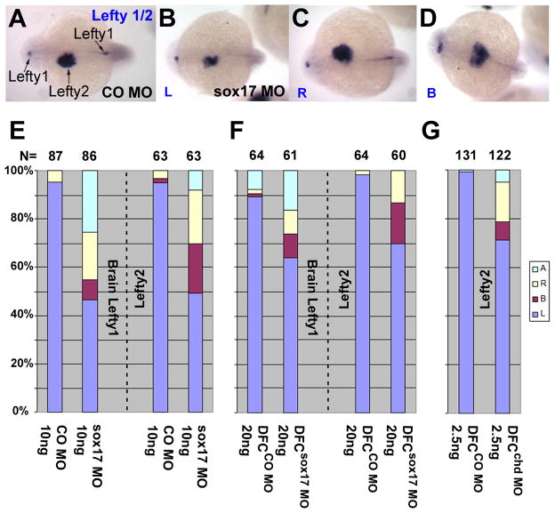
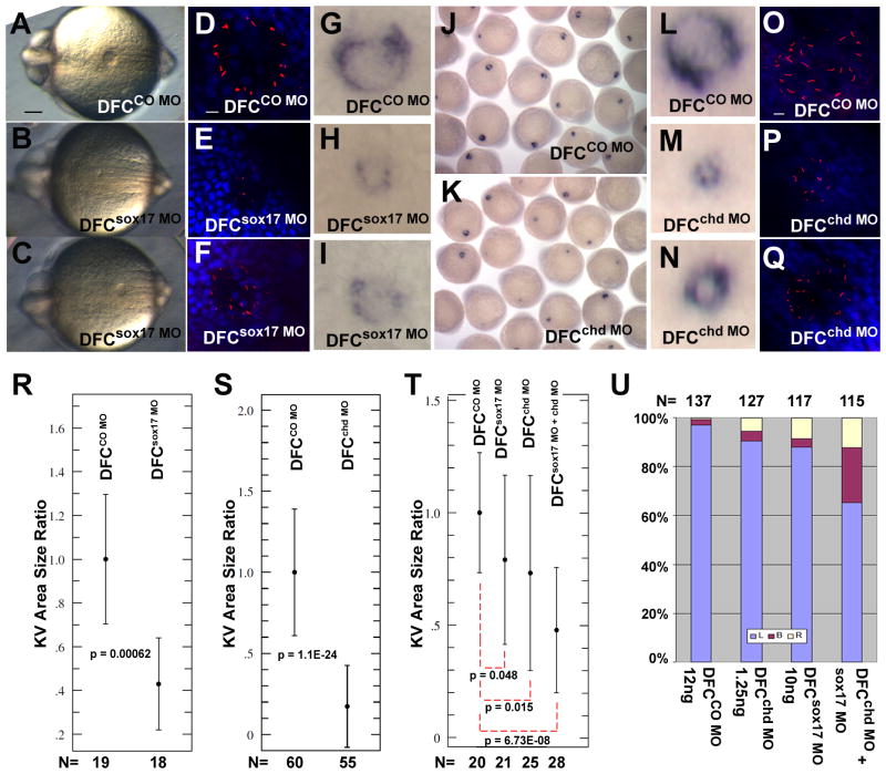
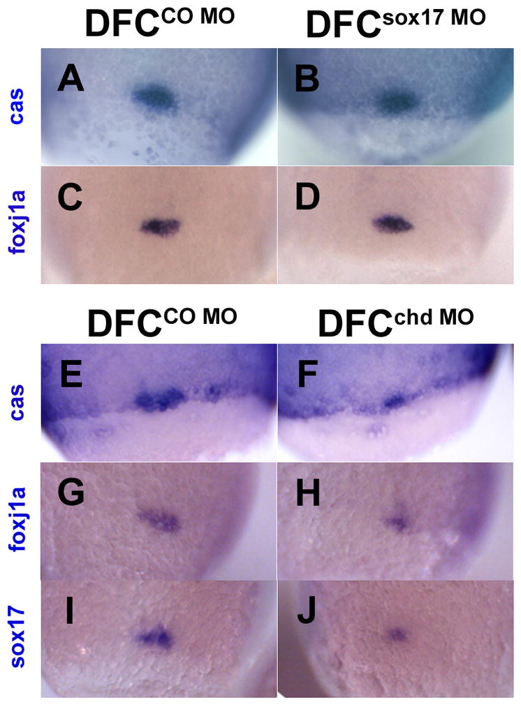
Kupffer's vesicle (KV), a ciliated fluid-filled sphere in the zebrafish embryo with a critical role in laterality determination, is derived from a group of superficial cells in the organizer region of the gastrula named the dorsal forerunner cells (DFC). We have examined the role of the expression of sox17 and chordin (chd) in the DFC in KV formation and laterality determination. Whereas sox17 was known to be expressed in DFC, its function in these cells was not studied before. Further, expression of chd in these cells has not been reported previously. Targeted knockdown of Sox17 and Chd in DFC led to aberrant Left-Right (L-R) asymmetry establishment, as visualized by the expression of southpaw and lefty, and heart and pancreas placement in the embryo. These defects correlated with the formation of small KVs with apparently diminished cilia, consistent with the known requirement for ciliary function in the laterality organ for the establishment of L-R asymmetry.


Figures

Fig. 1
Fig. 1
Global phenotypes arise in sox17 morphants, but not in DFCsox17 MO embryos. (A) Location of the sox17 MO. 3’-Fluorescein tagged sox17 MO was widely distributed in the embryos injected at the one-cell stage (B, shield; C, late somite stage). (D,E) 22hpf old embryos injected at the one-cell stage with 10ng control (CO) MO, and sox17 MO, respectively. (F–I) Injection into the yolk at the midblastula stage excludes the MO from most embryonic cells except the DFC (F, arrow. 80% epiboly,) and a few cells in the tail (G, arrows. 1dpf). (H–K) Embryos injected into the yolk at the midblastula stage with 20ng CO MO (H,J) or sox17 MO (I,K) were similar. Pictures taken under fluorescence illumination at about 8-somite stage (H,I), and in visible light at 2dpf (J,K). (L) The efficiency of sox17 MO, 10ng, injected at the one-cell stage, was assessed at 60–80% epiboly by RT-PCR with primers shown in (A); β-act was used as control. All pictures are lateral view, except vegetal view with dorsal on top in F. C–E and H–I anterior to the left, G anterior to the right.
Fig. 2
Fig. 2
Injection of sox17 MO leads to abnormal pancreas placement, which is rescued by sox17 RNA co-injection. Embryos injected at the one-cell stage with 10ng CO MO, or 10ng sox17 MO alone or combined with sox17 RNA as indicated, were in situ hybridized with insulin at 3dpf. (A–C) Sox17 MO injected embryos showed normal expression on the right side (A), and abnormal opposite expression on the left (B); dorsal views, anterior to left. Some embryos showed fragmented ins-positive domains (C left panel, arrows), with an example of an intact domain in the right panel (lateral views, anterior to right). (D) Localization of ins expression after injection of MOs with or without RNA, as indicated. R, Right; M, Middle; L, Left; f in Rf, Mf or Lf refers to fragmented ins-positive domains. Number of embryos is shown at the top.
Fig. 3
Fig. 3
Chordin expression in the dorsal forerunner cells in zebrafish embryos and targeting of DFC by chd MO. (A,B) Whole mount in situ hybridization for chd at 50% epiboly (A), and double staining of chd (blue) and sox17 (red) (B, upper panel), and chd (red) and foxj1a (blue) (B, lower panel) at 60% epiboly; arrows point to DFC. Embryos were injected at the midblastula stage with 2.5ng CO MO (C,E) or chd MO (D,F), and photographed at the oblong (C,D) and 16–18 somite (E,F) stages. The embryos in (D,F) are also shown as fluorescent images (G,H respectively. Arrow in G, YSL). A,B, dorsal views; E,F,H, lateral views, with anterior to the left.
Fig. 4
Fig. 4
Sox17 and chd are required for left-right asymmetry determination in zebrafish. Embryos were injected into the yolk at one-cell (C) or midblastula (A,B,D,E) stages, with CO MO (B,C,E), sox17 MO (A and B,C), or chd MO (D and E) as indicated in the bar graphs. Whole mount in situ hybridization for spaw (A,D: arrow heads, 15–18 somite; dorsal view with anterior on top) showed aberrant expression when either sox17 MO or chd MO was injected, as summarized in B,C and E. L: Left, B: Bilateral, R: Right, A: absent. Number of embryos is shown at the top.
Fig. 5
Fig. 5
Lefty1,2 expression after suppression of sox17 and of chd in DFC. Embryos were injected into the yolk at one-cell (A–D,E) or midblastula (F,G) stages, with CO MO (A and E–G), sox17 MO (B–D and E–F), or chd MO (G) as indicated. Whole mount in situ hybridization for lefty1 and/or lefty2 at 22hpf (A–D: arrows; dorsal view with anterior to the left) showed aberrant expression when either sox17 MO or chd MO were injected, as summarized in E–G. Expression of lefty1 in the brain and lefty2 in the heart field was scored, as indicated (E, F) in sox17 MO injected embryos, but only lefty2 was scored after chd MO injection (G). L: Left, B: Bilateral, R: Right, A: absent. Number of embryos is shown at the top.
Fig. 6
Fig. 6
Sox17 and chd are required for proper formation of KV. CO MO (A,D,G: 20ng; R: 15ng; J,L,O,S: 2.5ng; T,U: 12ng), sox17 MO (B,C,E,F,H,I: 20ng; R: 15ng; T,U: 10ng) or chd MO (K,M,N,P,Q,S: 2.5ng; T,U: 1.25ng) were injected into the yolk at the midblastula stage, and embryos were examined at 6–8 somites. Sox17 MO and chd MO injected embryos developed small and abnormal KVs as seen in live embryos (A–C; posterior view), by whole mount in situ hybridization with charon (G–N), and by immunostaining for cilia using anti-acetylated tubulin antibody (red) and DAPI (blue) (D–F and O–Q). Scale bars in A, 0.1mm; in D and O, 10μm. (R,S) NIH ImageJ was used to measure the KV area in injected embryos, shown as ratio to the mean control area; error bars are standard deviations; p values are given in the panels. DFCsox17 MO (R) and DFCchd MO (S) embryos showed a significant reduction in KV size compared to controls. (T,U) Embryos injected at the midblastula stage with low levels of individual MO (12ng CO MO, 1.25ng chd MO, 10ng sox17 MO) or a combination of 1.25ng chd MO and 10ng sox17 MO. KV area was measured as above (T), or lefty2 expression was tested at the ~21 somite stage (U).
Fig. 7
Fig. 7
DFC formation in embryos after targeted injection of sox17 MO or chd MO. In situ staining of cas (A,B,E,F) foxj1a (C,D,G,H) and sox17 (I,J) at the 80–90% epiboly stage after midblastula injection of 20ng sox17 MO (B,D), 20ng CO MO (A,C), 2.5ng chd MO (F,H,J) or 2.5ng CO MO (E,G,I). B and D show no reduction in DFC size whereas F, H and J show size reduction; sox17 expression also appears reduced (J). All views are dorsal.

References

    1. Aamar E, Dawid IB. Isolation and expression analysis of foxj1 and foxj1.2 in zebrafish embryos. Int J Dev Biol. 2008;52:985–991. - PMC - PubMed
    1. Alexander J, Rothenberg M, Henry GL, Stainier DY. casanova plays an early and essential role in endoderm formation in zebrafish. Dev Biol. 1999;215:343–357. - PubMed
    1. Amack JD, Wang X, Yost HJ. Two T-box genes play independent and cooperative roles to regulate morphogenesis of ciliated Kupffer's vesicle in zebrafish. Dev Biol. 2007;310:196–210. - PubMed
    1. Amack JD, Yost HJ. The T box transcription factor no tail in ciliated cells controls zebrafish left-right asymmetry. Curr Biol. 2004;14:685–690. - PubMed
    1. Bachiller D, Klingensmith J, Kemp C, Belo JA, Anderson RM, May SR, McMahon JA, McMahon AP, Harland RM, Rossant J, De Robertis EM. The organizer factors Chordin and Noggin are required for mouse forebrain development. Nature. 2000;403:658–661. - PubMed

Publication types

MeSH terms

LinkOut - more resources