. 2010 Nov 17;21(11):2065-75.
doi: 10.1021/bc100288c. Epub 2010 Oct 6.

Bishydrazide glycoconjugates for lectin recognition and capture of bacterial pathogens

Affiliations

Bishydrazide glycoconjugates for lectin recognition and capture of bacterial pathogens

Avijit Kumar Adak et al. Bioconjug Chem. .

Abstract

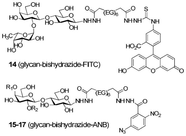
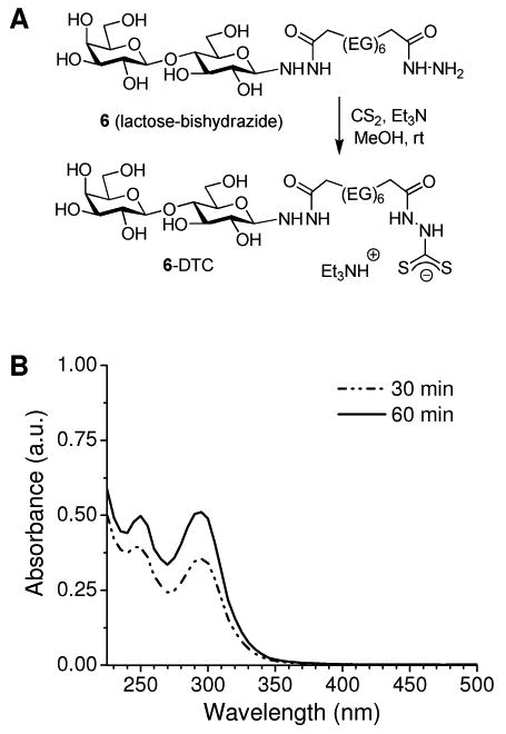
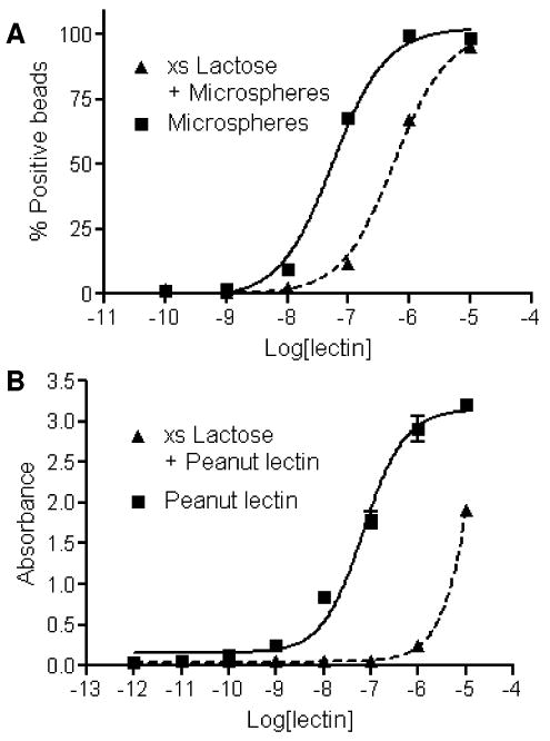
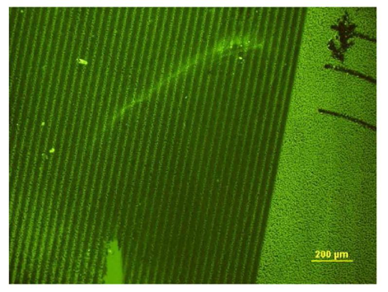
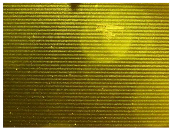
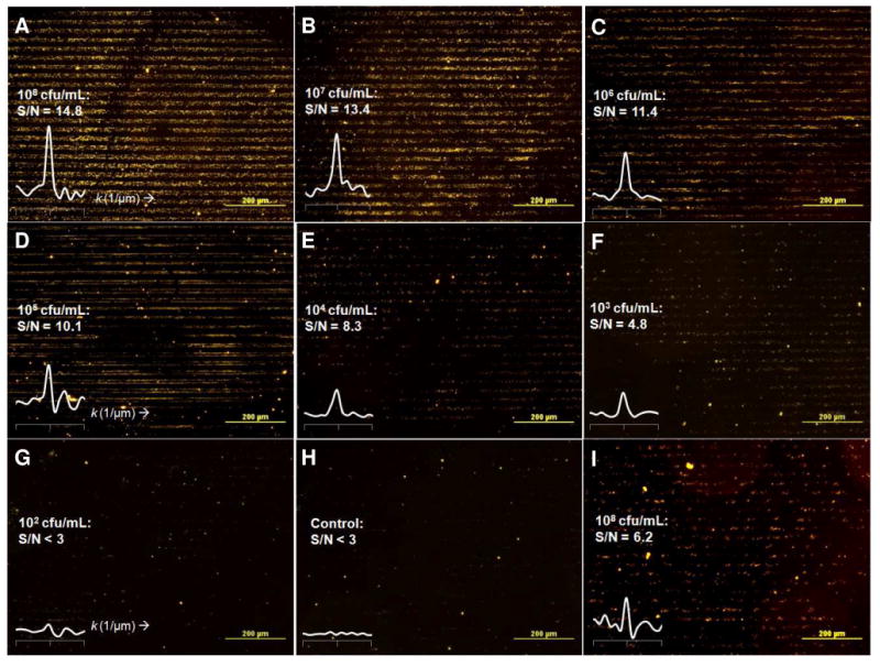
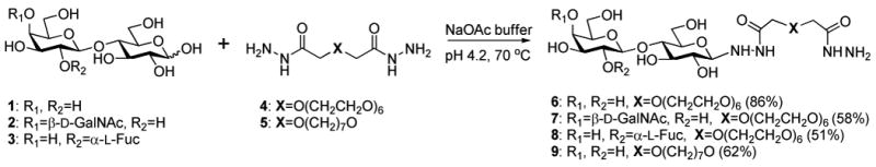
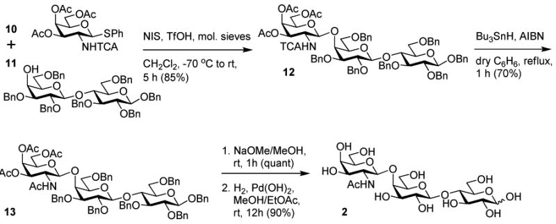
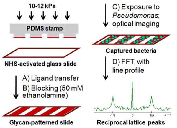
Bishydrazides are versatile linkers for attaching glycans to substrates for lectin binding and pathogen detection schemes. The α,ω-bishydrazides of carboxymethylated hexa(ethylene glycol) (4) can be conjugated at one end to unprotected oligosaccharides, then attached onto carrier proteins, tethered onto activated carboxyl-terminated surfaces, or functionalized with a photoactive cross-linking agent for lithographic patterning. Glycoconjugates of bishydrazide 4 can also be converted into dithiocarbamates (DTCs) by treatment with CS(2) under mild conditions, for attachment onto gold substrates. The immobilized glycans serve as recognition elements for cell-surface lectins and enable the detection and capture of bacterial pathogens such as Pseudomonas aeruginosa by their adsorption onto micropatterned substrates. A detection limit of 10³ cfu/mL is demonstrated, using a recently introduced method based on optical pattern recognition.


Figures

Figure 1
Figure 1
Glycan–bishydrazide conjugates labeled with fluorescent or photoreactive chromophores. (EG)6 = -O(CH2CH2O)6-. 15: R1, R2=H; 16: R1=β-d-GalNAc, R2=H; 17: R1=H, R2=α-l-Fuc.
Figure 2
Figure 2
(A) In situ DTC formation starting from lactose–bishydrazide conjugate 6; (EG)6 = O(CH2CH2O)6. (B) UV absorption spectra of 6–DTC in dilute aqueous solution, taken during in situ DTC formation. No further increases in peak intensities were observed after 60 min.
Figure 3
Figure 3
(A) Affinity and lactose competition assay of peanut lectin binding to microspheres conjugated with lactose–bishydrazide 6 using flow immunocytometry; (B) ELISA and competition assay of peanut lectin binding to immobilized 6–BSA.
Figure 4
Figure 4
Fluorescence immunostaining of peanut lectin bound to lactose–bishydrazide–ANB conjugate 15, photopatterned onto a BSA-coated substrate by UV irradiation (λ=254 nm) through a quartz mask.
Figure 5
Figure 5
Capture of Pseudomonas on BSA-coated substrates with photopatterned glycan–bishydrazide–ANB conjugate, imaged by darkfield microscopy. Bacterial capture mediated by 2′-fucosyllactose conjugate 17 at 108 cfu/mL; grating period a = 20 μm. Additional images in Supporting Information (Figures S2–S4).
Figure 6
Figure 6
Capture of live Pseudomonas on glass substrates patterned with BSA–glycan bishydrazide conjugates using μCP, as imaged by darkfield microscopy. (A–G) Substrates patterned with pulmonary trisaccharide conjugate 7–BSA after a 1-hour exposure to Pseudomonas, at concentrations ranging from 108 to 102 cfu/mL. Pattern contrast correlates with signal-to-noise (S/N) ratio defined by reciprocal lattice peak produced by FFT (k=0.05 μm-1). (H) Substrate patterned with 7–BSA without exposure to bacteria (control). (I) Pseudomonas capture mediated by 2′-fucosyllactose conjugate 8–BSA, in the presence of choline (108 cfu/mL).
Figure 7
Figure 7
(A) Fluorescence immunostaining of peanut lectin bound to lactose conjugate 6–DTC, presented as DAMs on Au substrates by μCP. (B) Stability profile of hydrazide–DTC patterns on roughened Au in PBS containing ME (10 or 100 μM), based on changes in relative luminosity.
Scheme 1
Scheme 1
Synthesis of glycan–bishydrazide conjugates.
Scheme 2
Scheme 2
Synthesis of pulmonary trisaccharide. TCA = trichloroacetyl.
Scheme 3
Scheme 3
(A, B) Glycan-patterned slides by microcontact printing of BSA glycoconjugates onto NHS-activated glass substrates, followed by a blocking step. (C) Patterned capture slides were exposed to Pseudomonas at variable concentrations, then imaged under darkfield condtions. (D) Image processing by FFT analysis produced reciprocal lattice peaks at k=±1/a in spectral format; central peak (k=0) scales with spatially averaged intensity of original image; noise from nonspecific binding is dispersed throughout k-space.

References

    1. Stevens J, Blixt O, Paulson JC, Wilson IA. Glycan microarray technologies: tools to survey host specificity of influenza viruses. Nat Rev Microbiol. 2006;4:857–864. - PMC - PubMed
    1. Wang HF, Gu LR, Lin Y, Lu FS, Meziani MJ, Luo PGJ, Wang W, Cao L, Sun YP. Unique aggregation of anthrax (Bacillus anthracis) spores by sugarcoated single-walled carbon nanotubes. J Am Chem Soc. 2006;128:13364–13365. - PubMed
    1. El-Boubbou K, Gruden C, Huang X. Magnetic glyco-nanoparticles: A unique tool for rapid pathogen detection, decontamination, and strain differentiation. J Am Chem Soc. 2007;129:13392–13393. - PubMed
    1. Kale RR, Mukundan H, Price DN, Harris JF, Lewallen DM, Swanson BI, Schmidt GJ, Iyer SS. Detection of intact influenza viruses using biotinylated biantennary S-sialosides. J Am Chem Soc. 2008;130:8169–8171. - PubMed
    1. Li-Hong Liu LH, Dietsch H, Schurtenberger P, Yan M. Photoinitiated coupling of unmodified monosaccharides to iron oxide nanoparticles for sensing proteins and bacteria. Bioconjugate Chem. 2009;20:1349–1355. - PMC - PubMed

Publication types

MeSH terms