Clinical Trial
. 2010 Oct 7;363(15):1429-37.
doi: 10.1056/NEJMoa1000228.

Dystrophin immunity in Duchenne's muscular dystrophy

Affiliations
Clinical Trial

Dystrophin immunity in Duchenne's muscular dystrophy

Jerry R Mendell et al. N Engl J Med. .

Abstract

We report on delivery of a functional dystrophin transgene to skeletal muscle in six patients with Duchenne's muscular dystrophy. Dystrophin-specific T cells were detected after treatment, providing evidence of transgene expression even when the functional protein was not visualized in skeletal muscle. Circulating dystrophin-specific T cells were unexpectedly detected in two patients before vector treatment. Revertant dystrophin fibers, which expressed functional, truncated dystrophin from the deleted endogenous gene after spontaneous in-frame splicing, contained epitopes targeted by the autoreactive T cells. The potential for T-cell immunity to self and nonself dystrophin epitopes should be considered in designing and monitoring experimental therapies for this disease. (Funded by the Muscular Dystrophy Association and others; ClinicalTrials.gov number, NCT00428935.).


Figures
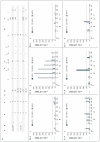
Figure 1
Figure 1. Dystrophin-Specifc Immune Responses in the Six Study Patients
Panel A shows the vector dose (in vector genomes [vg] per kilogram of body weight) received by the patients, their age, exon deletions, and glucocorticoid regimens. Deflazacort, used by Patient 1, was not prescribed, because it is not approved by the Food and Drug Administration for use in the United States. It was obtained by the patient’s family directly from Masters Pharmaceuticals in the United Kingdom. Dystrophin exons detected in revertant fibers with exon-specific antibodies are noted with a plus sign. T-cell responses are shown for Patients 1 through 6 in Panels B through G, respectively. Peripheral-blood mononuclear cells (PBMCs) collected at the indicated times were cultured with three pools of synthetic peptides (MDP1, MDP2, and MDP3) spanning mini-dystrophin domains (see the Supplementary Appendix); interferon-γ production was assessed 36 hours later with the use of an interferon-γ enzyme-linked immunosorbent spot (ELISPOT) assay. Data are presented as interferon-γ spot-forming cells (SFCs) per 1 million PBMCs. The dashed lines represent the threshold for a positive assay response (50 SFCs per 1 million PBMCs).
Figure 2
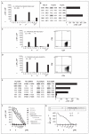
Figure 2. Characterization of Dystrophin-Specific Cellular Immune Responses in Patient 5
CD8+ T cells that were specific to the MDP1 pool of synthetic peptides and were detected after vector treatment, on day 60, were tested for recognition of intersecting mapping subpools (see the Supplementary Appendix). Only MDP1 subpools C and K stimulated interferon-γ production by the CD8+ T cells, localizing the epitope to shared peptide p19 that spanned amino acids (aa) 181 through 200, which were encoded by dystrophin exon 7 (Panel A). CD8+ T cells recognized only semimatched target cells that shared HLA-B*1801 expression with Patient 5 (Panel B). Two other dystrophin-specific T-cell populations expressed the CD4 protein, as assessed by flow cytometry (Panels C and D), and targeted peptide p23 contained in subpools D and H (amino acids 221 through 240, exon 8) (Panel C) or peptide p17 contained in subpools C and I (amino acids 161 through 180, exon 6) (Panel D). The p17 epitope was presented by the HLA-DQA1*0505 and DQB1*0301 class II molecules, as assessed by interferon-γ ELISPOT assay with the use of a panel of HLA-semimatched antigen-presenting cells (Panel E). The p17 epitope was further mapped to amino acids 161 through 180 of exon 6 (WSDGLALNAL) with the use of varying concentrations of the indicated synthetic peptides (Panel F). CD4+ T cells were stimulated with varying concentrations of dystrophin peptide p17 or the indicated β-spectrin peptide in an interferon-γ ELISPOT assay (Panel G). CP denotes control peptide pool, PBMCs peripheral-blood mononuclear cells, and SFCs spot-forming cells.
Figure 3
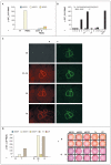
Figure 3. Mini-Dystrophin Cellular Immune Responses in Patient 2
Whole peripheral-blood mononuclear cells (PBMCs) or CD4+ T-cell–depleted PBMCs were assessed on day 60 for interferon-γ spot-forming cells (SFCs) with the indicated dystrophin peptide pools (Panel A). Intersecting MDP2 subpools E and H that stimulated an interferon-γ response contained a single common peptide, peptide p74 (amino acids [aa] 2809 through 2829, exon 57, rod domain 23) (Panel B). Revertant fibers in the muscle-biopsy specimen obtained on day 90 were detected by means of staining with the MANDYS2 antibody, which recognizes the C-terminal of dystrophin (Panel C, images on the right), or with antibodies specific for epitopes encoded by the indicated dystrophin exons (Panel C, images on the left). Interferon-γ T-cell responses were compared 1 week before treatment and on day 30 after gene transfer with the use of the three pools of synthetic peptides (MDP1, MDP2, and MDP3), peptide p74, and the control peptide pool (CP), consisting of enhanced green fluorescent protein (Panel D, left). Images of interferon-γ-producing SFCs before and after vector treatment are shown (Panel D, right). Each dot represents an interferon-γ SFC after stimulation of duplicate PBMC cultures with the indicated dystrophin peptide pools, peptide p74, or control peptides. The experiment was repeated three times with the same outcome. Antibodies used in this experiment were provided by Professor G.E. Morris.

Comment in

References

    1. Emery AE. The muscular dystrophies. Lancet. 2002;359:687–95. - PubMed
    1. Gregorevic P, Allen JM, Minami E, et al. rAAV6-microdystrophin preserves muscle function and extends lifespan in severely dystrophic mice. Nat Med. 2006;12:787–9. - PMC - PubMed
    1. Fabb SA, Wells DJ, Serpente P, Dickson G. Adeno-associated virus vector gene transfer and sarcolemmal expression of a 144 kDa micro-dystrophin effectively restores the dystrophin-associated protein complex and inhibits myofibre degeneration in nude/mdx mice. Hum Mol Genet. 2002;11:733–41. - PubMed
    1. Wang B, Li J, Xiao X. Adeno-associated virus vector carrying human minidystrophin genes effectively ameliorates muscular dystrophy in mdx mouse model. Proc Natl Acad Sci U S A. 2000;97:13714–9. - PMC - PubMed
    1. Mendell JR, Moxley RT, Griggs RC, et al. Randomized, double-blind six-month trial of prednisone in Duchenne’s muscular dystrophy. N Engl J Med. 1989;320:1592–7. - PubMed

Publication types

MeSH terms

Associated data