. 2010 Oct;26(5):407-19.
doi: 10.1089/jop.2010.0099.

Doxycycline hydrogels as a potential therapy for ocular vesicant injury

Affiliations

Doxycycline hydrogels as a potential therapy for ocular vesicant injury

Marion K Gordon et al. J Ocul Pharmacol Ther. 2010 Oct.

Abstract

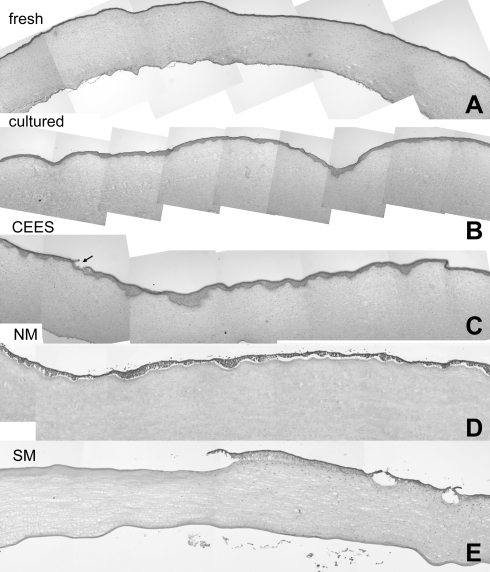
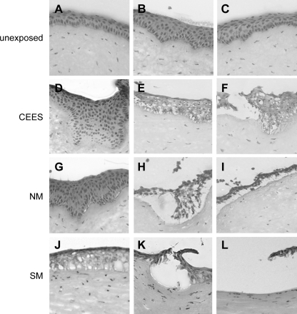
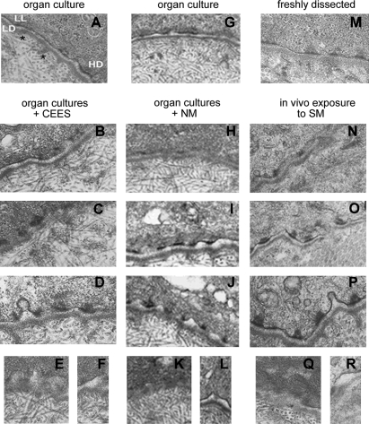
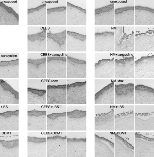
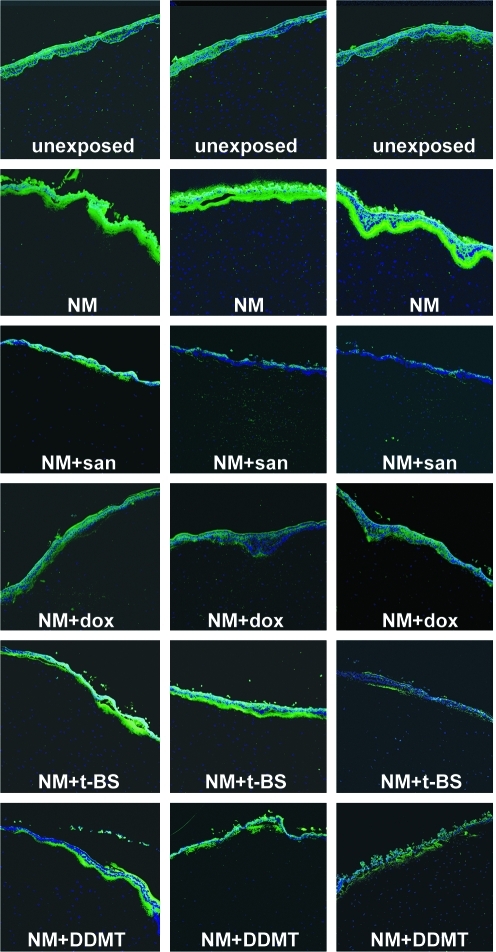
Purpose: The goals of this study were (1) to compare the injury at the basement membrane zone (BMZ) of rabbit corneal organ cultures exposed to half mustard (2 chloroethyl ethyl sulfide, CEES) and nitrogen mustard with that of in vivo rabbit eyes exposed to sulfur mustard (SM); (2) to test the efficacy of 4 tetracycline derivatives in attenuating vesicant-induced BMZ disruption in the 24-h period postexposure; and (3) to use the most effective tetracycline derivative to compare the improvement of injury when the drug is delivered as drops or hydrogels to eyes exposed in vivo to SM.

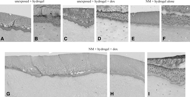
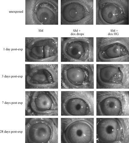
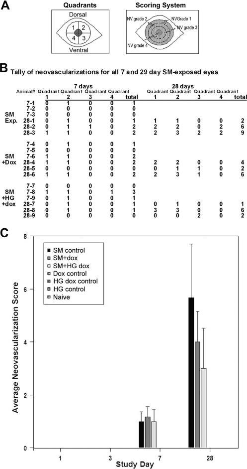
Methods: Histological analysis of hematoxylin and eosin–stained sections was performed; the ultrastructure of the corneal BMZ was evaluated by transmission electron microscopy; matrix metalloproteinase-9 was assessed by immunofluorescence; doxycycline as drops or a hydrogel was applied daily for 28 days to eyes exposed in vivo to SM. Corneal edema was assessed by pachymetry and the extent of neovascularization was graded by length of longest vessel in each quadrant.

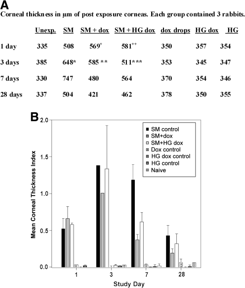
Results: Injury to the BMZ was highly similar with all vesicants, but varied in degree of severity. The effectiveness of the 4 drugs in retaining BMZ integrity did not correlate with their ability to attenuate matrix metalloproteinase-9 expression at the epithelial–stromal border. Doxycycline was most effective on organ cultures; therefore, it was applied as drops or a hydrogel to rabbit corneas exposed in vivo to SM. Eyes were examined at 1, 3, 7, and 28 days after exposure. At 7 and 28 days after SM exposure, eyes treated with doxycycline were greatly improved over those that received no therapy. Corneal thickness decreased somewhat faster using doxycycline drops, whereas the hydrogel formulation decreased the incidence of neovascularization.

Conclusions: Corneal cultures exposed to 2-chloroethyl ethyl sulfide and nitrogen mustard were effective models to simulate in vivo SM exposures. Doxycycline as drops and hydrogels ameliorated vesicant injury. With in vivo exposed animals, the drops reduced edema faster than the hydrogels, but use of the hydrogels significantly reduced neovascularization. The data provide proof of principle that a hydrogel formulation of doxycycline as a daily therapy for ocular vesicant injury should be further investigated.


Figures

FIG. 1.
FIG. 1.
(A) Diagram of an organ culture with delivery to the central cornea. Gray indicates the scleral rim. (B) Photograph of cultures on a black background. The corneas remain transparent in culture as evidenced by the white scleral rim.
FIG. 2.
FIG. 2.
Comparison of composites of hematoxylin and eosin–stained corneas plus and minus vesicants. (A) Sections of a cornea freshly dissected from a rabbit eye; (B) a composite of a corneal culture equilibrated at 37°C overnight and then incubated 24 h at 37°C as an unexposed control; (C) sections from a corneal culture exposed to CEES for 2 h and then incubated 24 h before analysis; (D) a composite of a culture exposed to NM for 2 h and then incubated for 24 h; (E) a composite of sections from a rabbit eye at 24 h after exposure to SM in vivo. CEES, 2-chloroethyl ethyl sulfide; NM, nitrogen mustard; SM, sulfur mustard. The arrow in 2C indicates a focal area of epithelial detachment.
FIG. 3.
FIG. 3.
Variability of vesicant exposures. (A–C) High-magnification images of unexposed cultured corneal sections stained with hematoxylin and eosin; (D–F) corneas at 24 h post-CEES exposure; (G–I) corneas at 24 h after NM exposures; (J–L) corneas at 24 h after an in vivo exposure to SM.
FIG. 4.
FIG. 4.
Vesicants altered BMZ structures. TEM of unexposed (A, G, M) and exposed corneal BMZ. (A, G) Unexposed organ cultures; (M) freshly dissected rabbit cornea. The range of BMZ phenotypes at 24 h after CEES exposure (B–F), NM exposure (H–L), and SM exposure (N–R) are from nearly normal to extremely disrupted. BMZ features include LL, lamina lucida; LD, lamina densa; HD, hemidesmosome (A). Asterisks appear just under 2 bundles of anchoring fibrils. BMZ, basement membrane zone.
FIG. 5.
FIG. 5.
Corneal epithelia are damaged by CEES and NM, and tetracycline derivatives decrease damage. Corneas that were treated for 24 h with tetracycline derivatives after a 2-h vesicant exposure were compared with unexposed and tetracycline derivative-only samples. Top row: sections from unexposed control corneas. Left column: unexposed corneas that received a 24-h treatment with tetracycline derivatives (drug-only controls). Middle column set: CEES exposure, untreated (row 2) or treated for 24 h with tetracycline derivatives (rows 3–6). Right column set: NM exposure, untreated (row 2) or treated for 24 h with tetracycline derivatives (rows 3–6). DDMT, dedimethylamino tetracycline; dox, doxycycline; t-BS, 9-t-butyl sancycline.
FIG. 6.
FIG. 6.
The increase in MMP-9 in the BMZ induced by NM is attenuated by tetracycline derivatives. Three sections illustrating MMP-9 distribution patterns in each treatment group from a typical NM exposure experiment. Top row: unexposed controls had low levels of MMP-9 and background staining in the epithelium; row 2: NM greatly increased MMP-9; rows 3–6: corneas exposed to NM, followed by treatment with a tetracycline derivative for 24 h. Green: MMP-9 antibody staining; blue: DAPI-stained nuclei. MMP, matrix metalloproteinase; san, sancycline.
FIG. 7.
FIG. 7.
Hydrogels designed to release doxycycline over 24 h improved the BMZ of NM-exposed corneal cultures. (A, B) Different magnifications of unexposed corneas, showing normal morphology after hydrogels treatment for 24 h; (C, D) low and high magnifications of corneas treated with hydrogel containing doxycycline; (E, F) low and high magnification of corneas treated with hydrogel for 24 h after a 2-h NM exposure; (G–I) low and high magnifications of corneas exposed to NM, followed by treatment with a hydrogel containing doxycycline, which have an improved BMZ phenotype compared with those that received hydrogel without the drug (E, F).
FIG. 8.
FIG. 8.
Doxycycline in drops or hydrogels facilitate wound healing of rabbit corneas exposed in vivo to SM. Rows: representative unexposed eyes (row 1) and eyes taken at the indicated time after exposure to SM. Left column: unexposed eye compared with eyes exposed to SM, on days 1, 3, 7, and 28 postexposure. Middle column: unexposed eye and SM-exposed eyes that received 3 dropwise treatments of doxycycline over a 24-h period for every day until sacrifice. Right column: unexposed eye and eyes exposed to SM, followed by daily application of a hydrogel containing doxycycline for the number of days indicated.
FIG. 9.
FIG. 9.
Doxycycline ameliorated the increase in corneal thickness (measured by pachymetry) induced by SM exposure. Corneal thickness was read at 5 areas in each cornea (left, right, top, bottom, and center). Values were averaged to obtain the mean thickness for each cornea. (A) Preexposure, the average of the 64 corneas was 353.5 ± 19.3 μm (standard deviation). Mean values of exposed eye corneal thickness at indicated times postexposure without taking into account each cornea's preexposure thickness. When damage is severe, pachymetery cannot be read. Therefore, 5 readings were not possible in the 1 and 3 days postexposure samples. The value for the 1 day postexposure thickness for the SM + dox corneas (indicated by “ + ”) was calculated from 4 readings from 1 animal, 3 readings from the second, and 5 readings from the third; the SM + hydrogel containing dox (HG dox) value (indicated by “ + + ”) was averaged from 5 readings from the first animal, 4 from the second, and 4 from the third. The value for the 3 days postexposure thickness for the SM-exposed corneas (indicated by *) was calculated from 1 reading from the first animal, 0 readings from the second, and 4 readings from the third; the value for the thickness of SM + dox corneas (indicated by **) was calculated from 1 reading from the first animal, 4 readings from the second, and 1 readings from the third; the SM + HG dox value (indicated by ***) was averaged from 1 reading from the first animal, 1 from the second, and 2 from the third. (B) Mean corneal thickness index for each group, shown in histogram form, correcting for the initial thickness of corneas before exposure. The equation for this calculation is “right eye postexposure” minus “right eye preexposure” divided by “right eye preexposure” for individual corneas. Next, the mean of the 3 corneas in the group was determined. This removes bias in the postexposure thickness that might be due to individual variations in the thickness of the preexposed corneas. At day 3 the damage was severe, and thickness due to edema could be reliably determined for only 1 cornea in the SM group and 1 cornea in the SM + dox group, and thus these histograms have no error bars. The 3 days SM + HG dox group had edema measurable in 2 corneas; therefore, an error bar is shown. The P values from statistical analysis (analysis of variance) of all groups were 0.0049 for 1 day, 0.1029 for 3 days, <0.0001 for 7 days, and 0.1053 for 28 days. The 3-day value includes all corneas in the group, that is, those that were of larger than preexposure thickness from edema, and those thinner from other SM damage. With a Tukey analysis, there was a significant difference between SM-challenged groups at 7 days: the corneal thickness index in the SM group was significantly greater than that for the SM + dox group (difference = 0.81, P value = 0.0015) and the SM + HG dox group (difference = 0.57, P value = 0.0156). A P value of <0.05 was considered significant.
FIG. 10.
FIG. 10.
Neovascularization (NV) of corneas after SM with and without doxycycline. Neovascularizations were not seen in unexposed controls or in any SM-exposed corneas at days 1 and 3, but were present at days 7 and 28. (A) Quadrants and scoring criteria diagram. For scoring, 0 indicates no new vascularization present; 1 indicates the longest vessel's length is up to ∼25% of the radius of the cornea; 2, the longest vessel's length is up to ∼26%–50% of the radius of the cornea; 3, the longest vessel's length is up to ∼51%–75% of the radius of the cornea; and 4, the longest vessel's length is greater than ∼75% of the radius of the cornea. (B) Tally of days 7 and 28 neovascularizations for each SM-exposed eye. Note that there were 6 rabbits assessed at 7 days, 3 that were to be sacrificed at 7 days, and 3 that were to be sacrificed at 28 days. (C) Histogram depiction of the increase in neovascularization with time, showing that SM-exposed animals had the greatest extent of angiogenesis (black bars). Applying doxycycline drops 3 times/day for 28 days (dark gray bars) attenuated neovascularization by ∼30%, and applying hydrogels with doxycycline (light gray bars) daily for 28 days attenuated it by ∼48%. Statistics: Kruskal–Wallis analysis was performed on total neovascularization grade scores for each day. P values for preexposure corneas, as well as 1 and 3 days postexposure corneas were 1.0000 because of all values being the same for all animals at these time points. The 7 days P value was 0.0328 and for 28 days it was 0.0465. A P value of <0.05 was considered significant.

References

    1. Smith W.J. Dunn M.A. Medical defense against blistering chemical warfare agents. Arch. Dermatol. 1991;127:1207–1213. - PubMed
    1. Papirmeister B. Feister A. Robinson S., et al. Medical Defense Against Mustard Gas: Toxic Mechanisms and Pharmacological Implications. Boca Raton, FL: CRC Press; 1991.
    1. Wattana M. Bey T. Mustard gas or sulfur mustard: an old chemical agent as a new terrorist threat. Prehosp. Disaster Med. 2009;24:19–29. discussion 30–31. - PubMed
    1. Balali-Mood M. Hefazi M. The pharmacology, toxicology, and medical treatment of sulphur mustard poisoning. Fundam. Clin. Pharmacol. 2005;19:297–315. - PubMed
    1. Balali-Mood M. Hefazi M. Comparison of early and late toxic effects of sulfur mustard in Iranian veterans. Basic Clin. Pharmacol. Toxicol. 2006;99:273–282. - PubMed

Publication types

MeSH terms